. 2023 May 25;16(6):788.
doi: 10.3390/ph16060788.

Innate Vascular Failure by Application of Neuroleptics, Amphetamine, and Domperidone Rapidly Induced Severe Occlusion/Occlusion-like Syndromes in Rats and Stable Gastric Pentadecapeptide BPC 157 as Therapy

Affiliations

Innate Vascular Failure by Application of Neuroleptics, Amphetamine, and Domperidone Rapidly Induced Severe Occlusion/Occlusion-like Syndromes in Rats and Stable Gastric Pentadecapeptide BPC 157 as Therapy

Sanja Strbe et al. Pharmaceuticals (Basel). .

Abstract

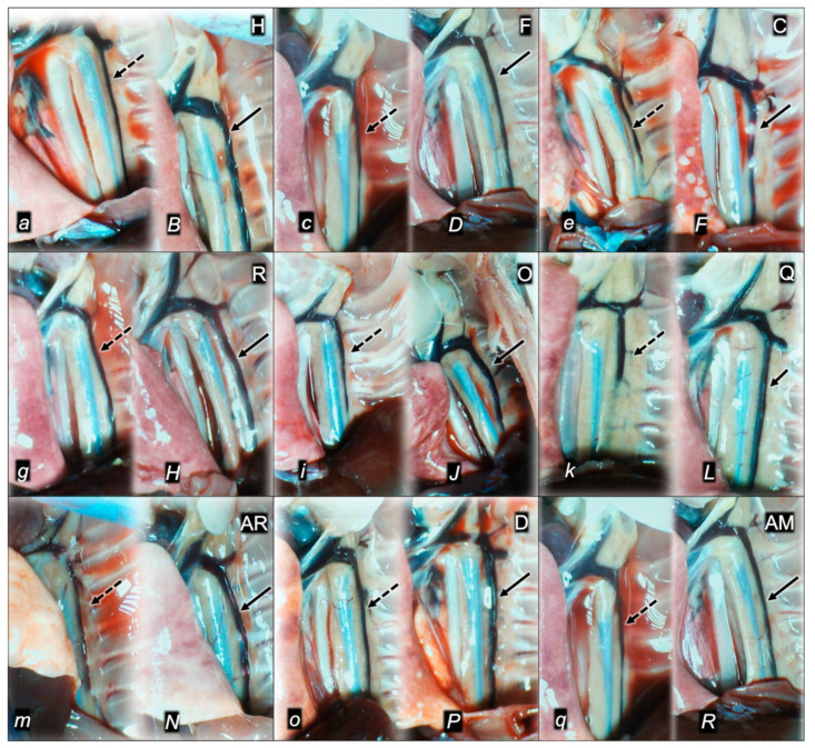
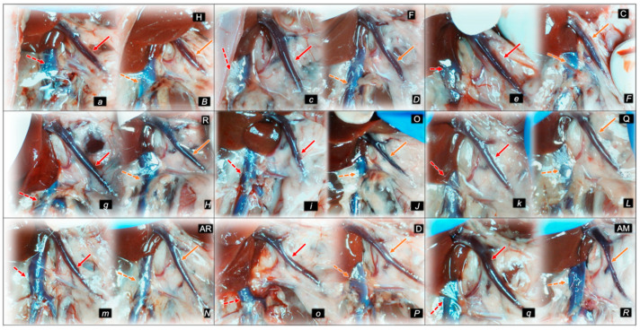
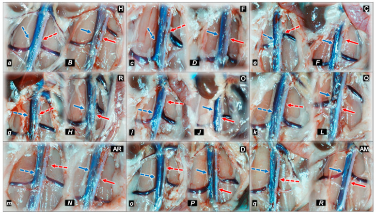
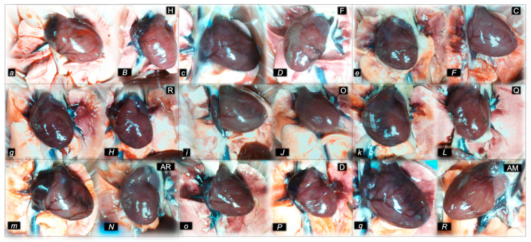
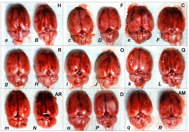
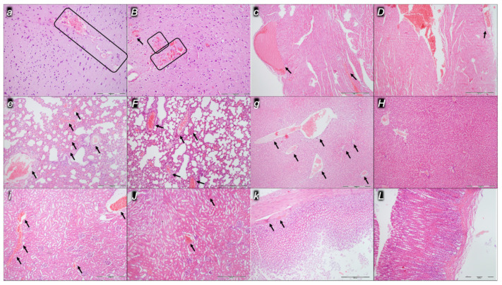
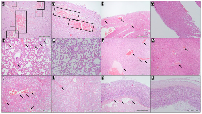
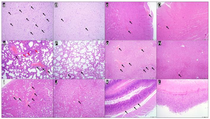
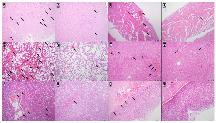
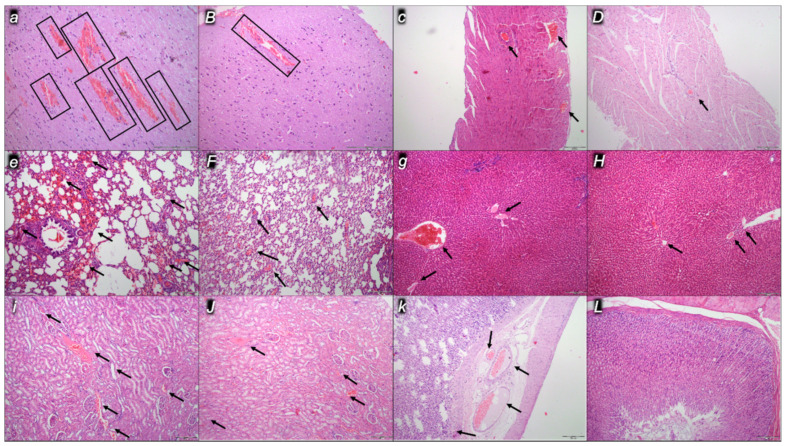
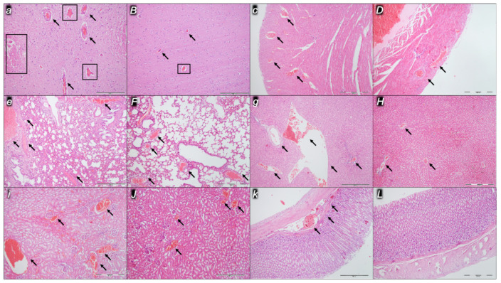
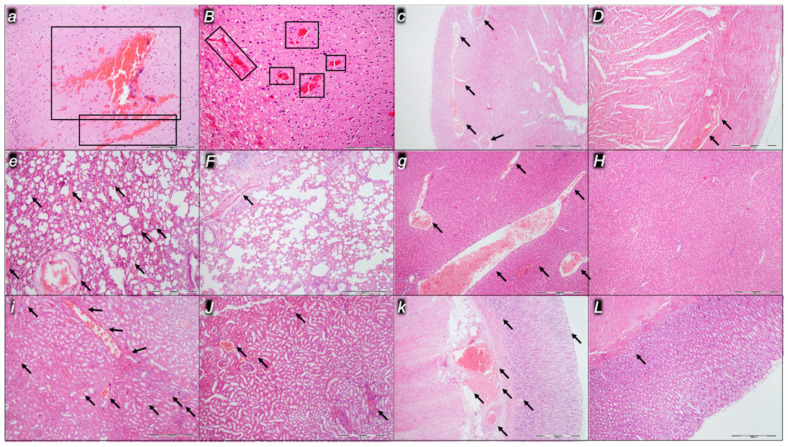
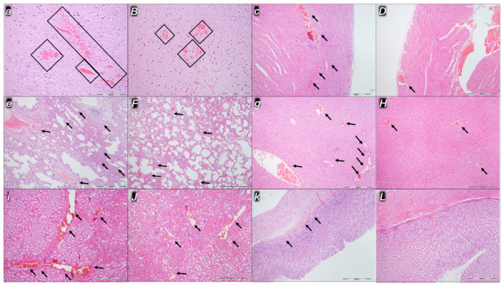
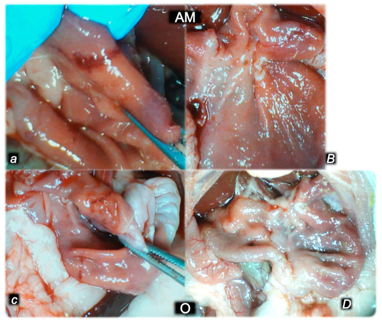
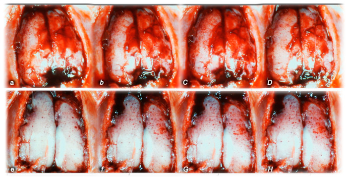
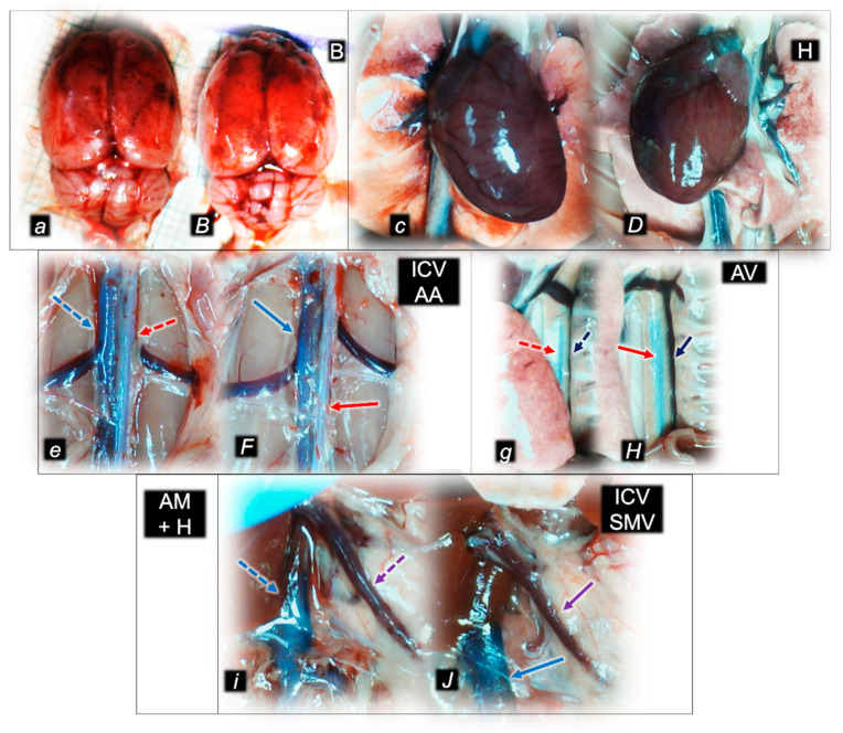
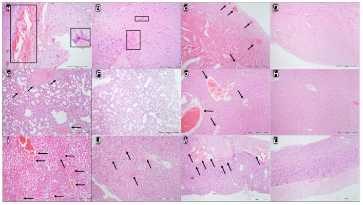
Even before behavioral disturbances, neuroleptics, amphetamine, and domperidone application rapidly emerged severe occlusion/occlusion-like syndrome, shared innate vascular and multiorgan failure in rats, comparable to occlusion/occlusion-like syndrome described with vessel(s) occlusion or similar noxious procedures application. As therapy, i.e., activation of the collateral pathways, "bypassing key" (activated azygos vein pathway, direct blood flow delivery), the stable gastric pentadecapeptide BPC 157 is a novel solution. Recently, BPC 157 therapy particularly counteracted neuroleptic- or L-NAME-induced catalepsy, lithium intoxication, and schizophrenia positive and negative symptoms (amphetamine/methamphetamine/apomorphine/ketamine). In rats with complete calvariectomy, medication (BPC 157 10 µg/kg, 10 ng/kg ip or ig) was given 5 min after distinctive dopamine agents (mg/kg ip) (haloperidol (5), fluphenazine (5), clozapine (10), risperidone (5), olanzapine (10), quetiapine (10), or aripiprazole (10), domperidone (25), amphetamine (10), and combined amphetamine and haloperidol) and assessed at 15 min thereafter. All neuroleptic-, domperidone-, and amphetamine-induced comparable vascular and multiorgan failure severe syndrome was alleviated with BPC 157 therapy as before major vessel(s) occlusion or other similar noxious procedures. Specifically, all severe lesions in the brain (i.e., immediate swelling, hemorrhage), heart (i.e., congestion, arrhythmias), and lung (i.e., congestion, hemorrhage), as well as congestion in the liver, kidney, and gastrointestinal (stomach) tract, were resolved. Intracranial (superior sagittal sinus), portal, and caval hypertension and aortal hypotension were attenuated or eliminated. BPC 157 therapy almost annihilated arterial and venous thrombosis, peripherally and centrally. Thus, rapidly acting Virchow triad circumstances that occur as dopamine central/peripheral antagonists and agonist essential class-points, fully reversed by BPC 157 therapy, might be overwhelming for both neuroleptics and amphetamine.

Keywords: amphetamine; domperidone; neuroleptics; occlusion/occlusion-like syndromes; pentadecapeptide BPC 157; rats; vascular failure.


Conflict of interest statement

The authors declare no conflict of interest.

Figures

Figure 1
Figure 1
Illustrative presentation of azygos vein in control rats (italic small letters, dashed arrows) and in BPC 157 treated rats (italic capital letters, full arrows) at 15 min following application of haloperidol (H) (a, B), fluphenazine (F) (c, D), clozapine (C) (e, F), risperidone (R) (g, H), olanzapine (O) (i, J), quetiapine (Q) (k, L), aripiprazole (AR) (m, N), domperidone (D) (o, P), and amphetamine (AM) (q, R).
Figure 2
Figure 2
Illustrative presentation of the control rats (italic small letters) and of the BPC 157-treated rats (italic capital letters) of the superior mesenteric vein (full arrows, red (controls), yellow (BPC 157)) and inferior caval vein (dashed arrows, red (controls), yellow (BPC 157)) at 15 min following application of haloperidol (H) (a, B), fluphenazine (F) (c, D), clozapine (C) (e, F), risperidone (R) (g, H), olanzapine (O) (i, J), quetiapine (Q) (k, L), aripiprazole (AR) (m, N), domperidone (D) (o, P), and amphetamine (AM) (q, R).
Figure 3
Figure 3
Illustrative presentation of inferior caval vein (blue arrows) and abdominal aorta (red arrows) in the control rats (italic small letters, dashed arrows) and in the BPC-157-treated rats (italic capital letters, full arrows) at 15 min following application of haloperidol (H) (a, B), fluphenazine (F) (c, D), clozapine (C) (e, F), risperidone (R) (g, H), olanzapine (O) (i, J), quetiapine (Q) (k, L), aripiprazole (AR) (m, N), domperidone (D) (o, P), and amphetamine (AM) (q, R).
Figure 4
Figure 4
Illustrative presentation of the heart in the control rats (italic small letters) and in the BPC 157 treated rats (italic capital letters) at 15 min following application of haloperidol (H) (a, B), fluphenazine (F) (c, D), clozapine (C) (e, F), risperidone (R) (g, H), olanzapine (O) (i, J), quetiapine (Q) (k, L), aripiprazole (AR) (m, N), domperidone (D) (o, P), and amphetamine (AM) (q, R).
Figure 5
Figure 5
Illustrative presentation of the brain in the control rats (italic small letters) and in the BPC 157 treated rats (italic capital letters) at 15 min following application of haloperidol (H) (a, B), fluphenazine (F) (c, D), clozapine (C) (e, F), risperidone (R) (g, H), olanzapine (O) (i, J), quetiapine (Q) (k, L), aripiprazole (AR) (m, N), domperidone (D) (o, P), and amphetamine (AM) (q, R).
Figure 6
Figure 6
Haloperidol-rats. Illustrative microscopic presentation of the brain (a, B), heart (c, D), lung (e, F), liver (g, H), kidney (i, J), and stomach (k, L) in the saline-treated rats (control, small italic letters) and BPC 157-treated rats (capital italic letters) at the end of the experiments at the 15 min. Brain (a, B). Control (a). A pronounced edema and congestion were visible affecting the cerebrum, and more prominent intracerebral cortical hemorrhage involving larger areas of cerebral brain tissue affecting the neocortex (rectangle area). BPC 157 (B). Only mild edema and congestion were found, with small, focal, and superficial areas of neocortical hemorrhage (rectangle areas and black arrow). Heart (c, D). Control (c). Moderated myocardial congestion (black arrows). BPC 157 (D). Only mild myocardial congestion (black arrow). Lung (e, F). Control (e). Moderate congestion of the lung parenchyma (black arrows). BPC 157 (F). Only mild lung congestion (black arrows). Liver (g, H). Control (g). A moderate dilatation and congestion of blood vessels in the portal tracts, central veins, and sinusoids (black arrows). BPC 157 (H). No changes were found. Kidney (i, J). Control (i). A moderate dilatation of blood vessels and congestion in the kidney tissue, as well as glomeruli, was found (black arrows). BPC 157 (J). Mild dilatation of blood vessels and congestion in the kidney tissue, as well as glomeruli (black arrows). Gastrointestinal lesion (k, L). Control (k). Moderate congestion of the stomach wall (black arrows). BPC 157 (L). No changes were found in the stomach wall. HE; magnification 100× (c, D, e, F, g, H, i, J, k, L), magnification 200× (a, B).
Figure 7
Figure 7
Fluphenazine-rats. Illustrative microscopic presentation of the brain (a, B), heart (c, D), lung (e, F), liver (g, H), kidney (i, J), and stomach (k, L) in the saline-treated rats (control, small italic letters) and BPC 157-treated rats (capital italic letters) at the end of the experiments at the 15 min. Brain (a, B). Control (a). A pronounced edema and congestion were visible affecting the cerebrum, and more prominent intracerebral cortical hemorrhage involving larger areas of cerebral brain tissue affecting the neocortex (rectangle areas). BPC 157 (B). Only mild edema and congestion were found, with small, focal, and superficial areas of neocortical hemorrhage (rectangle areas). Heart (c, D). Control (c). Marked myocardial congestion (black arrows). BPC 157 (D). No changes were found. Lung (e, F). Control (e). Moderate congestion of the lung parenchyma (black arrows). BPC 157 (F). No changes were found. Liver (g, H). Control (g). Marked dilatation and congestion of blood vessels in the portal tracts, central veins, and sinusoids (black arrows). BPC 157 (H). Only a mild dilatation and congestion of blood vessels in the portal tracts (black arrows). Kidney (i, J). Control (i). A moderate dilatation of blood vessels and congestion in the kidney tissue, as well as glomeruli, was found (black arrows). BPC 157 (J). Mild dilatation of blood vessels and congestion in the kidney tissue, as well as glomeruli (black arrows). Gastrointestinal lesion (k, L). Control (k). Mild congestion of the stomach wall(black arrows). BPC 157 (L). No changes were found in the stomach wall. HE; magnification 100× (c, D, e, F, g, H, i, J, k, L), magnification 200× (a, B).
Figure 8
Figure 8
Clozapine-rats. Illustrative microscopic presentation of the brain (a, B), heart (c, D), lung (e, F), liver (g, H), kidney (i, J), and stomach (k, L) in the saline-treated rats (control, small italic letters) and BPC 157-treated rats (capital italic letters) at the end of the experiments at the 15 min. Brain (a, B). Control (a). A pronounced edema (black arrows) and congestion were visible in the brain tissue, increased number of affected karyopyknotic cells in the cerebral cortex. BPC 157 (B). These were attenuated in BPC 157 treated rats (black arrows). Heart (c, D). Control (c). Moderate myocardial congestion (black arrows). BPC 157 (D). Mild myocardial congestion was found (black arrow). Lung (e, F). Control (e). Marked congestion (black arrows) of the lung parenchyma with intra-alveolar hemorrhage (red arrows). BPC 157 (F). Only mild lung congestion was found (black arrows). Liver (g, H). Control (g). A marked dilatation and congestion of blood vessels in the portal tracts, central veins, and sinusoids (black arrows). BPC 157 (H). Only a mild dilatation and congestion of blood vessels in the portal tracts (black arrow). Kidney (i, J). Control (i). A moderate dilatation of blood vessels and congestion in the kidney tissue, as well as glomeruli, was found (black arrows). BPC 157 (J). Mild dilatation of blood vessels and congestion in the kidney tissue, as well as glomeruli (black arrow). Gastrointestinal lesion (k, L). Control (k). Mild congestion of the stomach wall (black arrows). BPC 157 (L). No changes were found in the stomach wall. HE; magnification 100× (c, D, e, F, g, H, i, J, k, L), magnification 200× (a, B).
Figure 9
Figure 9
Risperidone-rats. Illustrative microscopic presentation of the brain (a, B), heart (c, D), lung (e, F), liver (g, H), kidney (i, J), and stomach (k, L) in the saline-treated rats (control, small italic letters) and BPC 157-treated rats (capital italic letters) at the end of the experiments at the 15 min. Brain (a, B). Control (a). A pronounced edema and congestion were visible in the brain tissue, increased number of karyopyknotic cells affecting the cerebral cortex (black arrows). BPC 157 (B). These were attenuated in BPC 157 treated rats (black arrows). Heart (c, D). Control (c). Moderate myocardial congestion (black arrows). BPC 157 (D). Mild myocardial congestion (black arrow). Lung (e, F). Control (e). A marked congestion of the lung parenchyma with intra-alveolar hemorrhage (black arrows). BPC 157 (F). Only mild lung congestion was found (black arrow). Liver (g, H). Control (g). A marked dilatation and congestion of blood vessels in the portal tracts, central veins, and sinusoids (black arrows). BPC 157 (H). Only a mild dilatation and congestion of blood vessels in the portal tracts (black arrows). Kidney (i, J). Control (i). A moderate dilatation of blood vessels and congestion in the kidney tissue, as well as glomeruli, was found (black arrows). BPC 157 (J). Mild dilatation of blood vessels and congestion in the kidney tissue, as well as glomeruli (black arrow). Gastrointestinal lesion (k, L). Control (k). Moderate congestion of the stomach wall (black arrows). BPC 157 (L). No changes were found in the stomach wall. HE; magnification 100× (c, D, e, F, g, H, i, J, k, L), magnification 200× (a, B).
Figure 10
Figure 10
Olanzapine-rats. Illustrative microscopic presentation of the brain (a, B), heart (c, D), lung (e, F), liver (g, H), kidney (i, J), and stomach (k, L) in the saline-treated rats (control, small italic letters) and BPC 157-treated rats (capital italic letters) at the end of the experiments at the 15 min. Brain (a, B). Control (a). A pronounced edema and congestion were visible in the brain tissue, an increased number of karyopyknotic cells affecting the cerebral cortex, and a prominent intracerebral cortical hemorrhage involving larger areas of brain tissue with transmural involvement of the neocortex (rectangular areas). BPC 157 (B). These were attenuated in BPC 157 treated rats. Only the superficial half of neocortical layers are affected by hemorrhage (rectangular area). Heart (c, D). Control (c). Moderate myocardial congestion (black arrows). BPC 157 (D). Mild myocardial congestion was found (black arrow). Lung (e, F). Control (e). Moderate congestion of the lung parenchyma (black arrows). BPC 157 (F). Only mild lung congestion was found (black arrows). Liver (g, H). Control (g). Marked dilatation and congestion of blood vessels in the portal tracts, central veins, and sinusoids (black arrows). BPC 157 (H). Only a mild dilatation and congestion of blood vessels in the portal tracts (black arrows). Kidney (i, J). Control (i). Moderate dilatation of blood vessels and congestion in the kidney tissue, as well as glomeruli, was found (black arrows). BPC 157 (J). Mild dilatation of blood vessels and congestion in the kidney tissue, as well as glomeruli (black arrows). Gastrointestinal lesion (k, L). Control (k). Moderate congestion of the stomach wall (black arrows). BPC 157 (L). No changes were found in the stomach wall. HE; magnification 100× (c, D, e, F, g, H, i, J, k, L), magnification 200× (a, B).
Figure 11
Figure 11
Quetiapine-rats. Illustrative microscopic presentation of the brain (a, B), heart (c, D), lung (e, F), liver (g, H), kidney (i, J), and stomach (k, L) in the saline-treated rats (control, small italic letters) and BPC 157-treated rats (capital italic letters) at the end of the experiments at the 15 min. Brain (a, B). Control (a). A pronounced edema and congestion were visible affecting the cerebrum (black arrows), and more prominent intracerebral cortical hemorrhage involving larger areas of cerebral brain tissue affecting the transmural neocortex (rectangular areas). BPC 157 (B). Only mild edema and congestion were found (black arrows), with small, focal, and superficial areas of neocortical hemorrhage (rectangular area). Heart (c, D). Control (c). Marked myocardial congestion (black arrows). BPC 157 (D). Moderate myocardial congestion (black arrows). Lung (e, F). Control (e). Moderate congestion of the lung parenchyma (black arrows). BPC 157 (F). Only mild lung congestion (black arrows). Liver (g, H). Control (g). A marked dilatation and congestion of blood vessels in the portal tracts, central veins, and sinusoids (black arrows). BPC 157 (H). Only a mild dilatation and congestion of blood vessels in the portal tracts (black arrows). Kidney (i, J). Control (i). A marked dilatation of blood vessels and congestion in the kidney tissue, as well as glomeruli, was found (black arrows). BPC 157 (J). Mild dilatation of blood vessels and congestion in the kidney tissue, as well as glomeruli (black arrows). Gastrointestinal lesion (k, L). Control (k). Moderate congestion of the stomach wall (black arrows). BPC 157 (L). No changes were found in the stomach wall. HE; magnification 100× (c, D, e, F, g, H, i, J, k, L), magnification 200× (a, B).
Figure 12
Figure 12
Aripiprazole-rats. Illustrative microscopic presentation of the brain (a, B), heart (c, D), lung (e, F), liver (g, H), kidney (i, J), and stomach (k, L) in the saline-treated rats (control, small italic letters) and BPC 157-treated rats (capital italic letters) at the end of the experiments at the 15 min. Brain (a, B). Control (a). A pronounced edema and congestion were visible affecting the cerebrum, and more prominent intracerebral cortical hemorrhage involving larger areas of cerebral brain tissue affecting the neocortex (rectangular areas). BPC 157 (B). Only mild edema and congestion were found, with small, focal, and superficial areas of neocortical hemorrhage (rectangular areas). Heart (c, D). Control (c). Moderated myocardial congestion (black arrows). BPC 157 (D). Only mild myocardial congestion (black arrows). Lung (e, F). Control (e). Moderate congestion of the lung parenchyma with intra-alveolar hemorrhage (black arrows). BPC 157 (F). Only mild lung congestion (black arrow). Liver (g, H). Control (g). A marked dilatation and congestion of blood vessels in the portal tracts, central veins, and sinusoids (black arrows). BPC 157 (H). No changes were found. Kidney (i, J). Control (i). A marked dilatation of blood vessels and congestion in the kidney tissue, as well as glomeruli, was found (black arrows). BPC 157 (J). Mild dilatation of blood vessels and congestion in the kidney tissue, as well as glomeruli (black arrows). Gastrointestinal lesion (k, L). Control (k). Moderate congestion of the stomach wall (black arrows). BPC 157 (L). No changes were found in the stomach wall. HE; magnification 100× (c, D, e, F, g, H, i, J, k, L), magnification 200× (a, B).
Figure 13
Figure 13
Domperidone-rats. Illustrative microscopic presentation of the brain (a, B), heart (c, D), lung (e, F), liver (g, H), kidney (i, J), and stomach (k, L) in the saline-treated rats (control, small italic letters) and BPC 157-treated rats (capital italic letters) at the end of the experiments at the 15 min. Brain (a, B). Control (a). A pronounced edema and congestion were visible affecting the cerebrum, and more prominent intracerebral cortical hemorrhage involving larger areas of cerebral brain tissue affecting the neocortex (rectangular areas). BPC 157 (B). Only mild edema and congestion were found, with small, focal, and superficial areas of neocortical hemorrhage (rectangular areas). Heart (c, D). Control (c). Marked myocardial congestion (black arrows). BPC 157 (D). Only mild myocardial congestion (black arrow). Lung (e, F). Control (e). Moderate congestion of the lung parenchyma (black arrows). BPC 157 (F). Only mild lung congestion (black arrows). Liver (g, H). Control (g). A moderate dilatation and congestion of blood vessels in the portal tracts, central veins, and sinusoids (black arrows). BPC 157 (H). Mild congestion was found (black arrows). Kidney (i, J). Control (i). Marked dilatation of blood vessels and congestion in the kidney tissue, as well as glomeruli, was found (black arrows). BPC 157 (J). Mild dilatation of blood vessels and congestion in the kidney tissue, as well as glomeruli (black arrows). Gastrointestinal lesion (k, L). Control (k). Moderate congestion of the stomach wall (black arrows). BPC 157 (L). No changes were found in the stomach wall. HE; magnification 100× (c, D, e, F, g, H, i, J, k, L), magnification 200× (a, B).
Figure 14
Figure 14
Amphetamine-rats. Illustrative microscopic presentation of the brain (a, B), heart (c, D), lung (e, F), liver (g, H), kidney (i, J), and stomach (k, L) in the saline-treated rats (control, small italic letters) and BPC 157-treated rats (capital italic letters) at the end of the experiments at the 15 min. Brain (a, B). Control (a). A pronounced edema and congestion were visible affecting the cerebrum, and more prominent intracerebral cortical hemorrhage involving larger areas of cerebral brain tissue affecting the neocortex (rectangular areas). BPC 157 (B). Only mild edema and congestion were found, with small, focal, and superficial areas of neocortical hemorrhage (rectangular areas). Heart (c, D). Control (c). Moderated myocardial congestion (black arrows). BPC 157 (D). No changes were found. Lung (e, F). Control (e). A marked congestion of the lung parenchyma with intra-alveolar hemorrhage (black arrows). BPC 157 (F). Only mild lung congestion (black arrow). Liver (g, H). Control (g). A moderate dilatation and congestion of blood vessels in the portal tracts, central veins, and sinusoids (black arrows). BPC 157 (H). No changes were found. Kidney (i, J). Control (i). A marked dilatation of blood vessels and congestion in the kidney tissue, as well as glomeruli, was found (black arrows). BPC 157 (J). Mild dilatation of blood vessels and congestion in the kidney tissue, as well as glomeruli (black arrows). Gastrointestinal lesion (k, L). Control (k). Mild congestion of the stomach wall (black arrows). BPC 157 (L). No changes were found in the stomach wall. HE; magnification 100× (c, D, e, F, g, H, i, J, k, L), magnification 200× (a, B).
Figure 15
Figure 15
Illustrative presentation of stomach lesions in the control rats (italic small letters) and lack of stomach lesions in the BPC 157-treated rats (italic capital letters) at 15 min following application of amphetamine (AM) (a,B) or olanzapine (O) (c,D).
Figure 16
Figure 16
Brain presentation before the challenge, after the challenge (haloperidol or amphetamine) application, and after the therapy application. Normal brain presentation (normal small letters) and brain swelling (italic small letters) upon noxious agent (haloperidol (upper) or amphetamine (lower)) application (italic small letters) and counteracted brain swelling after BPC 157 application (italic capital letters). Upper. Brain presentation in the normal healthy rat (a), brain swelling presentation immediately upon haloperidol application (b), decreased brain swelling immediately upon BPC 157 administration (C), and decreased brain swelling in BPC 157 treated rat immediately before sacrifice (D). Lower. Brain presentation in the normal healthy rat (e), brain swelling presentation immediately upon amphetamine application (f), decreased brain swelling immediately upon BPC 157 administration (G), and decreased brain swelling in BPC 157 treated rat immediately before sacrifice (H). A similar presentation occurred also with other dopamine agents’ applications and BPC 157 therapy.
Figure 17
Figure 17
Illustrative presentation of the unusual parallel activity of the haloperidol and amphetamine in the amphetamine and haloperidol combined challenged rats (AM+H), controls (italic small letters, dashed arrows) and BPC 157 treated (italic capital letters, full arrows) in the brain (B) (a, B), heart (H) (c, D), inferior caval vein (blue arrows) and abdominal aorta (red arrows) (ICVAA) (e, F), azygos vein (blue arrows) (AV) (g, H), and superior mesenteric vein (violet arrows) and inferior caval vein (blue arrows) (ICVSMV) (i, J). These changes occurred probably beyond the dopamine system since given together haloperidol and amphetamine did not antagonize each other effect, and the complete syndrome remained. However, it was consistently antagonized by BPC 157 application.
Figure 18
Figure 18
Amphetamine and haloperidol-rats (amphetamine+haloperidol). Illustrative microscopic presentation of the brain (a, B), heart (c, D), lung (e, F), liver (g, H), kidney (i, J), and stomach (k, L) in the saline-treated rats (control, italic small letters) and BPC 157-treated rats (italic capital letters) at the end of the experiments at the 15 min. Brain (a, B). Control (a). A pronounced edema and congestion were visible affecting the cerebrum, and more prominent intracerebral cortical hemorrhage involving larger areas of cerebral brain tissue affecting the periventricular area (rectangular areas). BPC 157 (B). Only mild edema and congestion were found, with small, focal, and superficial areas of periventricular hemorrhage (rectangular areas). Heart (c, D). Control (c). Marked myocardial congestion (black arrows). BPC 157 (D). No changes were found. Lung (e, F). Control (e). A marked congestion of the lung parenchyma with intra-alveolar hemorrhage (black arrows). BPC 157 (F). No changes were found. Liver (g, H). Control (g). A marked dilatation and congestion of blood vessels in the portal tracts, central veins, and sinusoids (black arrows). BPC 157 (H). No changes were found. Kidney (i, J). Control (i). A moderate dilatation of blood vessels and congestion in the kidney tissue, as well as glomeruli, was found (black arrows). BPC 157 (J). Mild dilatation of blood vessels and congestion in the kidney tissue, as well as glomeruli (black arrows). Gastrointestinal lesion (k, L). Control (k). A marked congestion of the stomach wall (black arrows). BPC 157 (L). No changes were found in the stomach wall. HE; magnification 100× (c, D, e, F, g, H, i, J, k, L), magnification 200× (a, B).

Similar articles

Cited by

References

    1. Sikiric P., Gojkovic S., Krezic I., Smoday I.M., Kalogjera L., Zizek H., Oroz K., Vranes H., Vukovic V., Labidi M., et al. Stable gastric pentadecapeptide BPC 157 may recover brain–gut axis and gut–brain axis function. Pharmaceuticals. 2023;16:676. doi: 10.3390/ph16050676. - DOI - PMC - PubMed
    1. Sikiric P., Gojkovic S., Knezevic M., Tepes M., Strbe S., Vukojevic J., Duzel A., Kralj T., Krezic I., Zizek H., et al. Stable gastric pentadecapeptide BPC 157: Prompt particular activation of the collateral pathways. Curr. Med. Chem. 2023;30:1568–1573. doi: 10.2174/0929867329666221005111553. - DOI - PubMed
    1. Sikiric P., Skrtic A., Gojkovic S., Krezic I., Zizek H., Lovric E., Sikiric S., Knezevic M., Strbe S., Milavic M., et al. Gastric pentadecapeptide BPC 157 in cytoprotection to resolve major vessel occlusion disturbances, ischemia-reperfusion injury following Pringle maneuver, and Budd-Chiari syndrome. World J. Gastroenterol. 2022;28:23–46. doi: 10.3748/wjg.v28.i1.23. - DOI - PMC - PubMed
    1. Sikiric P., Udovicic M., Barisic I., Balenovic D., Zivanovic Posilovic G., Strinic D., Uzun S., Sikiric S., Krezic I., Zizek H., et al. Stable gastric pentadecapeptide BPC 157 as useful cytoprotective peptide therapy in the hearth disturbances, myocardial infarction, heart failure, pulmonary hypertension, arrhythmias, and thrombosis presentation. Biomedicines. 2022;10:2696. doi: 10.3390/biomedicines10112696. - DOI - PMC - PubMed
    1. Staresinic M., Japjec M., Vranes H., Prtoric A., Zizek H., Krezic I., Gojkovic S., Smoday I.M., Oroz K., Staresinic E., et al. Stable gastric pentadecapeptide BPC 157 and striated, smooth, and heart muscle. Biomedicines. 2022;10:3221. doi: 10.3390/biomedicines10123221. - DOI - PMC - PubMed

Grants and funding