. 2023 Jun 2;16(6):833.
doi: 10.3390/ph16060833.

Carbon Dots-Biomembrane Interactions and Their Implications for Cellular Drug Delivery

Affiliations

Carbon Dots-Biomembrane Interactions and Their Implications for Cellular Drug Delivery

Barbara Mavroidi et al. Pharmaceuticals (Basel). .

Abstract

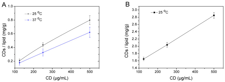
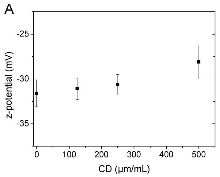
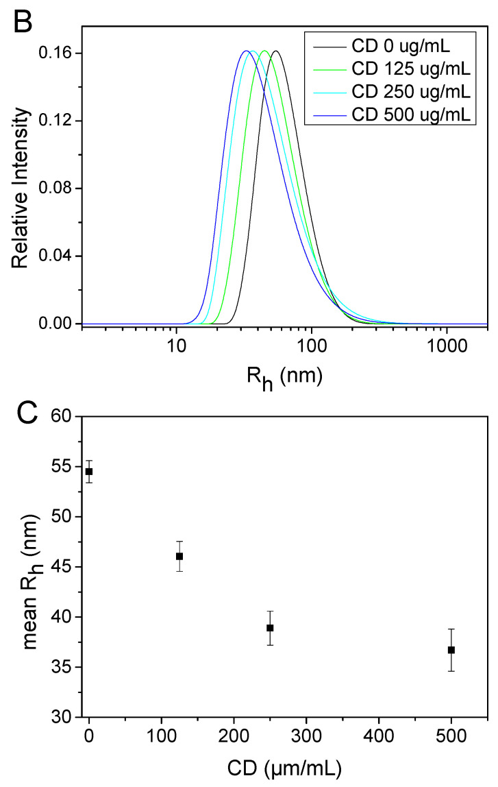
The effect of carbon dots (CDs) on a model blayer membrane was studied as a means of comprehending their ability to affect cell membranes. Initially, the interaction of N-doped carbon dots with a biophysical liposomal cell membrane model was investigated by dynamic light scattering, z-potential, temperature-modulated differential scanning calorimetry, and membrane permeability. CDs with a slightly positive charge interacted with the surface of the negative-charged liposomes and evidence indicated that the association of CDs with the membrane affects the structural and thermodynamic properties of the bilayer; most importantly, it enhances the bilayer's permeability against doxorubicin, a well-known anticancer drug. The results, like those of similar studies that surveyed the interaction of proteins with lipid membranes, suggest that carbon dots are partially embedded in the bilayer. In vitro experiments employing breast cancer cell lines and human healthy dermal cells corroborated the findings, as it was shown that the presence of CDs in the culture medium selectively enhanced cell internalization of doxorubicin and, subsequently, increased its cytotoxicity, acting as a drug sensitizer.

Keywords: biomembranes; carbon dots; doxorubicin; lipid bilayers; membrane permeability.


Conflict of interest statement

The authors declare no conflict of interest.

Figures

Figure 1
Figure 1
Carbon dots (CDs) adsorbed on DPPC:DPPG:DSPE-PEG liposomes in PBS (A) or water (B) as a function of their concentration in the outer medium. The DPPC:DPPG:DSPE-PEG liposomes after incubation with CDs in PBS were isolated by ultracentrifugation and the content of CDs was analyzed by fluorescence spectroscopy. Data were obtained from three different independent sets of experiments (the lines are only drawn as guides).
Figure 2
Figure 2
(A) Z-potential of DPPC:DPPG:DSPE-PEG liposomes in PBS as a function of CDs’ concentration in the outer medium. (B,C) Size variation of DPPC:DPPG:DSPE-PEG liposomes in PBS as a function of CDs’ concentration in the outer medium: (B) hydrodynamic radius distributions of unilamellar vesicles obtained by CONTIN analysis of DLS measurements and (C) apparent mean hydrodynamic radii vs. CDs’ concentration. All data were obtained from three different independent sets of experiments.
Figure 2
Figure 2
(A) Z-potential of DPPC:DPPG:DSPE-PEG liposomes in PBS as a function of CDs’ concentration in the outer medium. (B,C) Size variation of DPPC:DPPG:DSPE-PEG liposomes in PBS as a function of CDs’ concentration in the outer medium: (B) hydrodynamic radius distributions of unilamellar vesicles obtained by CONTIN analysis of DLS measurements and (C) apparent mean hydrodynamic radii vs. CDs’ concentration. All data were obtained from three different independent sets of experiments.
Figure 3
Figure 3
Temperature-modulated DSC analysis of the main phase transition of liposomal DPPC:DPPG:DSPE-PEG formulations in the presence of various CD concentrations in the outer PBS medium: (A) Total heat flow profiles of liposomal dispersions during the first heating cycle (endo down); (B) Total, reversing and non-reversing enthalpies of the main phase transition. Data were obtained from three different independent sets of experiments. (C) Reversing heat flow signals of liposomal dispersions during the first heating cycle (endo down). (D) Non-reversing heat flow signals of liposomal dispersions during the first heating cycle (endo down). The heating scan rate for all of the thermographs was 2 °C/min.
Figure 3
Figure 3
Temperature-modulated DSC analysis of the main phase transition of liposomal DPPC:DPPG:DSPE-PEG formulations in the presence of various CD concentrations in the outer PBS medium: (A) Total heat flow profiles of liposomal dispersions during the first heating cycle (endo down); (B) Total, reversing and non-reversing enthalpies of the main phase transition. Data were obtained from three different independent sets of experiments. (C) Reversing heat flow signals of liposomal dispersions during the first heating cycle (endo down). (D) Non-reversing heat flow signals of liposomal dispersions during the first heating cycle (endo down). The heating scan rate for all of the thermographs was 2 °C/min.
Figure 4
Figure 4
Time-dependent DOX release at 37 °C from DPPC:DPPG:DSPE-PEG unilamellar liposomes in the presence of various CD concentrations in the outer PBS medium. Measurements were taken every 0.1 min during the two minutes of incubation and every 0.5 min afterwards. Data were obtained from three different independent sets of experiments.
Figure 5
Figure 5
Doxorubicin internalization in MCF-7 cells (A) and human dermal fibroblasts (HDF, (B). Cells in 96-well plates were incubated at 37 °C and treated with CDs (500 μg/mL), DOX (3 μΜ), and increasing concentration of CDs (125, 250, and 500 μg/mL) for 1 h before adding DOX (3 μM). After 3 h, the wells were washed with RPMI without phenol red and DOX concentration was measured with an Infinite M200 plate reader (λex = 510 nm, λem = 580 nm) and expressed as fluorescence intensity in arbitrary units (a.u.). The results are shown as the mean ± SD for at least three independent experiments and were analyzed using a Student’s t-test (* p < 0.05, ** p < 0.01, **** p < 0.0001, ns not significant). Statistical analysis is not shown if it was not considered significant (n = 3).
Figure 6
Figure 6
Carbon dots sensitizes p53 WT (MCF-7) and mutant p53 breast cancer (MDA-MB-231) cell lines but not human dermal fibroblasts (HDF) to DOX. Cells were treated with CDs (500 μg/mL), DOX (3 μΜ), as well as with increasing concentrations of CDs (125, 250, and 500 μg/mL) for 1 h before adding DOX (3 μM). After 24 h, cell survival of MCF-7 (A), MDA-MB-231 (B) and HDF (C) cell lines was measured with MTS assay. Results are expressed as the mean ± SD for at least three independent experiments and analyzed using Student’s t-test (* p < 0.05, ** p < 0.01, *** p < 0.001, ns not significant).
Figure 6
Figure 6
Carbon dots sensitizes p53 WT (MCF-7) and mutant p53 breast cancer (MDA-MB-231) cell lines but not human dermal fibroblasts (HDF) to DOX. Cells were treated with CDs (500 μg/mL), DOX (3 μΜ), as well as with increasing concentrations of CDs (125, 250, and 500 μg/mL) for 1 h before adding DOX (3 μM). After 24 h, cell survival of MCF-7 (A), MDA-MB-231 (B) and HDF (C) cell lines was measured with MTS assay. Results are expressed as the mean ± SD for at least three independent experiments and analyzed using Student’s t-test (* p < 0.05, ** p < 0.01, *** p < 0.001, ns not significant).

Similar articles

Cited by

References

    1. Doane T.L., Burda C. The unique role of nanoparticles in nanomedicine: Imaging, drug delivery and therapy. Chem. Soc. Rev. 2012;41:2885–2911. doi: 10.1039/c2cs15260f. - DOI - PubMed
    1. Beddoes C.M., Case C.P., Briscoe W.H. Understanding nanoparticle cellular entry: A physicochemical perspective. Adv. Colloid Interface Sci. 2015;218:48–68. doi: 10.1016/j.cis.2015.01.007. - DOI - PubMed
    1. Rascol E., Devoisselle J.M., Chopineau J. The relevance of membrane models to understand nanoparticles–cell membrane interactions. Nanoscale. 2016;8:4780–4798. doi: 10.1039/C5NR07954C. - DOI - PubMed
    1. Mintz K.J., Zhou Y., Leblanc R.M. Recent development of carbon quantum dots regarding their optical properties, photoluminescence mechanism, and core structure. Nanoscale. 2019;11:4634–4652. doi: 10.1039/C8NR10059D. - DOI - PMC - PubMed
    1. Lim S.Y., Shen W., Gao Z. Carbon quantum dots and their applications. Chem. Soc. Rev. 2015;44:362–381. doi: 10.1039/C4CS00269E. - DOI - PubMed

LinkOut - more resources