Review
. 2023 Jun 12;16(6):870.
doi: 10.3390/ph16060870.

Elevated Intraocular Pressure and Glaucomatous Optic Neuropathy: Genes to Disease Mechanisms, Therapeutic Drugs, and Gene Therapies

Affiliations
Review

Elevated Intraocular Pressure and Glaucomatous Optic Neuropathy: Genes to Disease Mechanisms, Therapeutic Drugs, and Gene Therapies

Najam A Sharif. Pharmaceuticals (Basel). .

Abstract

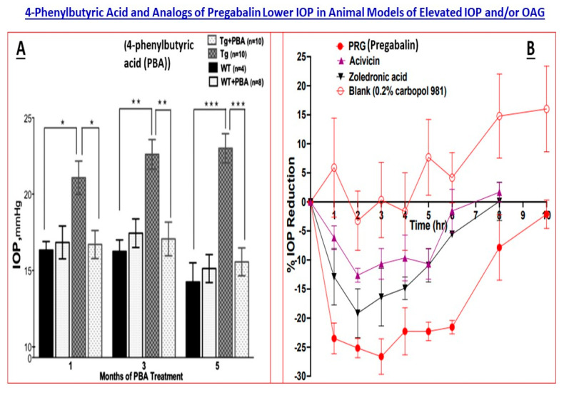
This review article focuses on the pathogenesis of and genetic defects linked with chronic ocular hypertension (cOHT) and glaucoma. The latter ocular disease constitutes a group of ocular degenerative diseases whose hallmark features are damage to the optic nerve, apoptotic demise of retinal ganglion cells, disturbances within the brain regions involved in visual perception and considerable visual impairment that can lead to blindness. Even though a number of pharmaceuticals, surgical and device-based treatments already exist addressing cOHT associated with the most prevalent of the glaucoma types, primary open-angle glaucoma (POAG), they can be improved upon in terms of superior efficacy with reduced side-effects and with longer duration of activity. The linkage of disease pathology to certain genes via genome-wide associated studies are illuminating new approaches to finding novel treatment options for the aforementioned ocular disorders. Gene replacement, gene editing via CRISPR-Cas9, and the use of optogenetic technologies may replace traditional drug-based therapies and/or they may augment existing therapeutics for the treatment of cOHT and POAG in the future.

Keywords: CRISPR-Cas9; genome-wide associated studies (GWAS); ocular hypertension; optogenetics; primary open-angle glaucoma.


Conflict of interest statement

The author declares no conflict of interest.

Figures

Figure 1
Figure 1
High-level anatomy of the human eye anterior chamber and the pathways involved in aqueous humor drainage to reduce IOP are shown in (A). (B) illustrates the IOP-lowering effects of a topical ocularly delivered novel non-prostaglandin EP2-receptor agonist, omidenepag isopropyl ester (OMDI), in ocular hypertensive Cynomolgus monkey eyes. (C) shows the promotion of AQH outflow by OMDI via both the conventional (Y) and uveoscleral (Z) routes without influencing the inflow of AQH (X) in the latter monkey’s eyes. All figures are adapted from [15,16] under open-access terms and from author’s own publications. The statistical significance is shown amongst the different groups. * p < 0.05, ** p < 0.01; *** p < 0.001.
Figure 2
Figure 2
The many sites of dysfunction within the visual axis as a result of elevated IOP/ocular hypertension and glaucomatous optic neuropathy are shown in this schematic. These tissues and the cells therein, and the chemicals and factors shown, represent opportunities for intervention to mitigate the pathogenesis and progression of the cOHT, POAG and NTG. The Figure is adapted from a recent publication of the author [24] under the terms and conditions of the (Open Access) Creative Commons Attribution (CC BY) license (https://creativecommons.org/licenses/by/4.0/). Additional references include author’s own publications as in [15,16].
Figure 3
Figure 3
Displayed in this figure are a typical testing funnel and the components therein for synthesis of test compounds and/or gene therapy capsids and their characterization, followed by their evaluation in vitro and in vivo to eventually produce suitable compounds for progression towards Investigational New Drug (IND)-enabling studies (A). A more comprehensive screening paradigm for discovering and analyzing the pharmacological and biochemical features of test agents in a variety of in vitro assay systems, followed by screening in animal models of increasing complexity, is shown in (B). Examples of certain stage-gate passing criteria for progression down the testing funnel are also included for illustration purposes. Once the preferred compound/gene therapy has met all the prescribed criteria, it can undergo IND-enabling studies and then into proof-of-concept clinical trials for the target disease (e.g., for elevated IOP; slowing RGC/optic nerve damage). Figures are adapted from [15,16], the author’s own recent publications.
Figure 4
Figure 4
The varied mechanisms of action and pathways engagement by various drugs to promote egress of AQH from the anterior chamber of the eye via the conventional TM/SC pathway and/or via the uveoscleral pathway to relieve the elevated IOP are depicted in Figure 4A. Moreover, drugs that inhibit the production of AQH (inflow inhibitors) are also shown in (A). (B) displays a list of genes that are impacted and/or are implicated in the etiology of glaucoma (POAG/cOHT) via TM, SC and/or both tissues. (A) is modified from Figure 1A, which was adapted from Refs. 99 and 100 (the author’s own recent publications). The small tabulate list shown in (B) is derived with gratitude from [25] under Creative Commons Attribution 4.0 International License and which is a Nature Communications article.
Figure 5
Figure 5
The 4-phenylbutyric acid (PBA)-induced ocular hypotension in a mouse model of myocilin-induced glaucoma (transgenic mice) is shown in (A) [57]. In the adjoining (B), the time-dependent IOP-lowering action of pregabalin and its analogs (CACNA2D1 inhibitors) is displayed [59]. Both Figures are adapted from the aforementioned references under the terms and conditions of the (Open Access) Creative Commons Attribution (CC BY) license (https://creativecommons.org/licenses/by/4.0/). The statistical significance is shown amongst the different groups. * p < 0.05, ** p < 0.01; *** p < 0.001.

References

    1. Saldanha I.J., Lindsley K., Do D.V., Chuck R.S., Meyerle C., Jones L.S., Coleman A.L., Jampel H.D., Dickersin K., Virgili G. Comparison of clinical trial and systematic review outcomes for the 4 most prevalent eye diseases. JAMA Ophthalmol. 2017;135:933–940. doi: 10.1001/jamaophthalmol.2017.2583. - DOI - PMC - PubMed
    1. Assi L., Chamseddine F., Ibrahim P., Sabbagh H., Rosman L., Congdon N., Evans J., Ramke J., Kuper H., Burton M.J., et al. A global assessment of eye health and quality of life: A systematic review of systematic reviews. JAMA Ophthalmol. 2021;139:526–541. doi: 10.1001/jamaophthalmol.2021.0146. - DOI - PMC - PubMed
    1. Weinreb R.N., Aung T., Medeiros F.A. The pathophysiology and treatment of glaucoma: A review. J. Am. Med. Assoc. 2014;311:1901–1911. doi: 10.1001/jama.2014.3192. - DOI - PMC - PubMed
    1. Sharif N.A. Ocular hypertension and glaucoma: A review and current perspectives. Int. J. Ophthalmol. Vis. Sci. 2017;2:22–36.
    1. Tham Y.C., Li X., Wong T.Y., Quigley H.A., Aung T., Cheng C.Y. Global prevalence of glaucoma and projections of glaucoma burden through 2040. Ophthalmology. 2014;121:2081–2090. doi: 10.1016/j.ophtha.2014.05.013. - DOI - PubMed

LinkOut - more resources