. 2023 Jun 8;12(12):2247.
doi: 10.3390/plants12122247.

Tracking Permeation of Dimethyl Sulfoxide (DMSO) in Mentha × piperita Shoot Tips Using Coherent Raman Microscopy

Affiliations

Tracking Permeation of Dimethyl Sulfoxide (DMSO) in Mentha × piperita Shoot Tips Using Coherent Raman Microscopy

Heidi D Kreckel et al. Plants (Basel). .

Abstract

Cryopreservation has emerged as a low-maintenance, cost-effective solution for the long-term preservation of vegetatively propagated crops. Shoot tip cryopreservation often makes use of vitrification methods that employ highly concentrated mixtures of cryoprotecting agents; however, little is understood as to how these cryoprotecting agents protect cells and tissues from freezing. In this study, we use coherent anti-Stokes Raman scattering microscopy to directly visualize where dimethyl sulfoxide (DMSO) localizes within Mentha × piperita shoot tips. We find that DMSO fully penetrates the shoot tip tissue within 10 min of exposure. Variations in signal intensities across images suggest that DMSO may interact with cellular components, leading to its accumulation in specific regions.

Keywords: CARS microscopy; Raman; brightfield microscopy; cryoprotectant distribution; peppermint; plant cryopreservation.


Conflict of interest statement

The authors declare no conflict of interest.

Figures

Figure 1
Figure 1
Brightfield microscopy images of peppermint shoot tip (A) prior to exposure and (B) 21 min after exposure to 15% (w/v) DMSO in aqueous media. LP = leaf primordia region, M = meristem region. The black line indicates the initial edge of the shoot tip prior to exposure. Images were collected using a 40×, 0.60 NA air-immersion objective. Image size: 1600 × 1200 pixels. Pixel size: 0.11 μm/pixel.
Figure 2
Figure 2
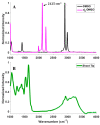
(A) Spontaneous Raman spectra of pure d6−DMSO (magenta) and DMSO (black). (B) IR absorption spectrum of a peppermint shoot tip (green).
Figure 3
Figure 3
CARS montage for a z-stack of a peppermint shoot tip after >10 min exposure to 15% (w/v) d6-DMSO. Top left: brightfield image of the shoot tip identifying leaf primordia (LP) and meristem (M) regions. All others: CARS images acquired from the same shoot tip at depths of 0, 3, 6, 9, 12, 15, 18, 21, 24, and 27 µm. The second panel from the left in the top row represents an image taken from the top of the shoot tip; subsequent images are presented, in order, from the top to the bottom of the shoot tip. Right: Calibration bar generated by a calibration curve correlating mean pixel intensity to aqueous d6-DMSO concentration (see Figure S2). Images were collected using a 20×, 0.75 NA air immersion objective. Vibrational frequency: 2125 cm−1. Image size: 800 × 800 pixels. Pixel size: 0.12 μm/pixel Scale bar = 10 μm.
Figure 4
Figure 4
(A) Representative CARS z-stack image (depth of 15 µm, Figure 3) of a peppermint shoot tip following exposure to 15% (w/v) d6-DMSO. Colored boxes, each containing a unique symbol, represent selected regions of interest (ROIs). Black ROIs = background regions, or regions without plant tissue; green ROIs = leaf primordia; cyan ROIs = meristem. The unique symbols in each ROI correspond to data points shown in B and Figure 5. Each ROI contains 5400 pixels. (B) Mean pixel intensity recorded for each ROI indicated in A for each tissue thickness (corresponding images in Figure 3). Shaded regions represent areas of low signal intensity due to technical aspects of CARS imaging (see Supplementary Materials) and are not considered in our analyses. Images were collected using a 20×, 0.75 NA air immersion objective. Vibrational frequency: 2125 cm−1. Image size: 800 × 800 pixels. Pixel size: 0.12 μm/pixel.
Figure 5
Figure 5
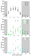
Mean (solid symbols) and maximum (open symbols) pixel intensities of 5400 data points over tissue thickness (shown in Figure 3) for (A) background (black), (B) leaf primordia (green), and (C) meristem (cyan) regions of interest (ROIs) identified in Figure 4A. Error bars represent standard deviations in pixel intensity for each ROI. Shaded regions represent areas of low signal intensity due to technical aspects of CARS imaging (see Supplementary Materials) and are therefore eliminated from our analyses.
Figure 6
Figure 6
Box and whisker plots for the spread of pixel intensity for each ROI (representative image shown in Figure 4A) at 12 µm tissue thickness (image shown in Figure 3). Each data set contains 5400 data points, corresponding to the 5400 pixel intensities within each ROI; outliers are reported as individual data points. The reported number of outliers for Bkgd1, Bkgd2, LP1, LP2, and meristem regions of interest is 5, 16, 21, 36, and 21, respectively.

References

    1. Engelmann F. Use of Biotechnologies for the Conservation of Plant Biodiversity. Vitro Cell. Dev. Biol. Plant. 2011;47:5–16. doi: 10.1007/s11627-010-9327-2. - DOI
    1. Li D.-Z., Pritchard H.W. The Science and Economics of Ex Situ Plant Conservation. Trends Plant Sci. 2009;14:614–621. doi: 10.1016/j.tplants.2009.09.005. - DOI - PubMed
    1. Jenderek M.M., Reed B.M. Cryopreserved Storage of Clonal Germplasm in the USDA National Plant Germplasm System. Vitro Cell. Dev. Biol. Plant. 2017;53:299–308. doi: 10.1007/s11627-017-9828-3. - DOI
    1. Engelmann F. Plant Cryopreservation: Progress and Prospects. Vitro Cell. Dev. Biol. Plant. 2004;40:427–433. doi: 10.1079/IVP2004541. - DOI
    1. Bettoni J.C., Bonnart R., Volk G.M. Challenges in Implementing Plant Shoot Tip Cryopreservation Technologies. Plant Cell Tissue Organ Cult. 2021;144:21–34. doi: 10.1007/s11240-020-01846-x. - DOI

LinkOut - more resources