

Mesenchymal Stem Cell Membrane-Coated TPCS2a-Loaded Nanoparticles for Breast Cancer Photodynamic Therapy
- PMID: 37376102
- PMCID: PMC10302938
- DOI: 10.3390/pharmaceutics15061654
Mesenchymal Stem Cell Membrane-Coated TPCS2a-Loaded Nanoparticles for Breast Cancer Photodynamic Therapy
Abstract
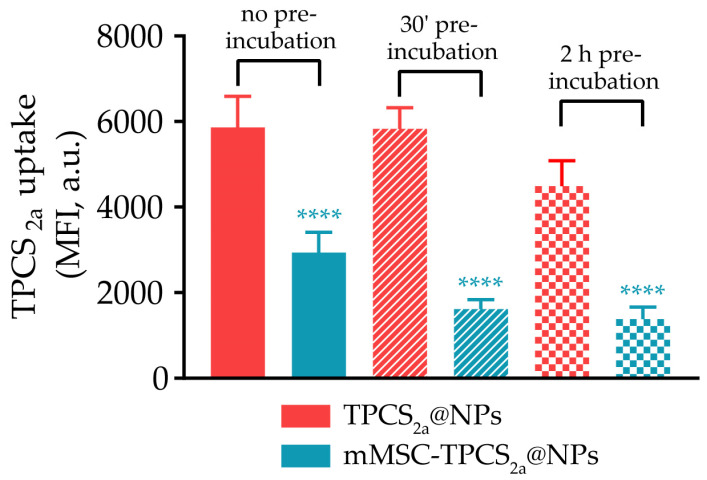
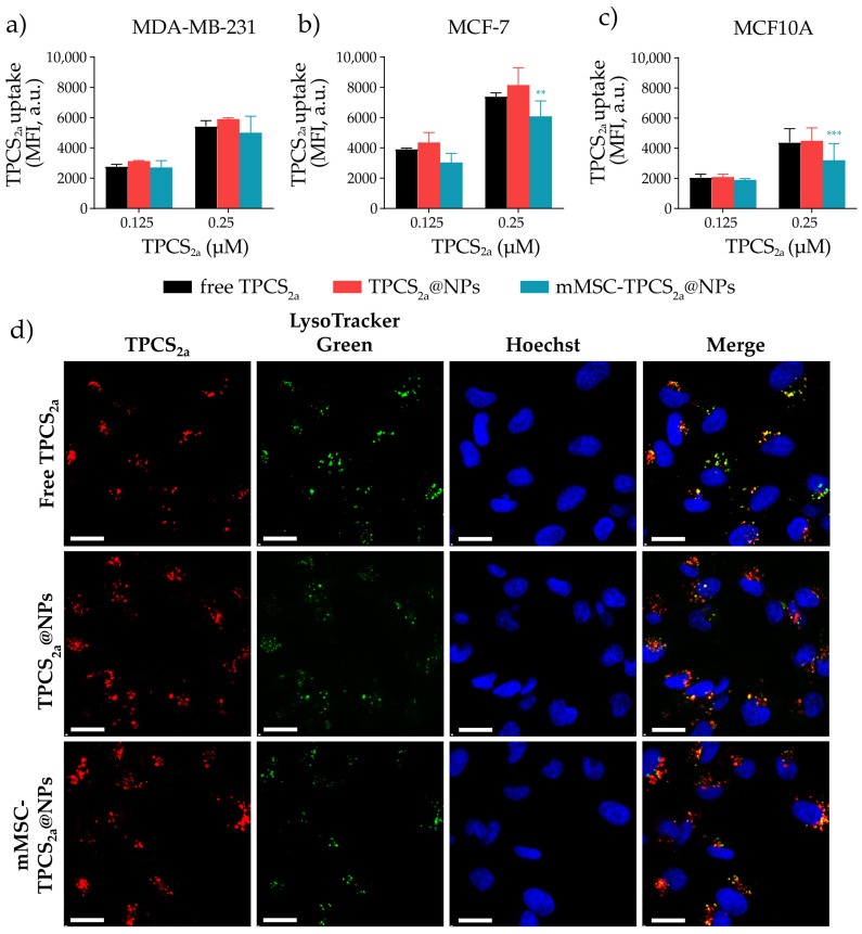
Despite substantial improvements in breast cancer (BC) treatment there is still an urgent need to find alternative treatment options to improve the outcomes for patients with advanced-stage disease. Photodynamic therapy (PDT) is gaining a lot of attention as a BC therapeutic option because of its selectivity and low off-target effects. However, the hydrophobicity of photosensitizers (PSs) impairs their solubility and limits the circulation in the bloodstream, thus representing a major challenge. The use of polymeric nanoparticles (NPs) to encapsulate the PS may represent a valuable strategy to overcome these issues. Herein, we developed a novel biomimetic PDT nanoplatform (NPs) based on a polymeric core of poly(lactic-co-glycolic)acid (PLGA) loaded with the PS meso-tetraphenylchlorin disulfonate (TPCS2a). TPCS2a@NPs of 98.89 ± 18.56 nm with an encapsulation efficiency percentage (EE%) of 81.9 ± 7.92% were obtained and coated with mesenchymal stem cells-derived plasma membranes (mMSCs) (mMSC-TPCS2a@NPs, size of 139.31 ± 12.94 nm). The mMSC coating armed NPs with biomimetic features to impart long circulation times and tumor-homing capabilities. In vitro, biomimetic mMSC-TPCS2a@NPs showed a decrease in macrophage uptake of 54% to 70%, depending on the conditions applied, as compared to uncoated TPCS2a@NPs. Both NP formulations efficiently accumulated in MCF7 and MDA-MB-231 BC cells, while the uptake was significantly lower in normal breast epithelial MCF10A cells with respect to tumor cells. Moreover, encapsulation of TPCS2a in mMSC-TPCS2a@NPs effectively prevents its aggregation, ensuring efficient singlet oxygen (1O2) production after red light irradiation, which resulted in a considerable in vitro anticancer effect in both BC cell monolayers (IC50 < 0.15 µM) and three-dimensional spheroids.
Keywords: biomimetic nanoparticles; breast cancer; photodynamic therapy.
Conflict of interest statement
The authors declare no conflict of interest.
Figures


Similar articles
-
Co-delivery of Docetaxel and Disulfonate Tetraphenyl Chlorin in One Nanoparticle Produces Strong Synergism between Chemo- and Photodynamic Therapy in Drug-Sensitive and -Resistant Cancer Cells.Mol Pharm. 2018 Oct 1;15(10):4599-4611. doi: 10.1021/acs.molpharmaceut.8b00597. Epub 2018 Sep 7. Mol Pharm. 2018. PMID: 30148955
-
CD44 Targeting Mediated by Polymeric Nanoparticles and Combination of Chlorine TPCS2a-PDT and Docetaxel-Chemotherapy for Efficient Killing of Breast Differentiated and Stem Cancer Cells In Vitro.Cancers (Basel). 2020 Jan 23;12(2):278. doi: 10.3390/cancers12020278. Cancers (Basel). 2020. PMID: 31979218 Free PMC article.
-
Coating of chitosan on poly D,L-lactic-co-glycolic acid thymoquinone nanoparticles enhances the anti-tumor activity in triple-negative breast cancer.Front Chem. 2023 Feb 8;11:1044953. doi: 10.3389/fchem.2023.1044953. eCollection 2023. Front Chem. 2023. PMID: 36846852 Free PMC article.
-
Biomimetic cell membrane-coated poly(lactic-co-glycolic acid) nanoparticles for biomedical applications.Bioeng Transl Med. 2022 Nov 2;8(2):e10441. doi: 10.1002/btm2.10441. eCollection 2023 Mar. Bioeng Transl Med. 2022. PMID: 36925703 Free PMC article. Review.
-
Polymeric Nanoparticles for Cancer Photodynamic Therapy.Top Curr Chem. 2016;370:61-112. doi: 10.1007/978-3-319-22942-3_3. Top Curr Chem. 2016. PMID: 26589506 Review.
Cited by
-
Advancements in Cell Membrane-Derived Biomimetic Nanotherapeutics for Breast Cancer.Int J Nanomedicine. 2025 May 12;20:6059-6083. doi: 10.2147/IJN.S502144. eCollection 2025. Int J Nanomedicine. 2025. PMID: 40385497 Free PMC article. Review.
References
-
- Kassam F., Enright K., Dent R., Dranitsaris G., Myers J., Flynn C., Fralick M., Kumar R., Clemons M. Survival Outcomes for Patients with Metastatic Triple-Negative Breast Cancer: Implications for Clinical Practice and Trial Design. Clin. Breast Cancer. 2009;9:29–33. doi: 10.3816/CBC.2009.n.005. - DOI - PubMed
LinkOut - more resources
Full Text Sources
Miscellaneous
