. 2023 May 29;15(6):1269.
doi: 10.3390/v15061269.

Complement Activation-Independent Attenuation of SARS-CoV-2 Infection by C1q and C4b-Binding Protein

Affiliations

Complement Activation-Independent Attenuation of SARS-CoV-2 Infection by C1q and C4b-Binding Protein

Nazar Beirag et al. Viruses. .

Abstract

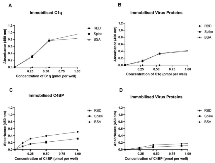
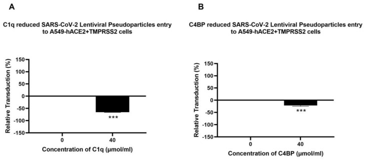
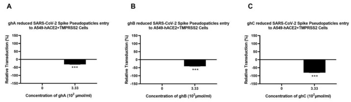
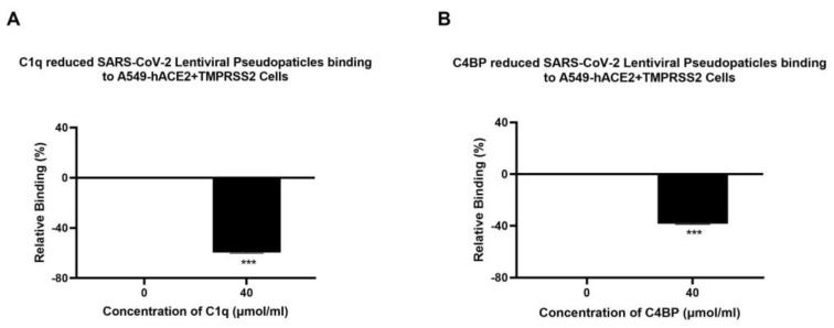
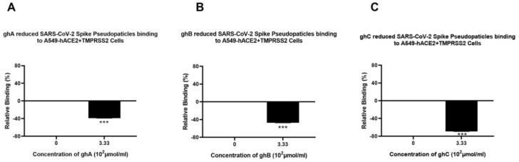
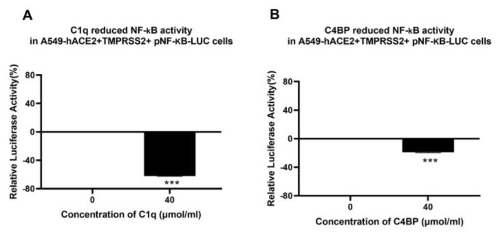
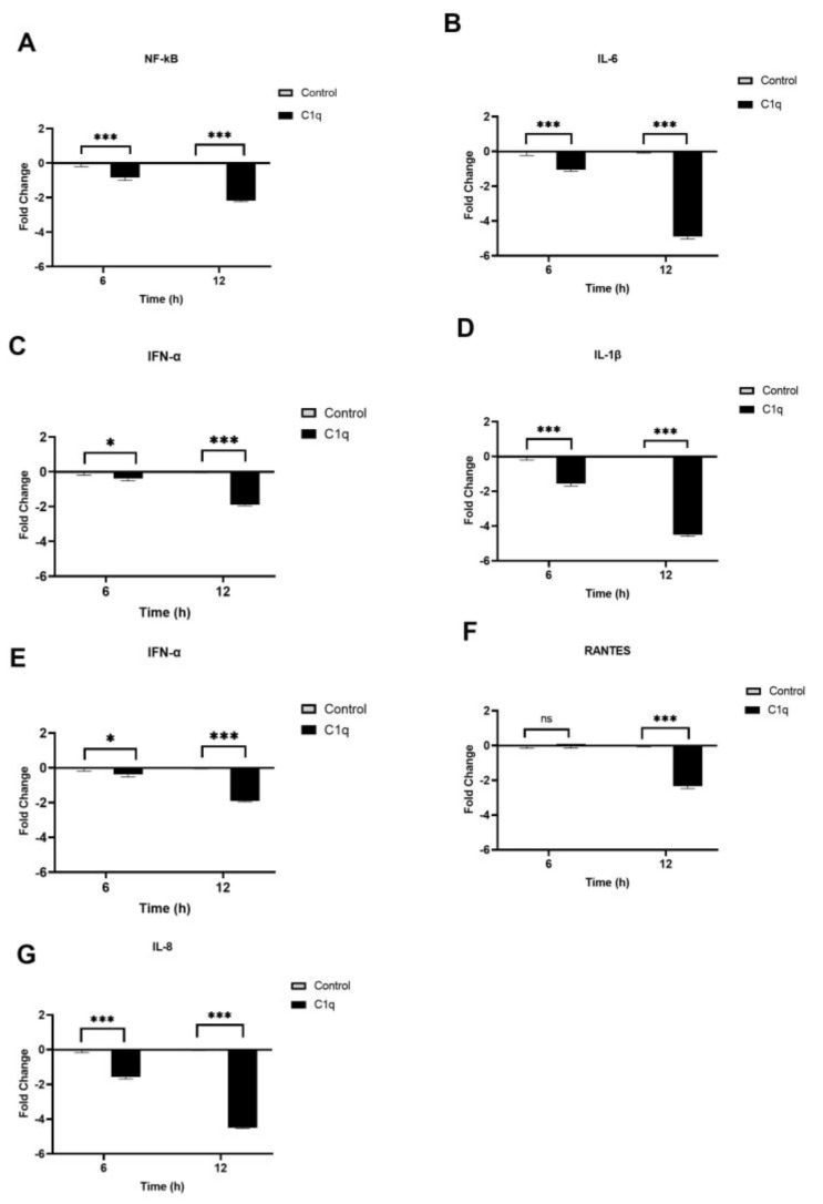
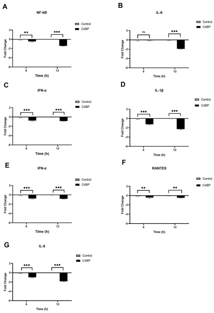
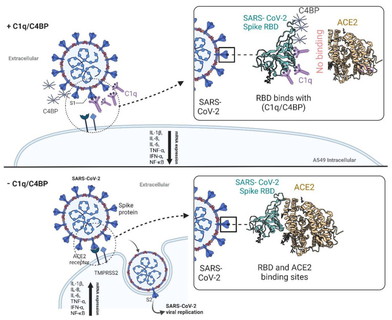
The complement system is a key component of the innate immune response to viruses and proinflammatory events. Exaggerated complement activation has been attributed to the induction of a cytokine storm in severe SARS-CoV-2 infection. However, there is also an argument for the protective role of complement proteins, given their local synthesis or activation at the site of viral infection. This study investigated the complement activation-independent role of C1q and C4b-binding protein (C4BP) against SARS-CoV-2 infection. The interactions of C1q, its recombinant globular heads, and C4BP with the SARS-CoV-2 spike and receptor binding domain (RBD) were examined using direct ELISA. In addition, RT-qPCR was used to evaluate the modulatory effect of these complement proteins on the SARS-CoV-2-mediated immune response. Cell binding and luciferase-based viral entry assays were utilised to assess the effects of C1q, its recombinant globular heads, and C4BP on SARS-CoV-2 cell entry. C1q and C4BP bound directly to SARS-CoV-2 pseudotype particles via the RBD domain of the spike protein. C1q via its globular heads and C4BP were found to reduce binding as well as viral transduction of SARS-CoV-2 spike protein expressing lentiviral pseudotypes into transfected A549 cells expressing human ACE2 and TMPRSS2. Furthermore, the treatment of the SARS-CoV-2 spike, envelope, nucleoprotein, and membrane protein expressing alphaviral pseudotypes with C1q, its recombinant globular heads, or C4BP triggered a reduction in mRNA levels of proinflammatory cytokines and chemokines such as IL-1β, IL-8, IL-6, TNF-α, IFN-α, and RANTES (as well as NF-κB) in A549 cells expressing human ACE2 and TMPRSS2. In addition, C1q and C4BP treatment also reduced SARS-CoV-2 pseudotype infection-mediated NF-κB activation in A549 cells expressing human ACE2 and TMPRSS2. C1q and C4BP are synthesised primarily by hepatocytes; however, they are also produced by macrophages, and alveolar type II cells, respectively, locally at the pulmonary site. These findings support the notion that the locally produced C1q and C4BP can be protective against SARS-CoV-2 infection in a complement activation-independent manner, offering immune resistance by inhibiting virus binding to target host cells and attenuating the infection-associated inflammatory response.

Keywords: C1q; C4BP; COVID-19; SARS-CoV-2; classical pathway; complement; innate immunity.


Conflict of interest statement

The authors declare no conflict of interest.

Figures

Figure 1
Figure 1
SARS-CoV-2 directly interacts with C1q and C4BP. The decreasing concentration of immobilized C1q (1, 0.5, 0.125, and 0 pmol/well) (A,C) or constant concentrations of viral proteins (spike 3 pmol/well or RBD 30 pmol/well) (B,D) were coated in a 96-well plate using a carbonate–bicarbonate (CBC) buffer, pH 9.6, at 4 °C overnight. A constant concentration of viral proteins (1 pmol/well) (A) or a decreasing amount of C1q/C4BP (1, 0.5, 0.125, and 0 pmol/well) (B) was added to the corresponding wells, and incubated at 37 °C for 2 h. After washing step, the wells were probed with primary antibodies (1:5000; 100 µL/well), i.e., rabbit anti-SARS-CoV-2 spike or rabbit anti-human C1q/C4BP. BSA was used as a negative control. The data are presented as a mean of three independent experiments carried out in triplicates ± SEM.
Figure 2
Figure 2
C1q (A) and C4BP (B) attenuate SARS-CoV-2 pseudoparticle entry into A549-hACE2+TMPRSS2 cells. Luciferase reporter activity of A549-hACE2+TMPRSS2 cells transduced with either treated or untreated SARS-CoV-2 lentiviral pseudoparticles pre-treated with C1q or C4BP (40 μmol/mL) was utilised to assess if the treatment by complement proteins interfered with the lentiviral pseudoparticles’ ability to enter the cells. The background was subtracted from all data points. The data obtained were normalised with 0% luciferase activity defined as the mean of the relative luminescence units recorded from the control sample (A549-hACE2+TMPRSS2 cells + SARS-CoV-2 lentiviral pseudoparticles). Pseudoparticles pre-treated with C1q and C4BP, blocked viral transduction. Data are shown as the normalized mean of three independent experiments carried out in triplicates ± SEM. Significance was determined using the two-way ANOVA test (*** p < 0.001) (n = 3).
Figure 3
Figure 3
Recombinant ghA, ghB, and ghC modules of human C1q block SARS-CoV-2 pseudoparticle entry into A549-hACE2+TMPRSS2 cells. SARS-CoV-2 pseudoparticles were pre-treated with ghA (A), ghB (B), or ghC (C) (3.33 × 10² μmol/mL) to determine if the recombinant modules of human C1q interfered with the ability of the pseudoparticles to enter the target cells. Luciferase reporter activity in A549-hACE2+TMPRSS2 cells that were transduced with pseudoparticles (and pre-treated with ghA, ghB, or ghC) was used. The background was subtracted from all data points. The data obtained were normalised with 0% luciferase activity defined as the mean of the relative luminescence units recorded from the control sample (A549-hACE2+TMPRSS2 cells + MBP + SARS-CoV-2 pseudoparticles). Data are shown as the normalized mean of three independent experiments carried out in triplicates ± SEM. Significance was determined using the two-way ANOVA test (*** p < 0.001) (n = 3).
Figure 4
Figure 4
Binding of C1q (A) or C4BP treated (B) SARS-CoV-2 pseudoparticles to A549-hACE2+TMPRSS2 cells. SARS-CoV-2 lentiviral pseudoparticles were used to transduce A549-hACE2+TMPRSS2 cells, which were pre-incubated with C1q or C4BP (40 µmol/mL). The wells were probed with rabbit anti-SARS-CoV-2 spike (1:200) polyclonal antibodies after being washed and fixed with 1% v/v paraformaldehyde for 1 min. The data obtained were normalised with 0% fluorescence defined as the mean of the relative fluorescence units recorded from the control sample (A549-hACE2+TMPRSS2 cells + SARS-CoV-2 lentiviral pseudoparticles). Three independent experiments were carried out in triplicates, and error bars are expressed as ± SEM. Significance was determined using the two-way ANOVA test (*** p < 0.001) (n = 3).
Figure 5
Figure 5
Binding of recombinant ghA- (A), ghB- (B), or ghC-treated (C) SARS-CoV-2 lentivirus pseudoparticles to A549-hACE2+TMPRSS2 cells. A549-hACE2+TMPRSS2 cells were transduced with SARS-CoV-2 pseudoparticles following pre-incubation with/without ghA, ghB, or ghC (3.33 × 10² μmol/mL). After removing unbound protein and viral particles, the wells were fixed with 1% v/v paraformaldehyde for 1 min and probed with rabbit anti-SARS-CoV-2 spike (1:200) polyclonal antibodies. The data obtained were normalised with 0% fluorescence defined as the mean of the relative fluorescence units recorded from the control sample (cells + MBP + pseudoparticles). Three independent experiments were carried out in triplicates, and error bars express ± SEM. Significance was determined using the two-way ANOVA test (*** p < 0.001) (n = 3).
Figure 6
Figure 6
C1q and C4BP inhibit NF-κB activation in SARS-CoV-2 spike-challenged A549-hACE2-TMPRSS2 cells. A549-hACE2+TMPRSS2 cells transfected with pNF-κB-LUC were challenged with SARS-CoV-2 spike protein (1.4 µmol/mL) was pre-treated with C1q (A) or C4BP (B) (40 µmol/mL). The cells were incubated for 24 h and examined for luciferase reporter activity. The background was subtracted from all data points. The data obtained were normalised with 0% luciferase activity defined as the mean of the relative luminescence units recorded from the control sample (A549-hACE2 + TMPRSS2 cells + SARS-CoV-2 spike protein). Data are shown as the normalized mean of three independent experiments carried out in triplicates ± SEM. Significance was determined using the two-way ANOVA test (*** p < 0.001) (n = 3).
Figure 7
Figure 7
C1q reduces the inflammatory response in SARS-CoV-2 pseudoparticle-challenged A549-hACE2+TMPRSS2 cells. SARS-CoV-2 alphaviral pseudoparticles, pre-incubated with 40 µmol/mL of C1q, were utilised to challenge A549-hACE2+TMPRSS2 cells. The cells were harvested at 6 h and 12 h to measure the mRNA levels of proinflammatory cytokines and chemokines. Cells were lysed, and purified RNA was converted into cDNA. The mRNA levels of NF-κB (A), IL-6 (B), IFN-α (C), IL-1β (D), TNF-α (E), RANTES (F), and IL-8 (G) were measured using RT-qPCR; the data were normalised against 18S rRNA expression as a control. The relative expression (RQ) was calculated using A549-hACE2+TMPRSS2 cells challenged with SARS-CoV-2 alphaviral pseudoparticles alone as the calibrator. RQ = 2−∆∆Ct was used to calculate the RQ value. Experiments were carried out in triplicates, and error bars represent ± SEM. Significance was determined using the two-way ANOVA test (* p < 0.05, *** p < 0.001, ns p > 0.05) (n = 3).
Figure 8
Figure 8
C4BP attenuates the inflammatory response in SARS-CoV-2 pseudotyped viral particle-challenged A549-hACE2+TMPRSS2 cells. The gene expression profile of cytokines and chemokines produced in A549-hACE2 +TMPRSS2 cells challenged with SARS-CoV-2 alphaviral pseudoparticles that were pre-treated with and without C4BP (40 µmol/mL) was examined. Expression levels of NF-κB (A), IL-6 (B), IFN-α (C), IL-1β (D), TNF-α (E), RANTES (F), and IL-8 (G) were measured using RT-qPCR at 6 h and 12 h. A549-hACE2+TMPRSS2 cells challenged with SARS-CoV-2 alphaviral pseudoparticles were used as a calibrator to calculate relative quantitation (RQ); RQ = 2−∆∆Ct. The experiments were conducted in triplicates, and error bars represent ± SEM. Additionally, 18S rRNA was used as an endogenous control. Significance was established using the two-way ANOVA test (** p < 0.01, *** p < 0.001, ns p > 0.05) (n = 3).
Figure 9
Figure 9
Complement independent attenuation of SARS-CoV-2 infection by C1q and C4BP. The activator of the classical pathway of the complement system, C1q, and the regulatory protein of the classical and lectin pathway of the complement system, C4BP, were found to interact with the receptor-binding domain (RBD) of the SARS-CoV-2 spike protein. This interaction helped reduce SARS-CoV-2 infection in A549 cells expressing human ACE2 and TMPRSS2 by preventing the virus from binding to its cell surface receptors, independent of complement activation. In addition, C1q and C4BP treatment decreased the mRNA levels of proinflammatory cytokines and chemokines (IL-1β, IL-8, IL-6, TNF-α, IFN-α, NF-kB, and RANTES), thereby attenuating infection-associated inflammation. These findings suggest that complement proteins have a novel role as soluble pattern recognition molecules, functioning as one of the first lines of defence against viral infections independent of their complement-related functions. C1q: Complement component 1q; C4BP: Complement component 4 binding protein; RBD: Receptor-binding domain; ACE2: Angiotensin-converting enzyme 2; TMPRSS2: Transmembrane protease serine 2.

References

    1. Cascella M., Rajnik M., Aleem A., Dulebohn S.C., Di Napoli R. StatPearls [Internet] StatPearls; Treasure Island, FL, USA: 2022. Features, evaluation, and treatment of coronavirus (COVID-19) - PubMed
    1. Gusev E., Sarapultsev A., Solomatina L., Chereshnev V. SARS-CoV-2-Specific immune response and the pathogenesis of COVID-19. Int. J. Mol. Sci. 2022;23:1716. doi: 10.3390/ijms23031716. - DOI - PMC - PubMed
    1. Varghese P.M., Tsolaki A.G., Yasmin H., Shastri A., Ferluga J., Vatish M., Madan T., Kishore U. Host-pathogen interaction in COVID-19: Pathogenesis, potential therapeutics and vaccination strategies. Immunobiology. 2020;225:152008. doi: 10.1016/j.imbio.2020.152008. - DOI - PMC - PubMed
    1. Merle N.S., Church S.E., Fremeaux-Bacchi V., Roumenina L.T. Complement system part I–molecular mechanisms of activation and regulation. Front. Immunol. 2015;6:262. doi: 10.3389/fimmu.2015.00262. - DOI - PMC - PubMed
    1. Murugaiah V., Varghese P.M., Beirag N., DeCordova S., Sim R.B., Kishore U. Complement proteins as soluble pattern recognition receptors for pathogenic viruses. Viruses. 2021;13:824. doi: 10.3390/v13050824. - DOI - PMC - PubMed

Publication types

MeSH terms