. 2023 May 31;15(6):1301.
doi: 10.3390/v15061301.

Effect of Intermediate Plus Vaccine and vvIBDV on Bursa Secretory Cells and Their Glycoprotein Production

Affiliations

Effect of Intermediate Plus Vaccine and vvIBDV on Bursa Secretory Cells and Their Glycoprotein Production

Imre Oláh et al. Viruses. .

Abstract

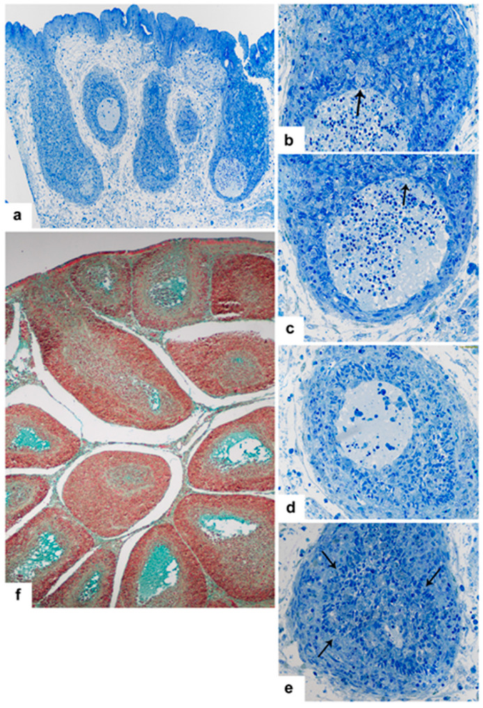
There are two types of secretory cells in the chicken bursa of Fabricius (BF): (a) interfollicular epithelial cells (IFE), and (b) bursal secretory dendritic cells (BSDC) in the medulla of bursal follicles. Both cells produce secretory granules, and the cells are highly susceptible to IBDV vaccination and infection. Before and during embryonic follicular bud formation, an electron-dense, scarlet-acid fuchsin positive substance emerges in the bursal lumen, the role of which is unknown. In IFE cells, IBDV infection may induce rapid granular discharge, and in several cells, peculiar granule formation, which suggests that the glycosylation of protein is injured in the Golgi complex. In control birds, the discharged BSDC granules appear in membrane-bound and subsequently solubilized, fine-flocculated forms. The solubilized, fine-flocculated substance is Movat-positive and can be a component of the medullary microenvironment, which prevents the medullary B lymphocytes from nascent apoptosis. Vaccination interferes with the solubilization of the membrane-bound substance, resulting in: (i) aggregation of a secreted substance around the BSDC, and (ii) solid lumps in the depleted medulla. The non-solubilized substance is possibly not "available" for B lymphocytes, resulting in apoptosis and immunosuppression. In IBDV infection, one part of the Movat-positive Mals fuse together to form a medullary, gp-containing "cyst". The other part of Mals migrate into the cortex, recruiting granulocytes and initiating inflammation. During recovery the Movat-positive substance appears as solid, extracellular lumps between the cells of FAE and Mals. Possibly the Mals and Movat-positive extracellular lumps glide into the bursal lumen via FAE to eliminate cell detritus from the medulla.

Keywords: bursa of Fabricius; chicken; effect of IBDV vaccination and infection; glycoprotein; microenvironment.


Conflict of interest statement

The authors declare no conflict of interest.

Figures

Figure 1
Figure 1
(a) 15-day old embryo. Semithin section (1.5 um thick), stained with toluidine blue, shows a homogenous material in the bursal lumen. (b) In 10-day old embryo, the transmission micrograph confirms the presence of an electron dense substance in the bursal lumen. (c) 12-day old embryonic bursa stained by Movat. In the bursal lumen, there is a scarlet acid fuchsin positive substance. (d) 15-day old embryo: Follicle formation stage. Scarlet acid fuchsin positive substance is still present in the bursal lumen. (e) BF from a five-week-old chicken, stained with Movat pentachrome staining. IFE cells are Movat-positive while the cells of FAE are negative. In the follicular medulla, the staining intensity is variable. (f) In several follicles, the Cy treatment of pullets resulted in an abundant dark extracellular substance. Cells with vesicle-like nucleus and pale-stained cytoplasm are embedded in a darkly stained substance in the medulla. Occasionally, the cell shows lymphocyte-like nucleus (arrow). (g) Mucin (gp) containing granules in the IFE cells of a pullet. (h) Three-days post-vaccination, the bursal lumen is full of Movat-positive gp.
Figure 2
Figure 2
(a) Five days post-vaccination. The chronic stage of virus replication. The Movat staining shows remarkable amounts of extracellular gp and very intense gp bodies inside and below the FAE. (b) Five dpi, the gp appears intracellularly in virus containing Mal. (c) Five dpi several cells of IFE show unique granular structure. (d) Other IFE cells, besides the larger normal and lamellated granules, reveal very small secretory granules. (e) Higher magnification shows the conical-shaped, lamellated granules. (f) Among the lamellated granules, moderately electron-dense granules, without translucent area, also occur (arrow).
Figure 3
Figure 3
(a) Transmission micrograph of a BSDC. The eccentrically located nucleus and the cytoplasmic granules endow with polarity to the cell. There is a fine-flocculated substance around the cell. (b) Vaccinated bird. Masses of solid, extracellular substance attaches to a BSDC (outlined). No fine-flocculated substance around the BSDC. (c) Higher magnification from Figure 3b. (d) A BSDC process is covered by a solid mass of extracellular substance (outlined). (e) Inside the medulla, plasma cells (PL) may be formed. The outlined areas show aggregated substance.
Figure 4
Figure 4
(a) Vaccinated bird. Mal with virus particles and numerous lipid droplets (L). The outlined areas show non-solubilized, solid, extracellular product of BSDC. The rectangular areas are shown on figures (b,c). (b) A BSDC process is firmly surrounded by solid, extracellular substance. (c) In the Mal, the virus particles are embedded in an electron-dense substance (d) In the depleted follicular medulla, the aggregated extracellular product of BSDC forms large irregular-shaped bodies (outlined).
Figure 5
Figure 5
vvIBDV infected bird. Toluidine blue stained semithin (1.5 µm) sections (ae). Four days post-infection, “cyst” formation takes place in the medulla. (a) The surface epithelium is wavy and in the medulla, “cysts” emerged. (b) The medullary “cyst” formation is created by fusion of Mal(s) (arrow). (c) The “cyst” has many apoptotic cells, Mals join to the “cyst” (arrow). (d) The cell number in the “cyst” dramatically decreased. (e) The gp from the “cyst” is absorbed, and the “cyst” collapsed. The cells of CMEA form a continuous layer around the medulla (arrows). (f) The Movat staining validates that the “cysts” contain gp.
Figure 6
Figure 6
vvIBDV infected bird. (a) In addition to the free virus particles the granules of Mal also enclose viruses. (b) Other part of a “cyst” contains only cell detritus and a few free virus particles. (c) Wall of a “cyst” is formed by stellate-shaped epithelial cells and Mal(s). Around the “cyst”, many solid, electron-dense bodies occur. (d) Immature BSDC in the “cyst”. (e) In close proximity of a “cyst”, a Mal is “isolated” by epithelial cells (E). The rectangular area is shown on (f). The dark, electron-dense area is the remaining fused BSDC granules. The virus particles are embedded in the granular substance, which is possibly eliminated together with the viruses.
Figure 7
Figure 7
vvIBDV infected bird 21 dpi. (a) Electron-dense, solidified, extracellular bodies among the cells of FAE. (b) The aggregated substance surrounds a BSDC process, like in vaccinated chicken (see Figure 3d and Figure 4b).

References

    1. Olah I., Glick B. Bursal secretory cells: An electron microscope study. Anat. Rec. 1987;219:268–274. doi: 10.1002/ar.1092190307. - DOI - PubMed
    1. Steinman R.M., Cohn Z.A. Identification of a novel cell type in peripheral lymphoid organs of mice. I. Morphology, quantitation, tissue distribution. J. Exp. Med. 1973;137:1142–1162. doi: 10.1084/jem.137.5.1142. - DOI - PMC - PubMed
    1. Brand A., Gilmour D.G., Goldstein G. Lymphocyte-differentiating hormone of bursa of fabricius. Science. 1976;193:319–321. doi: 10.1126/science.180600. - DOI - PubMed
    1. Olah I., Glick B. Secretory cell in the medulla of the bursa of Fabricius. Experientia. 1978;34:1642–1643. doi: 10.1007/BF02034727. - DOI - PubMed
    1. Olah I., Glick B., McCorkle F., Stinson R. Light and electron microscope structure of secretory cells in the medulla of bursal follicles of normal and cyclophosphamide treated chickens. Dev. Comp. Immunol. 1979;3:101–115. doi: 10.1016/S0145-305X(79)80010-5. - DOI - PubMed