. 2023 Jun 16;15(6):1381.
doi: 10.3390/v15061381.

m6A Regulates the Stability of Cellular Transcripts Required for Efficient KSHV Lytic Replication

Affiliations

m6A Regulates the Stability of Cellular Transcripts Required for Efficient KSHV Lytic Replication

Oliver Manners et al. Viruses. .

Abstract

The epitranscriptomic modification N6-methyladenosine (m6A) is a ubiquitous feature of the mammalian transcriptome. It modulates mRNA fate and dynamics to exert regulatory control over numerous cellular processes and disease pathways, including viral infection. Kaposi's sarcoma-associated herpesvirus (KSHV) reactivation from the latent phase leads to the redistribution of m6A topology upon both viral and cellular mRNAs within infected cells. Here we investigate the role of m6A in cellular transcripts upregulated during KSHV lytic replication. Our results show that m6A is crucial for the stability of the GPRC5A mRNA, whose expression is induced by the KSHV latent-lytic switch master regulator, the replication and transcription activator (RTA) protein. Moreover, we demonstrate that GPRC5A is essential for efficient KSHV lytic replication by directly regulating NFκB signalling. Overall, this work highlights the central importance of m6A in modulating cellular gene expression to influence viral infection.

Keywords: GPCR5A; KSHV; RNA modification; cell signalling; lytic replication; m6A methylation.


Conflict of interest statement

The authors declare no conflict of interest.

Figures
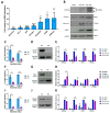
Figure 1
Figure 1
m6A affects the abundance of host transcripts upregulated during KSHV reactivation. (A) RT-qPCR analysis of cellular m6A-modified transcripts in TREX-BCBL1-Rta cells induced for 24 h compared to latent levels. (B) Representative Western blot of ZFP36L1 and GPRC5A protein levels in latent and reactivated TREX-BCBL1-Rta cells. (C) RT-qPCR analysis of WTAP RNA levels in latent and lytic WTAP shRNA-treated TREX-BCBL1-Rta cells. (D) Representative Western blot of WTAP protein levels in Scr- and WTAP-shRNA-treated latent TREX-BCBL1-Rta cells. (E) RNA levels of GPRC5A, STC1, FOSB and ZFP36L1 in Scr- or WTAP shRNA-treated latent and induced TREX-BCBL-1 cells. (F) RT-qPCR analysis of FTO RNA levels in latent and lytic FTO shRNA-treated TREX-BCBL1-Rta cells. (G) Representative Western blot of FTO protein levels in Scr- and FTO-shRNA-treated latent TREX-BCBL1-Rta cells. (H) RNA levels of GPRC5A, STC1, FOSB and ZFP36L1 in Scr- or FTO shRNA-treated latent and induced TREX-BCBL1-Rta cells. (I) RT-qPCR analysis of YTHDF1 RNA levels in latent and lytic YTHDF1 shRNA-treated TREX-BCBL1-Rta cells. (J) Representative Western blot of YTHDF1 protein levels in Scr- and YTHDF1-shRNA-treated latent TREX-BCBL1-Rta cells. (K) RNA levels of GPRC5A, STC1, FOSB and ZFP36L1 in Scr- or YTHDF1 shRNA-treated latent and induced TREx-BCBL1-Rta cells. In (A,C,E,F,H,I,K), data are presented as mean ± SD. * p < 0.05, ** p < 0.01, *** p < 0.001. All repeats are biological (n = 3).
Figure 2
Figure 2
m6A sites within the cellular GPRC5A transcript are essential for its stability. (A) m6A IP-qPCR analysis of cellular transcripts or control RNAs in latent TREX-BCBL1-Rta cells showing log2-fold enrichment of m6A-modified region relative to a non-methylated region in cis. Two regions were analysed within ZFP36L1, as this mRNA contains a wide flat m6A peak spanning several hundred bps. (B) m6A IP-qPCR analysis of GPRC5A or control RNAs in GPRC5A wild-type or Δm6A mutant-transfected HEK 293T cells showing fold enrichment of the m6A-modified region relative to a non-methylated region in cis. (C) RT-qPCR analysis of GPRC5A RNA in cells transfected with control plasmid, a wild type of GPRC5A-expressing constructs. (D) Representative Western blot of GPRC5A protein levels in cells transfected with no plasmid, wild-type or Δm6A GPRC5A-expressing constructs. (E) RT-qPCR analysis of wild-type or Δm6A GPRC5A RNA in cells treated with actinomycin D for 0, 4 or 8 h. In (A,B,C,E), data are presented as mean ± SD. * p < 0.05, ** p < 0.01, *** p < 0.001. All repeats are biological (n = 3).
Figure 3
Figure 3
GPRC5A is induced by the KSHV lytic transactivator, RTA. (A) Representative Western blot showing expression of transfected GFP-tagged viral proteins ORF50 and ORF57 in naïve uninfected HEK 293T cells. (B) RT-qPCR analysis of GPRC5A RNA levels in viral-protein-transfected HEK 293T cells. (C) RT-qPCR analysis of GPRC5A RNA levels in cells transfected with increasing concentrations of GFP-ORF50 DNA. (D) Representative Western blot of GPRC5A protein levels in cells transfected with a range of GFP-ORF50 DNA concentrations. (E) Densitometry quantification of immunoblots was performed using ImageJ software, and is shown as a percentage relative to the loading control, GAPDH. In (B,C,E), data are presented as mean ± SD. * p < 0.05, ** p < 0.01, *** p < 0.001. All repeats are biological (n = 3).
Figure 4
Figure 4
GPRC5A depletion reduces KSHV lytic replication. (A) RT-qPCR analysis of GPRC5A RNA levels in latent and lytic GPRC5A shRNA-treated TREX-BCBL1-Rta cells. (B) RT-qPCR analysis of viral RNAs ORF57 and ORF47 in latent and lytic GPRC5A-depleted cells. (C) Representative Western blot of viral proteins ORF57 and ORF65 in latent and lytic GPRC5A-depleted cells. (D) Densitometry quantification of immunoblots was performed using ImageJ software, and is shown as a percentage relative to the loading control, GAPDH. (E) RT-qPCR of ORF57 RNA levels in HEK 293T cells treated with virus-containing supernatant harvested from scrambled or GPRC5A shRNA-treated TREx-BCBL1-Rta cells. In (A,B,D,E), data are presented as mean ± SD. * p < 0.05, ** p < 0.01, *** p < 0.001. All repeats are biological (n = 3).
Figure 5
Figure 5
GPRC5A interacts with members of the Flotillin family and affects NFκB signalling. (A) Proteins with >1.5 enrichment in FLAG immunoprecipitates from GPRC5A-FLAG-overexpressing TREx-BCBL1-Rta cells compared with parental TREx-BCBL1-Rta cells after TMT mass spectrometry analysis (grey box: data points with less than 50 abundance in both latent and lytic cells were excluded). (B) STRING interaction map showing proteins with >1.4-fold interaction with GPRC5A in lytic replication compared with latency in TREx-BCBL1-Rta cells after TMT mass spectrometry analysis. (C) Immunoprecipitation of GFP or GPRC5A-GFP from transduced latent and lytic TREx-BCBL1-Rta cells showing increased co-immunoprecipitation of FLOT1 in lytic replication. (D) Immunofluorescence analysis of FLOT1 localisation in latent and lytic TREx-BCBL1-Rta cells transduced with a GFP control (upper two) or GFP-tagged (lower two) GPRC5A-expressing plasmid. (E) Representative Western blots of NFκB, pNFκB, pSTAT, pAKT, pERK and GAPDH protein expression in latent and lytic GPRC5A-depleted cells. (F) Densitometry quantification of NFκB and pNFκB immunoblots was performed using ImageJ software, and is shown as a percentage relative to the loading control, GAPDH. (G) Luciferase reporter assay from HEK 293Ts co-transfected with GFP or GPRC5A-GFP alongside various described signalling reporters with transcription factor binding sites attached to a luciferase reporter plasmid. Data presented are relative to an internal firefly control. In (F,G), data are presented as mean ± SD. * p < 0.01. All repeats are biological (n = 3).
Figure 6
Figure 6
Schematic showing potential regulation of GPRC5A gene expression during KSHV lytic replication. At the onset of KSHV lytic replication, the viral lytic master regulator RTA transactivates the GPRC5A promoter, increasing transcription of GPRC5A transcripts, which then become m6A-modified by the m6A methyltransferase complex. Methylation of GPRC5A mRNA increases its stability in cis, allowing translation of a larger pool of GPRC5A transcripts, and therefore, enhanced protein expression. Once translated, GPRC5A is transported to the cell membrane, where it is organised into lipid rafts composed of members of the flotillin family. As a membrane-bound GPCR, GPRC5A inhibits NFκB to enhance KSHV lytic replication.

References

    1. Ganem D. KSHV and the pathogenesis of Kaposi sarcoma: Listening to human biology and medicine. J. Clin. Investig. 2010;120:939–949. doi: 10.1172/JCI40567. - DOI - PMC - PubMed
    1. Dittmer D.P., Damania B. Kaposi’s Sarcoma-Associated Herpesvirus (KSHV)-Associated Disease in the AIDS Patient: An Update. Cancer Treat. Res. 2019;177:63–80. - PMC - PubMed
    1. Lange P., Damania B. Kaposi Sarcoma-Associated Herpesvirus (KSHV) Trends Microbiol. 2020;28:236–237. doi: 10.1016/j.tim.2019.10.006. - DOI - PubMed
    1. He M., Cheng F., da Silva S.R., Tan B., Sorel O., Gruffaz M., Li T., Gao S.J. Molecular Biology of KSHV in Relation to HIV/AIDS-Associated Oncogenesis. Cancer Treat. Res. 2019;177:23–62. - PMC - PubMed
    1. Dittmer D., Lagunoff M., Renne R., Staskus K., Haase A., Ganem D. A cluster of latently expressed genes in Kaposi’s sarcoma-associated herpesvirus. J. Virol. 1998;72:8309–8315. doi: 10.1128/JVI.72.10.8309-8315.1998. - DOI - PMC - PubMed

Publication types