. 2023 Aug;34(15-16):682-696.
doi: 10.1089/hum.2022.211.

An Engineered Adeno-Associated Virus Capsid Mediates Efficient Transduction of Pericytes and Smooth Muscle Cells of the Brain Vasculature

Affiliations

An Engineered Adeno-Associated Virus Capsid Mediates Efficient Transduction of Pericytes and Smooth Muscle Cells of the Brain Vasculature

Servio H Ramirez et al. Hum Gene Ther. 2023 Aug.

Erratum in

Abstract

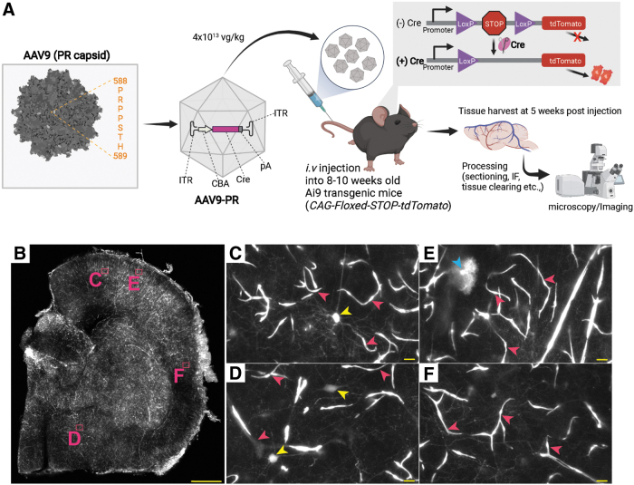
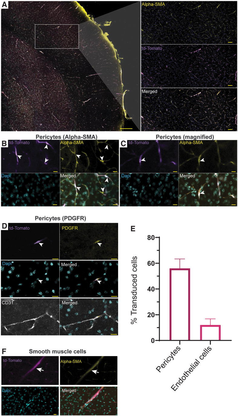
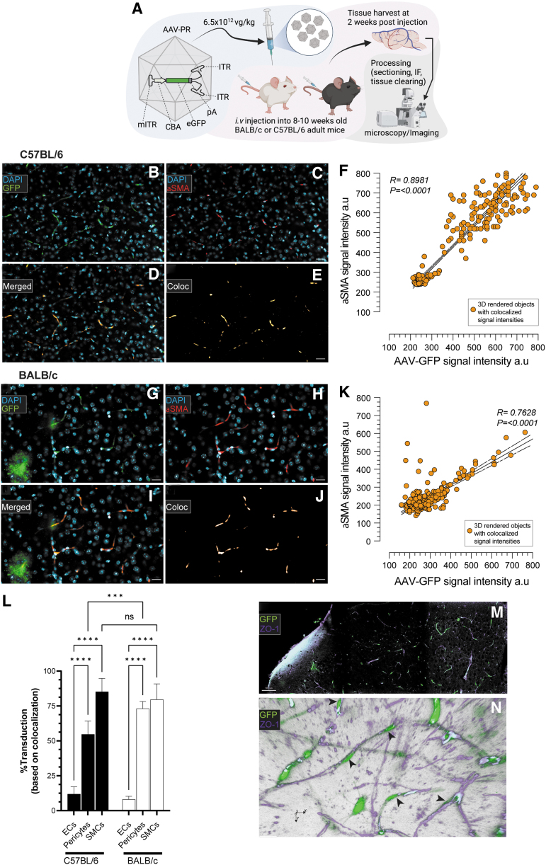
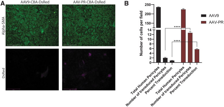
Neurodegeneration and cerebrovascular disease share an underlying microvascular dysfunction that may be remedied by selective transgene delivery. To date, limited options exist in which cellular components of the brain vasculature can be effectively targeted by viral vector therapeutics. In this study, we characterize the first engineered adeno-associated virus (AAV) capsid mediating high transduction of cerebral vascular pericytes and smooth muscle cells (SMCs). We performed two rounds of in vivo selection with an AAV capsid scaffold displaying a heptamer peptide library to isolate capsids that traffic to the brain after intravenous delivery. One identified capsid, termed AAV-PR, demonstrated high transduction of the brain vasculature, in contrast to the parental capsid, AAV9, which transduces mainly neurons and astrocytes. Further analysis using tissue clearing, volumetric rendering, and colocalization revealed that AAV-PR enabled high transduction of cerebral pericytes located on small-caliber vessels and SMCs in the larger arterioles and penetrating pial arteries. Analysis of tissues in the periphery indicated that AAV-PR also transduced SMCs in large vessels associated with the systemic vasculature. AAV-PR was also able to transduce primary human brain pericytes with higher efficiency than AAV9. Compared with previously published AAV capsids tropisms, AAV-PR represents the first capsid to allow for effective transduction of brain pericytes and SMCs and offers the possibility of genetically modulating these cell types in the context of neurodegeneration and other neurological diseases.

Keywords: adeno-associated virus vector; pericytes; smooth muscle cells.


Conflict of interest statement

C.A.M. has a financial interest in Sphere Gene Therapeutics, Inc., Chameleon Biosciences, Inc., and Skylark Bio, Inc., companies developing gene therapy platforms. C.A.M.'s interests were reviewed and are managed by MGH and Mass General Brigham in accordance with their conflict-of-interest policies. C.A.M., P.L.M., E.H., and K.S.H. have a filed patent application surrounding the AAV-PR capsid.

Figures

Figure 1.
Figure 1.
AAV-PR-CBA-Cre mediates vasculature-tropic transduction and gene modification in transgenic Ai9 mice (CAG-floxed-STOP-tdTomato). (A) AAV-PR peptide and insertion site. The peptide PRPPSTH is inserted after amino acid 588 of AAV9 VP1. A Cre-recombinase expression cassette is packaged inside AAV-PR and this is administered systemically to Ai9 mice, which contain a floxed-stop-tdTomato cassette in all cells. If AAV-PR mediates Cre expression, the loxP-stop is removed from the genome allowing tdTomato expression. (B) Half brain hemisphere of Ai9 mouse injected i.v. with AAV-PR-CBA-Cre showing bright intrinsic fluorescence (white signal). Letters (C–F) indicate areas of zoomed images in (B). The cells that were transduced had morphology consistent with the vasculature (pink arrowheads), and neurons (yellow and blue arrowheads). Scale bar in (B) = 500 μm. (C–F) = 10 μm. AAV, adeno-associated virus; CBA, hybrid CMV early/chicken β-actin promoter; i.v., intravenous.
Figure 2.
Figure 2.
Pericytes are efficiently transduced by AAV-PR after systemic injection. The identity of transduced vascular cells in Ai9 mice injected with AAV-PR-CBA-Cre was determined by immunofluorescence staining. (A) Low magnification image α-SMA staining (yellow) for pericytes and tdTomato (purple). Scale bar = 100 μm. Inset: higher magnification of the cortex showing colocalization. Scale bar = 50 μm (B, C). Higher magnification images of tdTomato/α-SMA colocalization. Scale bar = 10 μm (D). Colocalization of tdTomato with PDGFR-β. Bottom panel shows PDGFR (yellow), tdTomato (purple), DAPI (cyan), and CD31 endothelial cells (white). The colocalization of tdTomato was mainly with PDGFR and not CD31. (B–D) Arrows head point to transduced pericytes. (E) Quantitation of transduction efficiency in pericytes (PDGFR) and endothelial cells (CD31). Colocalization was based on a Pearson correlation of >0.35. (F) Transduction of smooth muscle cells on large caliber vessels. Arrow points to transduced smooth muscle cells. α-SMA, α-smooth muscle actin; DAPI, 4′,6-diamidino-2-phenylindole; PDGFR, platelet-derived growth factor receptor.
Figure 3.
Figure 3.
AAV-PR transduces aortic smooth muscle cells as well as multiple peripheral tissues. Ai9 mice were left untreated (noninjected) or were injected with AAV-PR-CBA-Cre and 5 weeks later, intrinsic tdTomato fluorescence observed. (A) Intrinsic tdTomato fluorescence (magenta) was readily detected in aorta of injected but not in untreated mice. Scale bar = 30 μm (B). Many of the transduced cells in aorta were smooth muscle cells as they colocalized with α-SMA and SM22α. Scale bar = 30 μm (C). Cell lining the coronary artery were transduced by AAV-PR. Scale bar = 100 μm (D). tdTomato expression in peripheral tissues of Ai9 mice injected with AAV-PR-CBA-Cre injected (left panels) and noninjected (right panels). Scale bars are as follows: liver, kidney = 500 μm; lungs, bladder, small intestine = 200 μm; large intestine = 100 μm.
Figure 4.
Figure 4.
AAV-PR transduces pericytes and smooth muscle cells in brain vasculature of C57BL/6 and BALB/c mice using a “gene addition” transgene expression cassette. (A) Overview of experiment. AAV-PR capsid packaged a sc cassette encoding GFP under the CBA promoter (AAV-PR-sc-CBA-GFP) was injected through the tail vein into adult C57BL/6 mice and BALB/c mice and 2 weeks later mice were sacrificed and processed for imaging of GFP fluorescence. (B) Transduction of α-SMA-positive pericytes in C57BL/6 mice. Scale bar = 25 μm. (C) Pericyte staining on small caliber vessels detected with α-SMA staining. (D) Merge of GFP (green) with α-SMA (red). (E) Rendering of GFP/α-SMA colocalization. (F) Correlation of α-SMA fluorescence signal intensity versus GFP signal intensity. (G–K) The same depiction as for C57BL/6, except in BALB/c mice. (L) Quantitation of endothelial cells, pericytes, and smooth muscle cells on large caliber vessels in both strains of mice. Values of significance indicated by asterisks compare transduction to endothelial cells. ****p < 0.0001; ***p = 0.0001; ns = not statistically significant. (M, N) An independent experiment was performed in C57BL/6 mice injected with AAV-PR-sc-CBA-GFP (6 × 1012 vg/kg) and 5 weeks later, brains were imaged for GFP (green) and ZO-1 to label endothelium (magenta). (M) Low magnification image of brain showing GFP cells transduced by AAV-PR and the endothelium labeled with ZO-1. Scale bar = 100 μm. (N) Volumetric rendering of stacked images shows spatial location of GFP-positive cells with pericyte morphology (black arrowheads) interacting with the ZO-1-positive cerebrum endothelium. GFP, green fluorescent protein; ITR, wild-type inverted terminal repeats; mITR, mutant inverted terminal repeats; pA, poly A signal sequence; sc, self-complementary; vg, vector genomes.
Figure 5.
Figure 5.
AAV-PR transduces primary human pericytes in culture. Human pericytes were transduced with equal doses of either AAV9 or AAV-PR both packaging a sc AAV-CBA-DsRed genome. Three days posttransduction cells were examined for α-SMA to determine culture purity and DsRed for transgene expression. (A) Representative images of transduced cells with either vector. DsRed fluorescence is shown in magenta. Scale bars = 275 μm. (B) Quantitation results from particle counting image analysis of transduced (DsRed positive) positive pericytes for AAV9 and AAV-PR. Shown are the number of cells/field counted, the number of DsRed-positive cells/field counted and the percentage of total cells expressing DsRed. Performed in triplicate, n = 8 averaged fields of view, ****p < 0.0001 by Student's unpaired t-test.

References

    1. Sweeney MD, Kisler K, Montagne A, et al. . The role of brain vasculature in neurodegenerative disorders. Nat Neurosci 2018;21(10):1318–1331. - PMC - PubMed
    1. Marini S, Anderson CD, Rosand J. Genetics of cerebral small vessel disease. Stroke 2020;51(1):12–20. - PMC - PubMed
    1. Chojdak-Łukasiewicz J, Dziadkowiak E, Budrewicz S. Monogenic causes of strokes. Genes (Basel) 2021;12(12):1855. - PMC - PubMed
    1. Lauer A, Speroni SL, Patel JB, et al. . Cerebrovascular disease progression in patients with ACTA2 Arg179 pathogenic variants. Neurology 2021;96(4): e538–e552. - PMC - PubMed
    1. Foust KD, Nurre E, Montgomery CL, et al. . Intravascular AAV9 preferentially targets neonatal neurons and adult astrocytes. Nat Biotechnol 2009;27(1):59–65. - PMC - PubMed

Publication types

Substances