. 2023 Aug 1;44(29):2746-2759.
doi: 10.1093/eurheartj/ehad381.

Sirtuin 2 deficiency aggravates ageing-induced vascular remodelling in humans and mice

Affiliations

Sirtuin 2 deficiency aggravates ageing-induced vascular remodelling in humans and mice

Yang Zhang et al. Eur Heart J. .

Abstract

Aims: The mechanisms underlying ageing-induced vascular remodelling remain unclear. This study investigates the role and underlying mechanisms of the cytoplasmic deacetylase sirtuin 2 (SIRT2) in ageing-induced vascular remodelling.

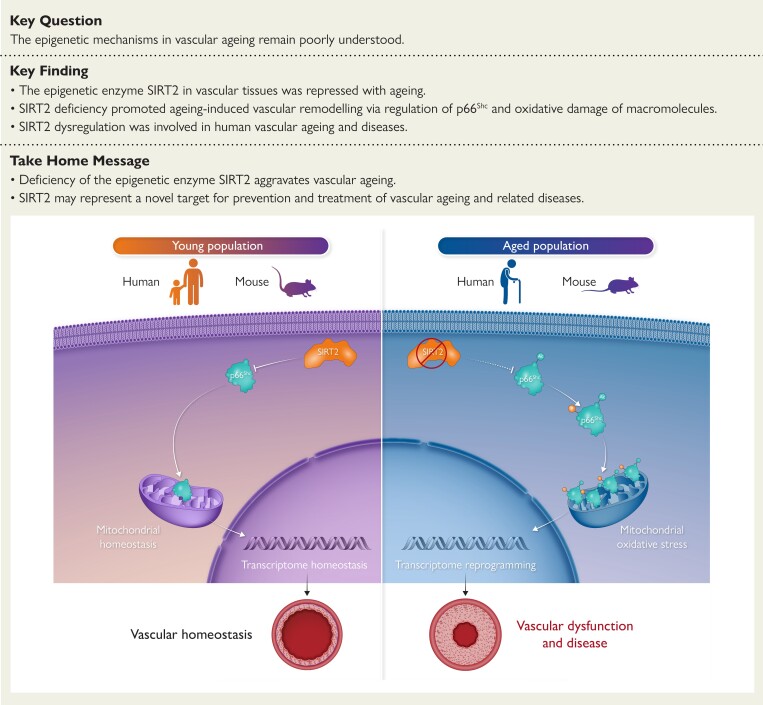
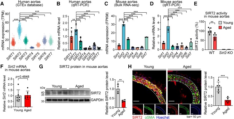
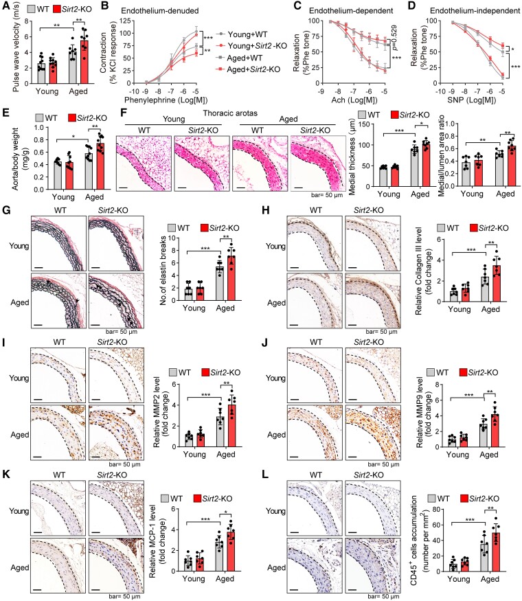
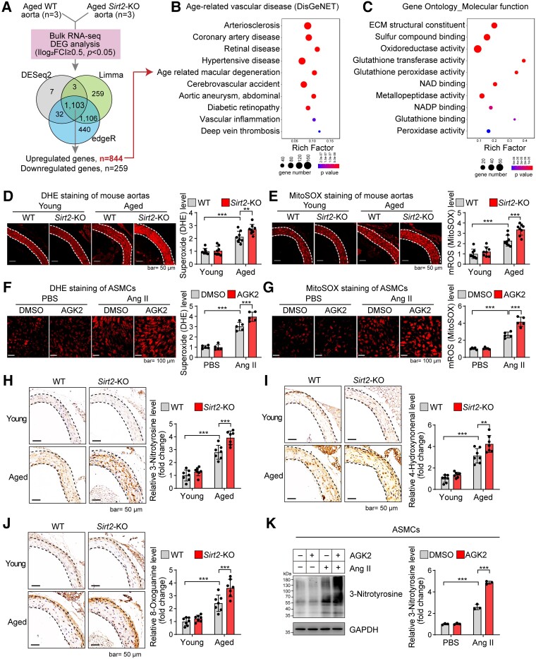
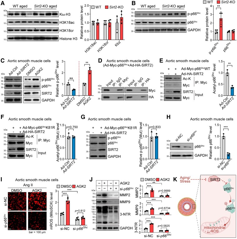
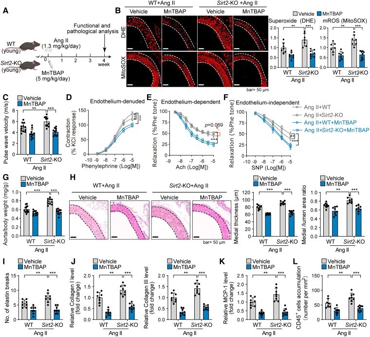
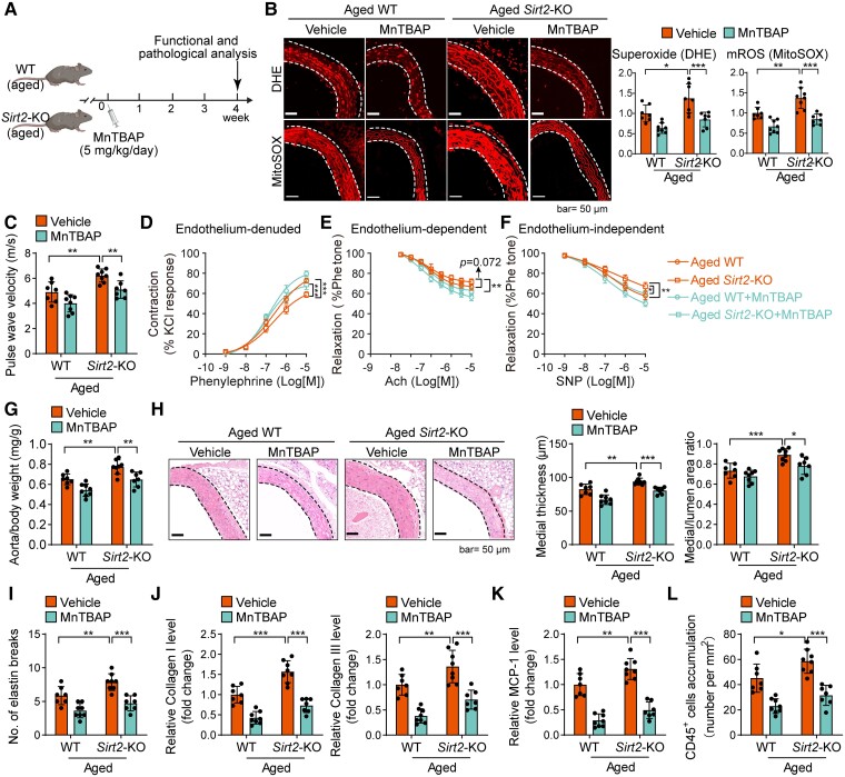
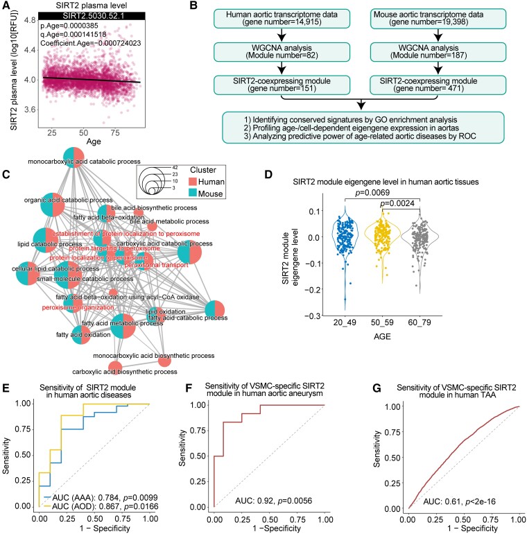
Methods and results: Transcriptome and quantitative real-time PCR data were used to analyse sirtuin expression. Young and old wild-type and Sirt2 knockout mice were used to explore vascular function and pathological remodelling. RNA-seq, histochemical staining, and biochemical assays were used to evaluate the effects of Sirt2 knockout on the vascular transcriptome and pathological remodelling and explore the underlying biochemical mechanisms. Among the sirtuins, SIRT2 had the highest levels in human and mouse aortas. Sirtuin 2 activity was reduced in aged aortas, and loss of SIRT2 accelerated vascular ageing. In old mice, SIRT2 deficiency aggravated ageing-induced arterial stiffness and constriction-relaxation dysfunction, accompanied by aortic remodelling (thickened vascular medial layers, breakage of elastin fibres, collagen deposition, and inflammation). Transcriptome and biochemical analyses revealed that the ageing-controlling protein p66Shc and metabolism of mitochondrial reactive oxygen species (mROS) contributed to SIRT2 function in vascular ageing. Sirtuin 2 repressed p66Shc activation and mROS production by deacetylating p66Shc at lysine 81. Elimination of reactive oxygen species by MnTBAP repressed the SIRT2 deficiency-mediated aggravation of vascular remodelling and dysfunction in angiotensin II-challenged and aged mice. The SIRT2 coexpression module in aortas was reduced with ageing across species and was a significant predictor of age-related aortic diseases in humans.

Conclusion: The deacetylase SIRT2 is a response to ageing that delays vascular ageing, and the cytoplasm-mitochondria axis (SIRT2-p66Shc-mROS) is important for vascular ageing. Therefore, SIRT2 may serve as a potential therapeutic target for vascular rejuvenation.

Keywords: Ageing; Arterial stiffness; SIRT2; Vascular remodelling; mROS; p66Shc.


Figures

Structured Graphical Abstract
The epigenetic regulator sirtuin 2 (SIRT2) governs a cytoplasm–mitochondria signal to repress vascular ageing. Ageing reduces SIRT2 protein levels and activity, which results in the hyperacetylation and activation of the adaptor protein p66Shc and production of mitochondrial reactive oxygen species (mROS), which subsequently reprogrammes the vascular transcriptome to aggravate ageing-induced vascular remodelling and diseases. The circulating SIRT2 may serve as a prognostic biomarker for ageing-related vascular diseases, and the cytoplasm–mitochondria axis SIRT2–p66Shc–mROS could be targeted for treating ageing-related vascular diseases.
Figure 1
Figure 1
Sirtuin 2 expression pattern in young and aged aortas. (A) Levels of SIRT1SIRT7 mRNAs in human aortas analysed using the Genotype-Tissue Expression database (n = 607). (B) Quantitative real-time PCR analysis of SIRT1SIRT7 mRNA levels in human aortas (n = 6). (C) The bulk RNA-seq analysis of the expression level of sirtuin members Sirt1Sirt7 in mouse aortas (n = 3). (D) Quantitative real-time PCR analysis of sirtuin members Sirt1Sirt7 in mouse aortas (n = 3). (E) Sirtuin 2 enzymatic activity was reduced in aged aortas (24 months old) compared with that in young aortas (4 months) of mice (n = 6). (F) mRNA level of Sirt2 in aortas from young and aged mice (n = 6). (G) Protein level of sirtuin 2 (both bands) in aortas from young and aged mice (n = 4). (H) Immunofluorescence staining revealed that the sirtuin 2 protein level was reduced in aged aortas compared with that in young aortas of mice. Representative images and quantitative results are shown (n = 5). αSMA, alpha-smooth muscle actin. Statistical analyses were performed using the Kruskal–Wallis test with Dunn’s post hoc test (A), one-way ANOVA with the Bonferroni post hoc test (B–D), unpaired Student’s t-test (E, G, H), and the Mann–Whitney U test (F). **P < .01, *** P < .001.
Figure 2
Figure 2
Sirtuin 2 deficiency aggravates ageing-induced vascular dysfunction and remodelling. (A) Pulse wave velocity measurements in young and aged mice revealed that Sirt2 knockout promoted an ageing-induced increase in arterial stiffness (n = 8–10). (B–D) Ex vivo analysis of the vascular constriction–relaxation function of aortas from mice (n = 5). (B) Arterial vessel contractions mediated through phenylephrine. (C) Endothelium-dependent relaxation in response to acetylcholine (Ach). (D) Endothelium-independent relaxation in response to nitric oxide donor sodium nitroprusside (NO-donor SNP), an endothelium-independent agonist. (E) Sirt2 knockout increased aged mice’s aorta weight-to-body weight ratio (n = 8–10). The aorta weight/body weight ratio of young and aged wild-type and Sirt2 knockout mice was analysed. (F) Sirt2 knockout enhanced vascular remodelling in aged mice. Haematoxylin–eosin staining of the thoracic aortas was performed, and the medial thickness and the ratio of the media area to the vessel lumen were quantified (n = 7–8). (G) Elastin van Gieson staining of elastin fibres in thoracic aortas. Representative images and quantitative results are shown (n = 7). Arrows denote broken elastin fibres. (H–L) Immunohistochemical staining of collagen III (H), matrix metallopeptidase 2 (I), matrix metallopeptidase 9 (J), monocyte chemotactic protein-1 (K), and CD45 (L). Representative images and quantitative results are shown (n = 7). Statistical analyses were performed using one-way ANOVA with the Bonferroni post hoc test. *P < .05, **P < .01, ***P < .001.
Figure 3
Figure 3
Sirtuin 2 regulates gene signatures associated with age-related arterial diseases and oxidative stress. (A) Bulk RNA-seq analysis of the transcriptome of aged aortas of wild-type and Sirt2 knockout mice (n = 3). Venn plot showing the overlapped genes among differentially expressed genes identified by three methods. A total of 844 genes were up-regulated, while 259 were down-regulated in aged Sirt2 knockout aortas (|log2 FC| ≥ 0.5, adjusted P < .05). (B) Sirt2 knockout led to the enrichment of genes involved in age-related arterial diseases. Human disease enrichment analysis (DisGeNET) was performed with the up-regulated genes using ToppGene Suite. (C) Sirt2 knockout led to the enrichment of genes involved in the oxidation–reduction process. Gene Ontology Molecular_function enrichment analysis was performed with the up-regulated genes using ToppGene Suite. (D, E) Sirt2 knockout increased ageing-induced oxidative stress in mouse aortas. Total cellular superoxide and mitochondrial reactive oxygen species were monitored by dihydroethidium and MitoSOX staining, respectively. Representative results and quantitative results of dihydroethidium (D) and MitoSOX staining (E) are shown (n = 8). (F, G) Sirtuin 2 inhibition promoted stress-induced oxidative stress in aortic smooth muscle cells. Aortic smooth muscle cells were treated with the sirtuin 2–specific inhibitor AGK2 (10 μM) with/without angiotensin II (1 μM) treatment for 24 h. Total cellular superoxide and mitochondrial reactive oxygen species were monitored by dihydroethidium and MitoSOX staining, respectively. Representative results and quantitative results of dihydroethidium (F) and MitoSOX staining (G) are shown (n = 5). (H–J) Sirt2 knockout increased ageing-induced oxidation of macromolecules in mouse aortas: (H) protein oxidation (3-nitrotyrosine); (I) lipid peroxidation (4-hydroxynonenal); and (J) DNA oxidation (8-oxoguanine). Representative results and quantitative results are shown (n = 7). (K) Sirtuin 2 inhibition promoted stress-induced protein oxidation in aortic smooth muscle cells. Aortic smooth muscle cells were treated with the sirtuin 2–specific inhibitor AGK2 (10 μM) with/without angiotensin II (1 μM) treatment for 24 h. Protein oxidation was analysed by western blot. Representative blots and quantitative results are shown (n = 3). Statistical analyses were performed using one-way ANOVA with the Bonferroni post hoc test (D–K). **P < .01, ***P < .001.
Figure 4
Figure 4
Sirtuin 2 regulates oxidative stress by deacetylating p66Shc. (A) Sirtuin 2 effects on histone modification in aortas from aged mice. Representative western blot and quantitative results are shown (n = 4). (B) Sirtuin 2 deficiency increased p66Shc phosphorylation in the aortas of aged mice. Representative and quantitative results are shown (n = 4). (C) Sirtuin 2 repressed p66Shc phosphorylation in aortic smooth muscle cells. Aortic smooth muscle cells were infected with an adenovirus carrying sirtuin 2/control adenovirus or treated with the sirtuin 2 inhibitor AGK2 (10 μM)/DMSO for 24 h, followed by western blot analysis. Representative and quantitative results are shown (n = 3). (D) Sirtuin 2 interacted with p66Shc in aortic smooth muscle cells. Aortic smooth muscle cells were infected with an adenovirus carrying Myc-tagged p66Shc and HA-tagged sirtuin 2, followed by immunoprecipitation with Myc and HA antibodies and western blot analysis (n = 3). (E) Sirtuin 2 deacetylated p66Shc in aortic smooth muscle cells. Aortic smooth muscle cells were infected with Ad-Myc-p66Shc with Ad-SIRT2 or Ad-Ctrl, followed by immunoprecipitation with anti-Myc antibodies and western blot analysis of acetylated lysine (Ac-K) on p66Shc (n = 3). (F, G) Sirtuin 2 deacetylated p66Shc at lysine 81 (K81) to inhibit p66Shc phosphorylation in aortic smooth muscle cells. Aortic smooth muscle cells were infected with Myc-tagged K81R (lysine to arginine) mutant p66Shc (Ad-Myc-p66ShcK81R) with Ad-SIRT2 or Ad-Ctrl, followed by immunoprecipitation with anti-Myc antibodies and western blot analysis of acetylated lysine on p66Shc (F) (n = 3) and phosphorylation of p66Shc (G) (n = 3). (H) siRNA-mediated knockdown of p66Shc in aortic smooth muscle cells. The cells were transfected with si-NC or si-p66Shc for 48 h. Then, a western blot was performed to analyse protein expression (n = 3). (I) p66Shc contributed to sirtuin 2 function in regulating mitochondrial reactive oxygen species in aortic smooth muscle cells. Aortic smooth muscle cells were transfected with si-p66Shc or control si-NC for 24 h, followed by treatment with/without the sirtuin 2–specific inhibitor AGK2 (10 μM) and angiotensin II (1 μM) for another 24 h. Mitochondrial reactive oxygen species were monitored by MitoSOX staining. Representative and quantitative results are shown (n = 5). (J) p66Shc contributed to sirtuin 2 function in regulating protein oxidation and matrix metalloproteinase expression in aortic smooth muscle cells. The cells were treated as in (I), and protein expression was analysed by western blot. Representative blots and quantitative results are shown (n = 3). (K) Illustration showing that sirtuin 2 regulates p66Shc acetylation and phosphorylation to repress mitochondrial reactive oxygen species in vascular cells. Statistical analyses were performed using unpaired Student’s t-test (A–H) and one-way ANOVA with the Bonferroni post hoc test (I, J). **P < .01, ***P < .001.
Figure 5
Figure 5
Sirtuin 2–reactive oxygen species axis contributes to vascular remodelling and dysfunction in angiotensin II–challenged mice. (A) Experimental design. Male young (4-month-old) wild-type and Sirt2 knockout mice were infused with angiotensin II (1.3 mg/kg/day) to replicate vascular ageing phenotypes. The mice were treated with/without the superoxide scavenger MnTBAP (5 mg/kg/day, i.p.) treatment for 4 weeks. (B) MnTBAP reduced reactive oxygen species in angiotensin II–infused wild-type and Sirt2 knockout mice. Young wild-type and Sirt2 knockout mice were infused with angiotensin II (1.3 mg/kg/day) and intraperitoneally treated with MnTBAP (5 mg/kg/day, daily) or saline (vehicle) for 4 weeks. Then, reactive oxygen species levels in mouse thoracic aortas were analysed by dihydroethidium (total superoxide) and MitoSOX (mitochondrial reactive oxygen species) staining (n = 7–8). (C) MnTBAP treatment reduced the angiotensin II–induced increase in pulse wave velocity (n = 10–12). (D–F) Ex vivo analysis of the vascular constriction–relaxation function of aortas from mice (n = 5). (D) Arterial vessel contractions mediated through phenylephrine. (E) Endothelium-dependent relaxation in response to acetylcholine. (F) Endothelium-independent relaxation in response to nitric oxide donor sodium nitroprusside. (G) MnTBAP treatment reduced the angiotensin II–induced increase in aorta weight (n = 10–12). (H) Representative and quantitative results showing MnTBAP treatment reduced angiotensin II–induced aortic remodelling (n = 8). (I) Elastin van Gieson staining of elastin fibres in thoracic aortas. Quantitative results are shown (n = 8). (J–L) Immunohistochemical staining of collagen I and III (J), monocyte chemotactic protein-1 (K), and CD45 (L). Quantitative results are shown (n = 7–8). Statistical analyses were performed using one-way ANOVA with the Bonferroni post hoc test. **P < .01, ***P < .001.
Figure 6
Figure 6
Reactive oxygen species contributes to sirtuin 2 deficiency–mediated aggravation of vascular remodelling and dysfunction in aged mice. (A) Experimental design. Aged wild-type and Sirt2 knockout mice received MnTBAP (5 mg/kg/day, i.p.) or vehicle therapy for 4 weeks. (B) MnTBAP reduced reactive oxygen species in aged wild-type and Sirt2 knockout mice. Aged wild-type and Sirt2 knockout mice were intraperitoneally treated with MnTBAP (5 mg/kg/day, daily) or saline (vehicle) for 4 weeks. Then, reactive oxygen species levels in mouse thoracic aortas were analysed by dihydroethidium (total superoxide) and MitoSOX (mitochondrial reactive oxygen species) staining (n = 7–8). (C) MnTBAP therapy mitigated sirtuin 2 effects on pulse wave velocity in aged mice (n = 7–8). (D–F) Ex vivo analysis of mouse aortas’ vascular constriction–relaxation function (n = 5). Arterial vessel contractions mediated through phenylephrine (D), endothelium-dependent relaxation in response to acetylcholine (E), endothelium-independent relaxation in response to nitric oxide donor sodium nitroprusside (F), and an endothelium-independent agonist. (G) MnTBAP therapy reduced sirtuin 2 effects on aorta weight in aged mice (n = 7–8). (H) Representative and quantitative results showing MnTBAP therapy reduced sirtuin 2 effects on aortic remodelling in aged mice (n = 7–8). (I) Elastin van Gieson staining of elastin fibres in thoracic aortas. Quantitative results are shown (n = 7–8). (J–L) Immunohistochemical staining of collagen I and III (J), monocyte chemotactic protein-1 (K), and CD45 (L). Quantitative results are shown (n = 7–8). Statistical analyses were performed using one-way ANOVA with the Bonferroni post hoc test. *P < .05, **P < .01, ***P < .001.
Figure 7
Figure 7
Sirtuin 2 in ageing-related arterial remodelling and diseases in humans. (A) Plasma sirtuin 2 level is decreased with ageing. Plasma sirtuin 2 level across the lifespan was analysed using a public proteome dataset, which involved plasma protein data from 4263 young adults to nonagenarians (18–95 years old). (B) Flowchart for the identification of the sirtuin 2 coexpression module in human and mouse aortas. (C) Network showing the gene ontology enrichment of the sirtuin 2 coexpression module in humans and mice. The dot size denotes the ratio of genes within the pathway, and the dot colour represents the species. (D) Comparison of the expression level of the sirtuin 2 coexpression module eigengene in human aortas across three age groups (Kruskal–Wallis test with Dunn’s post hoc test). (E–G) Receiver operating characteristic (ROC) curves of the sirtuin 2 module level show that the sirtuin 2 coexpression module is associated with age-related human aortic diseases. Area under the curve was calculated by receiver operating characteristic analysis. (E) Sirtuin 2 coexpression module exhibits a predictive significance for human abdominal aortic aneurysm and aortic occlusive disease. Gene expression data (GSE57691) of human abdominal aortic aneurysm and atherosclerosis (aortic occlusive disease) were used to analyse the level of the sirtuin 2 coexpression module, and subsequently, receiver operating characteristic analysis was performed. (F) Sirtuin 2 coexpression module in smooth muscle cells shows a predictive significance for aortic aneurysms. Gene expression data (GSE140947) of fresh human normal aortic media smooth muscle cells and aortic aneurysm media smooth muscle cells were subjected to evaluate the sirtuin 2 coexpression module, and subsequently, receiver operating characteristic analysis was performed. (G) Sirtuin 2 coexpression module in single smooth muscle cells exhibits a predictive significance for thoracic aortic aneurysms. scRNA-seq data of control and human thoracic aortic aneurysm tissues (GSE155468) were used to analyse the sirtuin 2 coexpression module level in smooth muscle cells, and then receiver operating characteristic analysis was performed at the single-cell level.

Comment in

References

    1. Roth GA, Forouzanfar MH, Moran AE, Barber R, Nguyen G, Feigin VL, et al. . Demographic and epidemiologic drivers of global cardiovascular mortality. N Engl J Med 2015;372:1333–1341. 10.1056/NEJMoa1406656 - DOI - PMC - PubMed
    1. Camici GG, Liberale L. Aging: the next cardiovascular disease? Eur Heart J 2017;38:1621–1623. 10.1093/eurheartj/ehx239 - DOI - PubMed
    1. Paneni F, Diaz Cañestro C, Libby P, Lüscher TF, Camici GG. The aging cardiovascular system: understanding it at the cellular and clinical levels. J Am Coll Cardiol 2017;69:1952–1967. 10.1016/j.jacc.2017.01.064 - DOI - PubMed
    1. North BJ, Sinclair DA. The intersection between aging and cardiovascular disease. Circ Res 2012;110:1097–1108. 10.1161/CIRCRESAHA.111.246876 - DOI - PMC - PubMed
    1. Grunewald M, Kumar S, Sharife H, Volinsky E, Gileles-Hillel A, Licht T, et al. . Counteracting age-related VEGF signaling insufficiency promotes healthy aging and extends life span. Science 2021;373:eabc8479. 10.1126/science.abc8479 - DOI - PubMed

Publication types

Substances