Review
. 2023 Jun 12:14:1184246.
doi: 10.3389/fneur.2023.1184246. eCollection 2023.

Nerve trunk healing and neuroma formation after nerve transection injury

Affiliations
Review

Nerve trunk healing and neuroma formation after nerve transection injury

Dong-Xu Huang et al. Front Neurol. .

Abstract

The nerve trunk healing process of a transected peripheral nerve trunk is composed of angiogenesis, nerve fiber regeneration, and scarring. Nerve trunk healing and neuroma formation probably share identical molecular mediators and similar regulations. At the nerve transection site, angiogenesis is sufficient and necessary for nerve fiber regeneration. Angiogenesis and nerve fiber regeneration reveal a positive correlation in the early time. Scarring and nerve fiber regeneration show a negative correlation in the late phase. We hypothesize that anti-angiogenesis suppresses neuromas. Subsequently, we provide potential protocols to test our hypothesis. Finally, we recommend employing anti-angiogenic small-molecule protein kinase inhibitors to investigate nerve transection injuries.

Keywords: angiogenesis; nerve fiber regeneration; peripheral nerve injury; protein kinase inhibitor; scarring; traumatic neuroma.


Conflict of interest statement

The authors declare that the research was conducted in the absence of any commercial or financial relationships that could be construed as a potential conflict of interest.

Figures

Figure 1
Figure 1
The peripheral nerve trunk consists of connective tissue layers, blood vessels, and nerve fibers (Created with BioRender.com). (A) Connective tissue components of the peripheral nerve trunk are arranged into three intimately connected layers from the outside to the inside: epineurium, perineurium, and endoneurium. (B) Extrafascicular and intrafascicular vessels spread throughout the epineurium and endoneurium, respectively. (C) Nerve fibers include myelinated axons and unmyelinated axons. (D) The peripheral nerve trunk is embedded in the surrounding tissue bed.
Figure 2
Figure 2
The change of a nerve trunk after transection injury (Created with BioRender.com). (A) The normal nerve trunk and extrafascicular blood vessels. (B) After nerve transection injury, the nerve trunk divides into the proximal and distal stumps. Two stumps retract because of the elasticity within the nerve trunk. An interstump gap inevitably forms.
Figure 3
Figure 3
A schematic illustration of a transected nerve trunk and its surrounding tissue bed (Created with BioRender.com). The nerve transection site has four individual compartments: the end of the proximal stump, the interstump gap, the end of the distal stump, and the surrounding tissue bed.
Figure 4
Figure 4
The nerve trunk healing process at the nerve transection site (Created with BioRender.com). (A,B) Angiogenesis occurs in the proximal stump, distal stump, interstump gap, and surrounding tissue bed. (C,D) Nerve fibers own a robust ability to regenerate from the proximal stump. Some nerve fibers cross the interstump gap and subsequently enter the distal stump where Wallerian degeneration occurs, but others enter the surrounding tissue bed. (E,F) Scarring by depositing collagen fibers occurs to reconstitute the interstump gap. DS, distal stump; PS, proximal stump.
Figure 5
Figure 5
The formation of terminal neuroma and suture line neuroma (Created with BioRender.com). A terminal neuroma revealing the chaos of regenerating nerve fibers, blood vessels, and collagen fibers grows at the end of the proximal stump when there is no distal stump. Similarly, a suture line neuroma forms at the nerve transection site even after the interstump gap is surgically repaired.
Figure 6
Figure 6
Relationships among angiogenesis, nerve fiber regeneration, and scarring at peripheral nerve transection sites at the tissue level (Created with BioRender.com). Angiogenesis at the nerve transection site is sufficient and necessary for nerve fiber regeneration in the early time. Angiogenesis and nerve fiber regeneration show a positive correlation in the early time. Scarring and nerve fiber regeneration reveal a negative correlation in the late time. The relationship between angiogenesis and scarring during nerve trunk healing is still unknown.
Figure 7
Figure 7
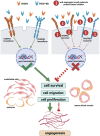
The main machinery of the angiogenic signaling pathway and its blocking with anti-angiogenic small-molecule protein kinase inhibitors (Created with BioRender.com). When a VEGFA dimer combines with two VEGFR2 monomers on the cell membrane, these VEGFR2 monomers dimerize and auto-phosphorylate, which initiates downstream signaling cascades leading to cell survival, migration, and proliferation. Similarly, following the combination of a PDGF-BB dimer and two PDGFRβ monomers, these PDGFRβ monomers dimerize and auto-phosphorylate. The VEGFA-VEGFR2 pathway is implicated in every aspect of angiogenesis. PDGFRβ regulates the recruitment of pericytes and the (PDGF-BB)-PDGFRβ pathway is essential for vascular maturation. The simultaneous blocking of the VEGFR and PDGFR with multi-target small-molecule protein kinase inhibitors is an effective anti-angiogenic strategy. VEGFA, vascular endothelial growth factor a; PDGF-BB, homodimer of platelet-derived growth factor b chain; VEGFR2, vascular endothelial growth factor receptor 2; PDGFRβ, platelet-derived growth factor receptor beta.

References

    1. Zochodne DW. The challenges and beauty of peripheral nerve regrowth. J Peripher Nerv Syst. (2012) 17:1–18. doi: 10.1111/j.1529-8027.2012.00378.x - DOI - PubMed
    1. Höke A, Brushart T. Introduction to special issue: challenges and opportunities for regeneration in the peripheral nervous system. Exp Neurol. (2010) 223:1–4. doi: 10.1016/j.expneurol.2009.12.001 - DOI - PMC - PubMed
    1. Fu SY, Gordon T. The cellular and molecular basis of peripheral nerve regeneration. Mol Neurobiol. (1997) 14:67–116. doi: 10.1007/bf02740621 - DOI - PubMed
    1. Scheib J, Höke A. Advances in peripheral nerve regeneration. Nat Rev Neurol. (2013) 9:668–76. doi: 10.1038/nrneurol.2013.227 - DOI - PubMed
    1. Lundborg G. Nerve injury and repair: regeneration, reconstruction, and cortical remodeling. 2nd ed. Philadelphia, PA: Elsevier; (2004). 248 p.

LinkOut - more resources