Case Reports
. 2023 Jun 12:13:1168963.
doi: 10.3389/fonc.2023.1168963. eCollection 2023.

Case report: Combined cervical incision with an intercostal uniportal video-assisted thoracoscopic surgery approach for mediastinal brachial plexus schwannoma

Affiliations
Case Reports

Case report: Combined cervical incision with an intercostal uniportal video-assisted thoracoscopic surgery approach for mediastinal brachial plexus schwannoma

Linlin Wang et al. Front Oncol. .

Abstract

Mediastinal neurogenic tumors primarily originate from the intercostal and sympathetic nerves, whereas schwannomas originating from the brachial plexus are rare. Surgical intervention for such tumors is complex and associated with the risk of postoperative upper limb dysfunction due to their unique anatomical location. In this report, we present the case of a 21-year-old female diagnosed with a mediastinal schwannoma, who underwent a novel surgical approach combining cervical incision and intercostal uniportal video-assisted thoracoscopic surgery (VATS). Our study reviewed the patient's clinical presentation, treatment approach, pathology, and prognosis. The findings of this study demonstrate that the cervical approach, combined with intercostal uniportal VATS, is a feasible surgical method for the removal of mediastinal schwannomas originating from the brachial plexus.

Keywords: brachial plexus; faster rehabilitation.; minimally invasive; schwannomas; uniportal video-assisted thoracoscopic surgery.


Conflict of interest statement

The authors declare that the research was conducted in the absence of any commercial or financial relationships that could be construed as a potential conflict of interest.

Figures

Figure 1
Figure 1
Imaging before surgery. (A) CT scan of the chest showing a 33 mm mass with clear boundaries located beside the mediastinum of the upper lobe of the left lung. Calcified nodules within the mass are slightly enhanced. Coronal (C) and sagittal (D) chest CT images reveal no abnormal changes in the adjacent thoracic vertebrae and ribs. (B) MRI of the thoracic vertebrae showing an equal-length T1, and long T2 mainly mixed signal shadows on the left side of the upper mediastinum (around the level of the T1-T2 vertebrae), low-signal shadows are observed on T2WI.
Figure 2
Figure 2
Three-dimensional reconstruction CT scans of tumor vessels were obtained preoperatively (A) and postoperatively (B).
Figure 3
Figure 3
(A) The cervical approach incision. (B–F) The procedure begins with a horizontal incision along the left supraclavicular area, followed by the sequential removal of tissue layers including the platysma muscle, deep cervical fascia, and carotid sheath, exposing the left recurrent laryngeal nerve, venous horn, lymphatic vessels, and left subclavian artery. The tumor was then carefully separated from the surrounding tissue and the upper pole of the tumor was excised. (G–I) The ultrasonic scalpel was utilized to carefully remove the mass from the root of the brachial plexus.
Figure 4
Figure 4
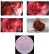
(A) The tumor’s anatomical relationship with the subclavian artery and surrounding tissues was visualized from the neck perspective. (B) The location of the lesion under uniportal VATS. (C) The superior aspect of the thoracic cavity following successful lesion resection. (D) The tumor measuring 3.3 × 3.3 cm in diameter after being removed. (E) Post-pathology results reveal that the mass was a mediastinal brachial plexus schwannoma. Immunohistochemical analysis revealed positive staining for CD34 (vessel), Ki-67 (approximately 1%), S-100, Vimentin, and SOX-10, whereas staining for CK-P, Desmin, and NSE was negative.

References

    1. Smahi M, Lakranbi M, Ouadnouni Y, Bouarhroum A, Sbai H, Znati K, et al. . Intrathoracic phrenic nerve neurofibroma. Ann Thorac Surg (2011) 91:e57–8. doi: 10.1016/j.athoracsur.2010.11.028 - DOI - PubMed
    1. D’Souza B, Lowe A, Stewart B, Roberts T. A rare case of schwannoma of the intrathoracic phrenic nerve. ANZ J Surg (2010) 80:841–2. doi: 10.1111/j.1445-2197.2010.05504.x - DOI - PubMed
    1. Roh JL. Function-preserving intracapsular enucleation of brachial plexus schwannomas: is it safe and effective? Oral Oncol (2022) 134:106102. doi: 10.1016/j.oraloncology.2022.106102 - DOI - PubMed
    1. Lee DY, Chi JY, Seok J, Han S, Lee MH, Jeong WJ, et al. . Feasibility of brachial plexus schwannoma enucleation with intraoperative neuromonitoring. Clin Exp Otorhinolaryngol (2020) 13:203–8. doi: 10.21053/ceo.2019.01207 - DOI - PMC - PubMed
    1. Ryu KH, Moon JI, Baek HJ, Cho SB, Choi BH, An HJ, et al. . Brachial plexus schwannoma mimicking cervical lymphadenopathy. Med (Baltimore) (2018) 97:e12880. doi: 10.1097/MD.0000000000012880 - DOI - PMC - PubMed

Publication types

LinkOut - more resources