An investigation of bovine papillomaviruses from ocular squamous cell carcinomas in cattle
- PMID: 37378381
- PMCID: PMC10291527
- DOI: 10.22099/IJVR.2022.43152.6282
An investigation of bovine papillomaviruses from ocular squamous cell carcinomas in cattle
Abstract
Background: Ocular squamous cell carcinomas (OSCCs) in cattle has been studied for many years, but no definite etiology has been established. Squamous cell carcinomas (SCCs) may occur in different body parts of cattle. Depending on the location, it can cause an economic loss of varying degrees.
Aims: The aim of this study was to investigate the causes of OSCCs in the eye region of cattle. Methods: Sixty tumoral masses taken form 60 cattle with proliferation in the eye region that were collected between the years 2012-2022 were used. These cases were admitted to our department for routine diagnosis. The tissues were diagnosed as OSCC using histopathological methods. The presence of bovine papillomavirus (BPV), one of the causative factors, was investigated using immunohistochemical and polymerase chain reaction (PCR).
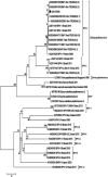
Results: Macroscopically masses were nodular or cauliflower-like and fragile and had hemorrhagic surfaces. Considering the keratin pearls, tumoral islands, and squamous differentiation, 20 out of 60 cases were classified as well, 20 as moderately, and 20 as poorly-differentiated OSCCs. 47 of the 60 cases were BPV positive using immunohistochemical methods. However, BPV nucleic acid was detected in only two cases with PCR. Only one of the cases could be sequenced. After phylogenetic analysis, virus strain was identified as BPV-1.
Conclusion: Our results indicated that papillomaviruses can contribute to the development of OSCCs, in both precursor lesions and also advanced stage OSCCs. We found that BPV-1 has a possible causative role; however, more studies are needed to investigate the role of other viral agents and their interaction with secondary factors.
Keywords: Bovine papillomavirus; Immunohistochemistry; Ocular squamous cell carcinoma; PCR.
Conflict of interest statement
The authors declare no competing interests.
Figures


References
-
- Altamura G, Cardeti G, Cersini A, Eleni C, Cocumelli C, Bartolomé Del Pino LE, Razzuoli E, Martano M, Maiolino P, Borzacchiello G. Detection of Felis catus papillomavirus type-2 DNA and viral gene expression suggest active infection in feline oral squamous cell carcinoma. Vet. Comp. Oncol. 2020;18:494–501. - PubMed
-
- Armando F, Mecocci S, Orlandi V, Porcellato I, Cappelli K, Mechelli L, Brachelente C, Pepe M, Gialletti R, Ghelardi A, Passeri B, Razzuoli E. Investigation of the epithelial to mesenchymal transition (EMT) process in Equine papillomavirus-2 (EcPV-2)-positive penile squamous cell carcinomas. Int. J. Mol. Sci. 2021;22 - PMC - PubMed
-
- Beytut E. Pathological and immunohistochemical evaluation of skin and teat papillomas in cattle. Turk. J. Vet. Anim. Sci. 2017;41:204–212.
LinkOut - more resources
Full Text Sources
