. 2023 Sep 1;11(9):1222-1236.
doi: 10.1158/2326-6066.CIR-22-0254.

AXL Inhibition Improves the Antitumor Activity of Chimeric Antigen Receptor T Cells

Affiliations

AXL Inhibition Improves the Antitumor Activity of Chimeric Antigen Receptor T Cells

R Leo Sakemura et al. Cancer Immunol Res. .

Abstract

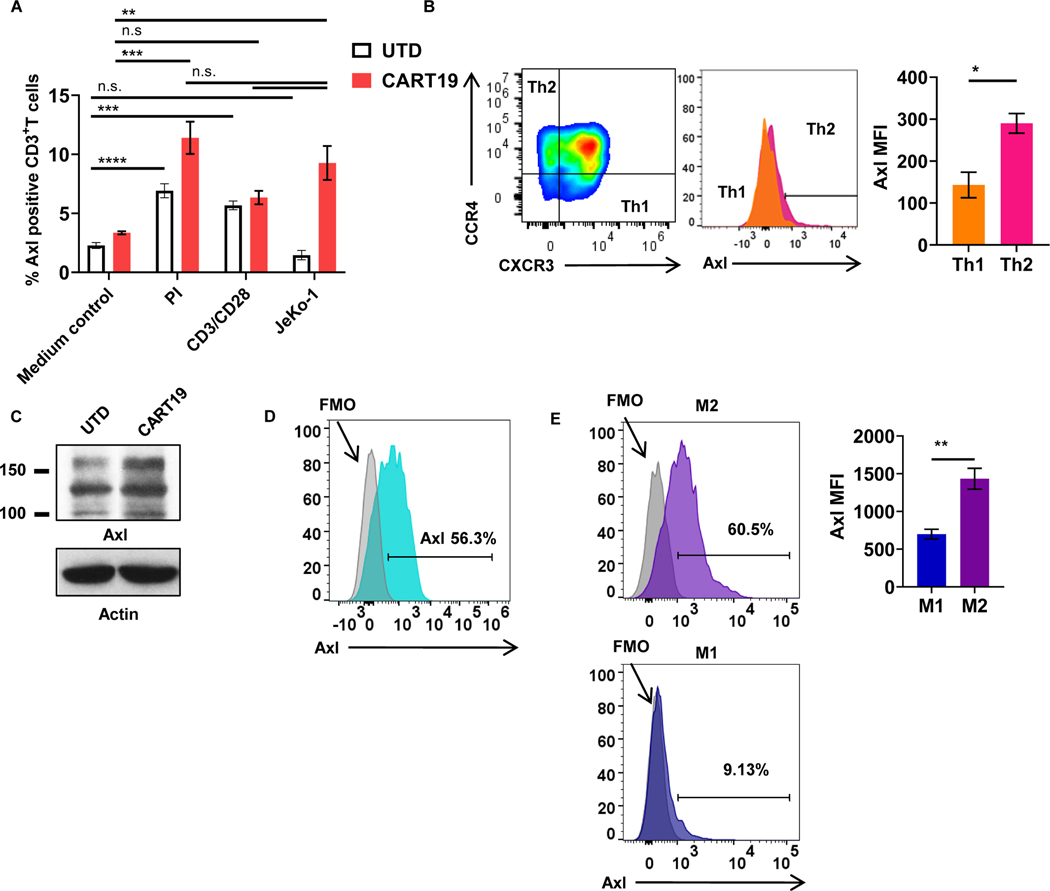
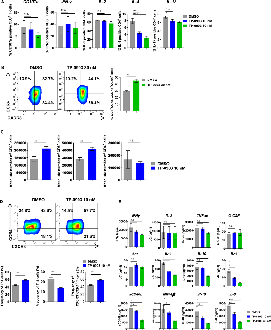
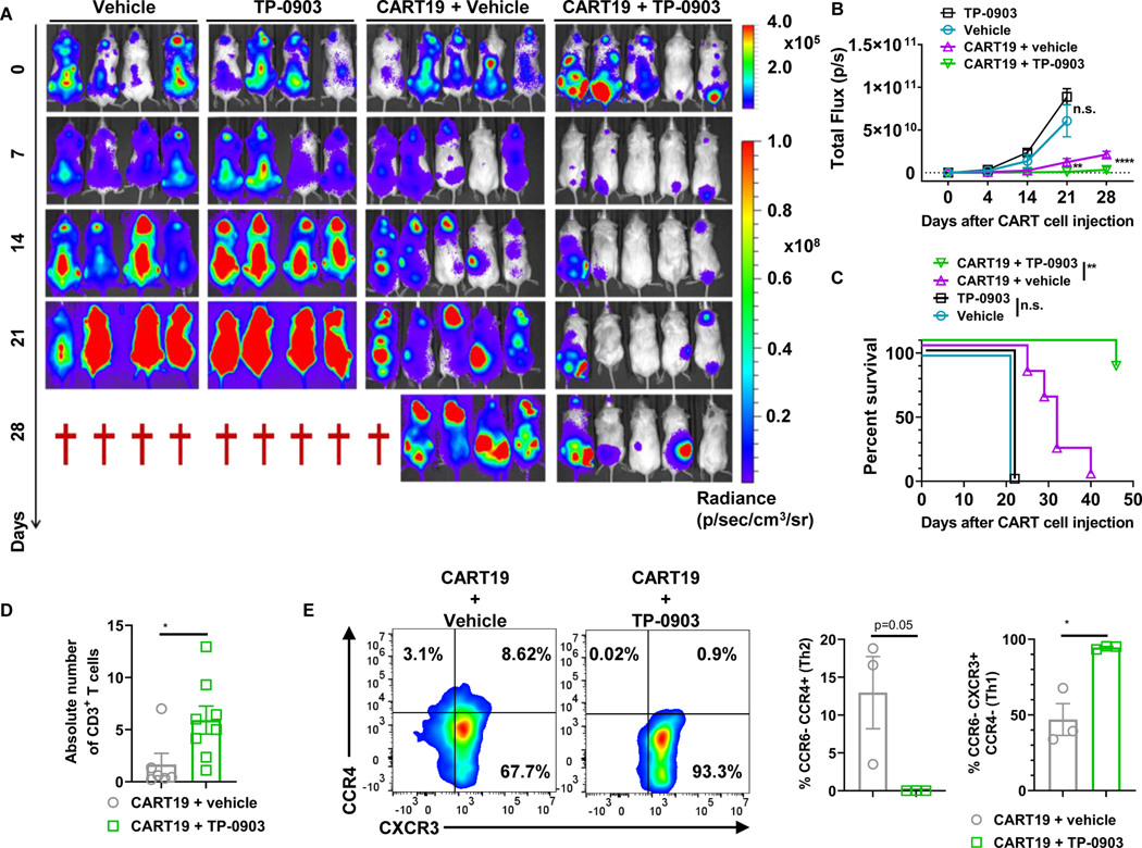
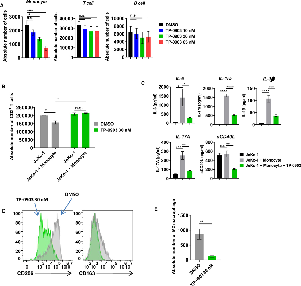
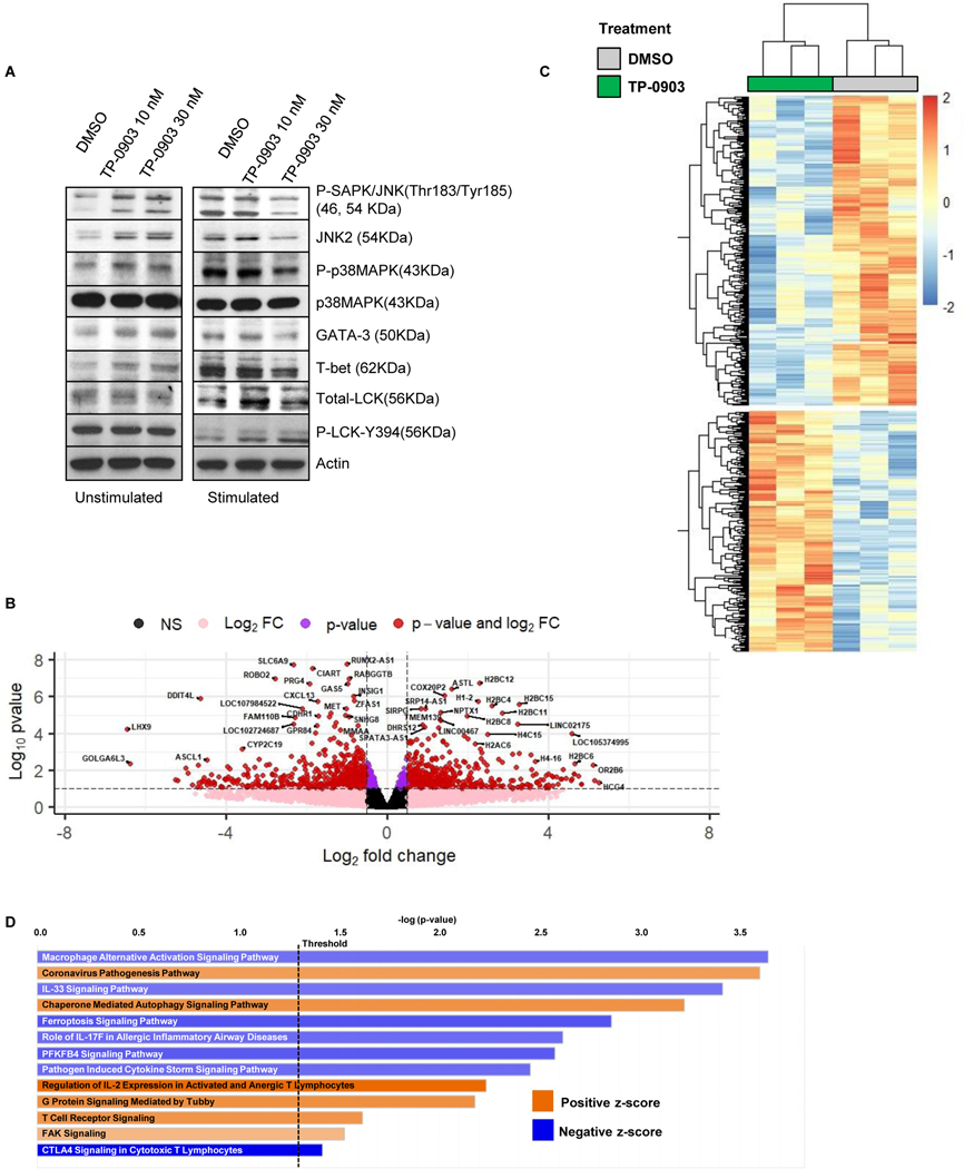
The receptor tyrosine kinase AXL is a member of the TYRO3, AXL, and proto-oncogene tyrosine-protein kinase MER family and plays pleiotropic roles in cancer progression. AXL is expressed in immunosuppressive cells, which contributes to decreased efficacy of immunotherapy. Therefore, we hypothesized that AXL inhibition could serve as a strategy to overcome resistance to chimeric antigen receptor T (CAR T)-cell therapy. To test this, we determined the impact of AXL inhibition on CD19-targeted CAR T (CART19)-cell functions. Our results demonstrate that T cells and CAR T cells express high levels of AXL. Specifically, higher levels of AXL on activated Th2 CAR T cells and M2-polarized macrophages were observed. AXL inhibition with small molecules or via genetic disruption in T cells demonstrated selective inhibition of Th2 CAR T cells, reduction of Th2 cytokines, reversal of CAR T-cell inhibition, and promotion of CAR T-cell effector functions. AXL inhibition is a novel strategy to enhance CAR T-cell functions through two independent, but complementary, mechanisms: targeting Th2 cells and reversing myeloid-induced CAR T-cell inhibition through selective targeting of M2-polarized macrophages.


Conflict of interest statement

SSK is an inventor on patents in the field of CAR immunotherapy that are licensed to Novartis (through an agreement between Mayo Clinic, University of Pennsylvania, and Novartis). RLS, MJC, and SSK are inventors on patents in the field of CAR immunotherapy that are licensed to Humanigen (through Mayo Clinic). SSK and MH are inventors on patents in the field of CAR immunotherapy that are licensed to Mettaforge (through Mayo Clinic). SSK and IC are inventors on patients that are licensed to Sendero (through Mayo Clinic). SSK is an inventor on intellectual property that is licensed to MustangBio (through Mayo Clinic). SSK receives research funding from Kite, Gilead, Juno, Celgene, Novartis, Humanigen, MorphoSys, Tolero, Sunesis, LeahLabs, and Lentigen. SSK has participated in advisory meetings of Juno, Celgene, Kite, Gilead, LeahLabs, CapstanBio, Torque, Luminary, and Humanigen. SSK has participated in data safety monitoring boards of Humanigen. RLS, LM, JF, SLW, NEK, and SSK are inventors on patents related to this work. NEK receives research funding from Acerta Pharma, BMS, Pharmacyclics, MEI Pharma, and Sunesis. NEK has participated in Advisory Board meetings of Cytomx Therapy, Janssen, Juno Therapeutics, Astra Zeneca, and Oncotracker; and on DSMC for Agios and Cytomx Therapeutics. SAP receives research funding from Pharmacyclics, MorphoSys, Janssen, AstraZeneca, TG Therapeutics, Bristol Myers Squibb, AbbVie, and Ascentage Pharma. SAP has participated in Advisory Board meetings of Pharmacyclics, AstraZeneca, Genentech, Gilead, GlaxoSmithKline, Verastem Oncology, and AbbVie (he was not personally compensated for his participation).

Figures

Figure 1:
Figure 1:. AXL is expressed on activated T cells, CART cells, and innate immune cells.
A, Untransduced T cells (UTDs) or CART19 cells were stimulated with 50 ng/mL PMA and 1.0 μg/mL of ionomycin, CD3+CD28+ Dynabeads (T cells:beads = 1:3), or CD19+ mantle cell lymphoma cell line, JeKo-1, for 24 hours and assessed for the surface expression of AXL (** p<0.01, *** p<0.001, **** p<0.0001, n.s. not significant, one-way ANOVA). B, T cells from experiment Fig.1A were further assessed for helper T-cell (Th) phenotypes. Th1 and Th2 cells were defined by CD4+CCR6CXCR3+CCR4 and CD4+CCR6CXCR3CCR4+, respectively. AXL expression was assessed by flow cytometry (*** p<0.0005, t-test, n=3). C, Rested UTD or CART19 cells after their manufacturing were assessed AXL with western blot. D, Monocytes were isolated by negative selection using the human classical monocyte isolation kit. Post monocyte isolation, AXL expression was assessed by flow cytometry (n=3). E, M1 or M2 induction was performed by the addition of LPS and IFNγ or IL4, respectively. Surface AXL expression of M1 and M2 macrophages were assessed by flow cytometry (*** p<0.01, t-test, n=3). Data are plotted as means ± SEM.
Figure 2:
Figure 2:. AXL inhibition with TP-0903 selectively reduces Th2 cells and cytokines and enhances CART19-cell proliferation.
A, Naïve T cells were stimulated with 50 ng/mL of PMA and 1 µg/mL of ionomycin for 4 hours in the presence of increasing doses of TP-0903 (10–30 nM). Flow cytometric analysis was used to assess activation of T cells by CD107a degranulation and intracytoplasmic cytokine production (IL2, IFNγ, IL4, IL13). n=3 biological replicates. B, Naïve T cells were stimulated with 5 ng/mL of PMA and 0.1 µg/mL of ionomycin for 3 days in the presence of TP-0903 (30 nM). Flow cytometric analysis was used to evaluate CD4+CXCR3+CCR4+ T cells (** p<0.01, t-test, n=3 biological replicates). C, CART19 cells were co-cultured with the CD19+ mantle cell lymphoma cell line JeKo-1 in the absence or in the presence of TP-0903 (10 nM) for 5 days. CART19 cell proliferation was then assessed for total, CD8+, and CD4+ T cells. n=3 biological replicates. D, CART19 cells were co-cultured with JeKo-1 in the presence of TP-0903 (10 nM) for 5 days and chemokine receptors were stained. Th1, Th2, and the CD4+CXCR3+CCR4+ fractions were assessed. ** p<0.005, t-test, n=3 biological replicates. E, CART19 cells were co-cultured with JeKo-1 cells for 3 days with/without TP-0903, and supernatants were analyzed for human cytokines and chemokines (38-multiplex). * p<0.05, ** p<0.01, *** p<0.001, n=2 biological replicates. Data are plotted as means ± SEM.
Figure 3:
Figure 3:. AXL inhibition with TP-0903 enhances CART19 anti-tumor activity in vivo.
A, 1.0 × 10⁶ luciferase+ JeKo-1 cells were injected via tail vein into NSG mice. Two weeks after the injection of JeKo-1 tumor burden was analyzed by bioluminescence imaging. Mice were then randomized into groups receiving vehicle (n=4), 20 mg/kg TP-0903 (p.o.) (n=4), 0.5 × 10⁶ of CART19 (i.v.) (n=5), or combination 20 mg/kg TP-0903 and 0.5 × 10⁶ of CART19 (n=5) groups. B, Tumor growth based on bioluminescent imaging. ** p<0.005 and **** p<0.0001, two-way ANOVA. C, Kaplan-Meier survival curve following different treatments. ** p<0.005, log-rank test, hazard ratio (HR) = 0.089 with 95% confidence interval (CI) (0.01595 to 0.5072), p=0.004. D, Mice were bled 17 days after the administration of CART19 cells. Absolute number of CART19 cells was determined via flow cytometry. * p<0.05, t-test, monotherapy group n=6; combination therapy group n=8. E, T-cell phenotype after CART19 cell administration was assessed in spleens of a subset of mice. * p<0.05, t-test, both groups n=3.
Figure 4:
Figure 4:. T-cell phenotype is altered following treatment with TP-0903 in a phase I clinical trial for patients with solid tumors (NCT02729298).
A, Peripheral blood-derived Th2 cells (CD4+CCR6CXCR3CCR4+) from patients were analyzed before and one day after treatment with TP-0903. Representative flow plot of 3 biological replicates is shown. B, Peripheral blood-derived Tregs (CD4+CD25+Foxp3+) from patients were analyzed before and one day after treatment with TP-0903. ** p<0.01, t-test, n=3 biological replicates. C, Tregs and carboxyfluorescein succinimidyl ester (CFSE) stained effector T cell (Teff) cells were co-cultured at the indicated ratio and co-cultured for 4 days in the presence or absence of 30 nM of TP-0903. At the end of the co-culture, the percent suppression of Teff was calculated using flow cytometry. ** p<0.01, two-way ANOVA, n=3, biological replicates.
Figure 5:
Figure 5:. AXL inhibition with TP-0903 preferentially targets monocytes and overcomes monocyte-induced CART-cell suppression.
A, Peripheral blood mononuclear cells derived from healthy donors were treated with increasing doses of TP-0903 (10–65 nM) for 24 hours, and the absolute number of cells was assessed by CountBright bead quantification with flow cytometry (n.s. not significant, ** p<0.01, **** p<0.001, one-way ANOVA, n=3 biological replicates). B, CART19 cells, JeKo-1 cells, and monocytes were co-cultured for 5 days and analyzed for the absolute number of CD3+ T cells by CountBright bead quantification (* p<0.05, t-test, n=3 biological replicates). C, Supernatants that were harvested from experiment in Fig. 5B were analyzed with 38-mulitplex. (* p<0.05, ** p<0.01, *** p<0.001, **** p<0.0001, one-way ANOVA, n=3 biological replicates). D, CD14+ cells were stained for M2-related markers CD206 and CD163 and analyzed via flow cytometry. E, CART19 cells, JeKo-1 cells, and M2-like macrophages were co-cultured for 5 days and analyzed for the absolute number of CD14+ cells by flow cytometry (** p<0.01, t-test, n=3 biological replicates). Data are plotted as means ± SEM.
Figure 6:
Figure 6:. CART19-cell transcriptomic changes following AXL inhibition.
A, CART19 cells were incubated with lethally irradiated JeKo-1 cells in the presence of TP-0903 (10–30 nM) at a 1:3 ratio for 24 hours. After the incubation, T cells were enriched and assessed via western blotting for changes in AXL downstream signaling pathways. B, Principal component analysis (PCA) of RNA-seq data from CART19 cells with/without 30 nM of TP-0903. C-D, Volcano plot and heatmap showing differential expression of transcripts in TP-0903-treated CART19 cells compared to untreated (DMSO) CART19 cells.

References

    1. van der Meer JHM, van der Poll T, van ‘t Veer C. TAM receptors, Gas6, and protein S: roles in inflammation and hemostasis. Blood 2014;123:2460–9 - PubMed
    1. Vouri M, Hafizi S. TAM Receptor Tyrosine Kinases in Cancer Drug Resistance. Cancer Res 2017;77:2775–8 - PubMed
    1. Myers KV, Amend SR, Pienta KJ. Targeting Tyro3, AXL and MerTK (TAM receptors): implications for macrophages in the tumor microenvironment. Molecular cancer 2019;18:94. - PMC - PubMed
    1. Seitz HM, Camenisch TD, Lemke G, Earp HS, Matsushima GK. Macrophages and dendritic cells use different AXL/Mertk/Tyro3 receptors in clearance of apoptotic cells. J Immunol 2007;178:5635–42 - PubMed
    1. Zhao GJ, Zheng JY, Bian JL, Chen LW, Dong N, Yu Y, et al. Growth Arrest-Specific 6 Enhances the Suppressive Function of CD4(+)CD25(+) Regulatory T Cells Mainly through AXL Receptor. Mediators Inflamm 2017;2017:6848430 - PMC - PubMed

Publication types

Substances