. 2024 Aug;15(4):714-728.
doi: 10.1007/s12975-023-01169-x. Epub 2023 Jun 28.

The Transcriptional Landscape of Pericytes in Acute Ischemic Stroke

Affiliations

The Transcriptional Landscape of Pericytes in Acute Ischemic Stroke

Carolina Buizza et al. Transl Stroke Res. 2024 Aug.

Abstract

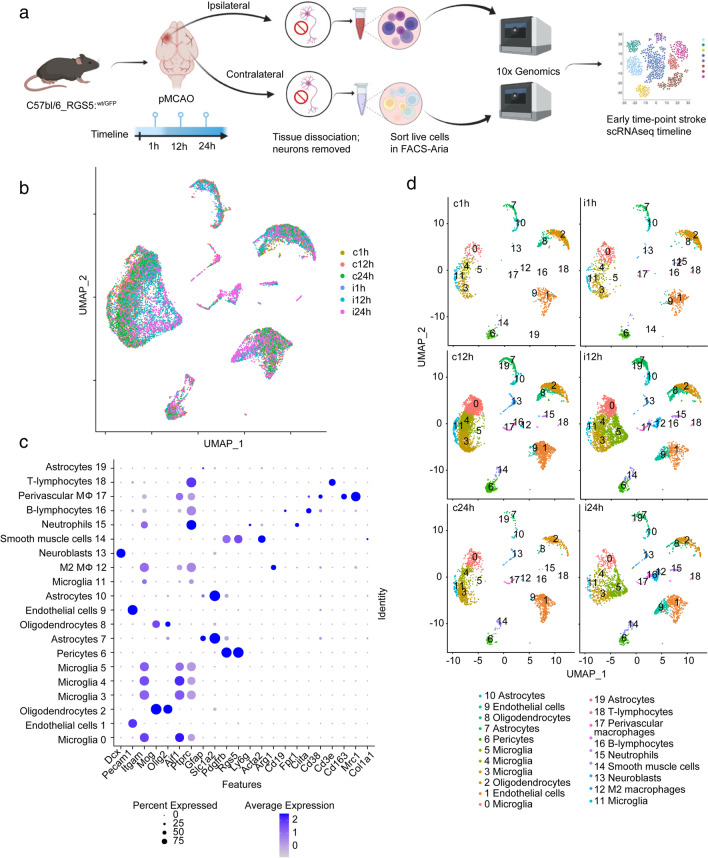
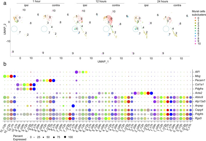
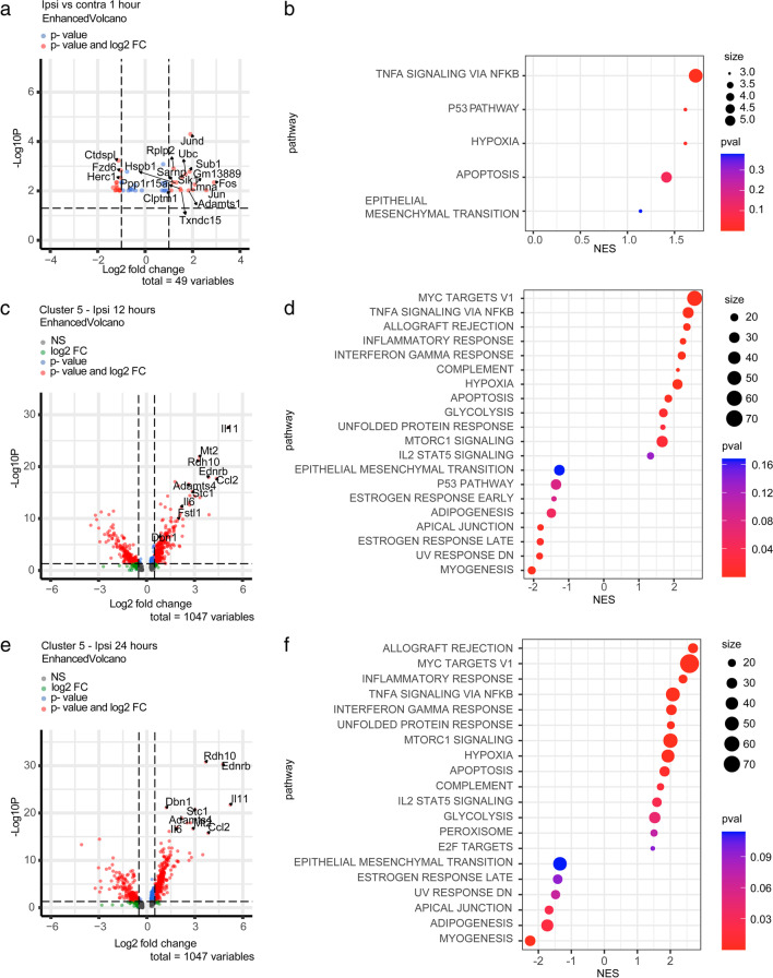
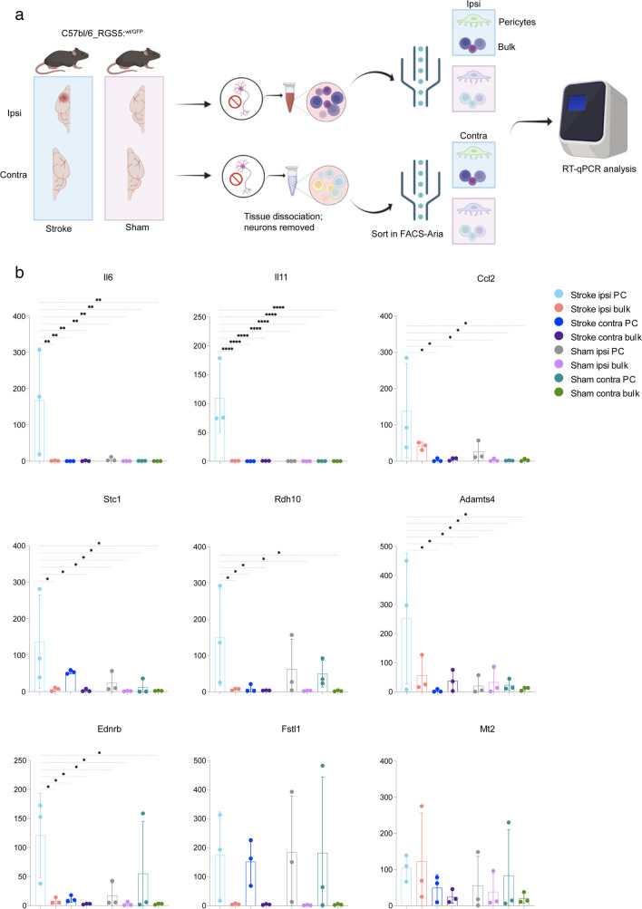
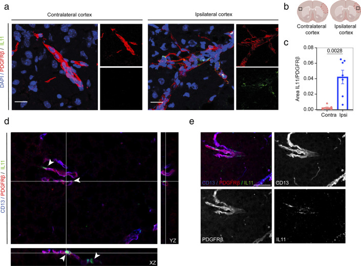
The current treatment options for ischemic stroke aim to achieve reperfusion but are time critical. Novel therapeutic approaches that can be given beyond the limited time window of 3-4.5 h are still an unmet need to be addressed to improve stroke outcomes. The lack of oxygen and glucose in the area of ischemic injury initiates a pathological cascade leading to blood-brain barrier (BBB) breakdown, inflammation, and neuronal cell death, a process that may be intercepted to limit stroke progression. Pericytes located at the blood/brain interface are one of the first responders to hypoxia in stroke and therefore a potential target cell for early stroke interventions. Using single-cell RNA sequencing in a mouse model of permanent middle cerebral artery occlusion, we investigated the temporal differences in transcriptomic signatures in pericytes at 1, 12, and 24 h after stroke. Our results reveal a stroke-specific subcluster of pericytes that is present at 12 and 24 h and characterized by the upregulation of genes mainly related to cytokine signaling and immune response. This study identifies temporal transcriptional changes in the acute phase of ischemic stroke that reflect the early response of pericytes to the ischemic insult and its secondary consequences and may constitute potential future therapeutic targets.

Keywords: Interleukin 11; Ischemic stroke; Pericytes; Single-cell RNA sequencing.


Conflict of interest statement

The authors declare no competing interests.

Figures

Fig. 1
Fig. 1
Single-cell sequencing of non-neuronal cells 1, 12, or 24 h after pMCAO stroke in mice. a Graphical diagram of the single-cell isolation and scRNA-seq experimental setup. b Visualization and clustering of the scRNA-seq data in Seurat from all the timepoints and areas analyzed in the study. c Cell types identified by clustering analysis and gene expression of the genes defining cell type identity. d UMAP plotted singularly for different timepoints and hemispheres. c contralateral; i ipsilateral; h hour
Fig. 2
Fig. 2
Heterogeneity of mural cells in the acute phase of ischemic stroke. a Re-clustering analysis on pericytes and smooth muscle cells reveals 8–10 different subclusters depending on the timepoint and experimental condition. The pericyte subcluster 5, stroke-specific, is highlighted. b Dot plot representing the marker expression of each mural cell subcluster. Different timepoints and hemispheres are denoted by colors. The size of the dots equals the percentage of the population expressing the marker, and the intensity of the color denotes the expression level, respectively. Pericytes are defined as Pdgfrβ+, RGS5+, Atp13a5+, Abcc9+, and Acta2-, SMCs Acta2+, fibroblasts Pdgfrβ+, Pdgfrα+. Clusters 6, 8, and 10 are pericytes co-expressing markers of pericytes and microglia (Aif1+), endothelial cells (Pecam1+), or oligodendrocytes (Mog+). c contralateral; i ipsilateral; h hour
Fig. 3
Fig. 3
DEG analysis and GSEA on the mural cells subclusters. a Volcano plot displaying DEGs in the pericyte subcluster 3 1 h after ischemic stroke. b Hallmark gene sets from MSigDB on the differentially expressed genes from subcluster 3 between ipsilateral and contralateral hemispheres 1 h after stroke. c Volcano plot displaying the genes enriched in cluster 5 at 12 h after stroke. d Hallmark gene sets on the differentially expressed genes from cluster 5 compared to the other mural cell subclusters 12 h after stroke. e Volcano plot displaying the genes enriched in cluster 5 compared to the other mural cell subclusters at 24 h after stroke. f Hallmark gene sets on the differentially expressed genes from cluster 5 24 h after stroke. Cut-off DEG p value < 0.05; fold change > 0.5
Fig. 4
Fig. 4
RT-qPCR validation of RNA sequencing data for the top 10 most differentially upregulated genes by the stroke-specific subcluster 5 of pericytes 24 h after stroke. a Graphical diagram of single-cell isolation and FACS sorting for RT-qPCR experiments. b Bar graphs show that mRNA values of Ednrb, Rdh10, Stc1, Il6, Il11, Ccl2, Mt2, Adamts4, and Fstl1 presented as mean of experimental replicates ± SD (n = 4 pooled mice per replicate, 3 replicates). * = 0.05 > P > 0.001; ** = 0.01 > P > 0.001; *** = 0.001 > P > 0.0001; **** = P <0.001
Fig. 5
Fig. 5
Validation of IL11 increased expression in the stroke ipsilateral hemisphere by immunohistochemistry. a IL11 is only detected in the ipsilateral hemisphere of mice subjected to stroke in the proximity of pericytes. b Location of the selected images. The infarct core is outlined. c Area fraction occupied by IL11 and PDGFR𝛽 signals. Data are presented as mean ± SEM. P = 0.028, paired t-test 95% confidence level (confidence intervals 0.01985 to 0.05979). Scale bar = 20 μm. d Orthogonal views showing IL11 expression in PDGFR𝛽 and CD13 expressing cells in the ipsilateral cortex and e 2D maximum projection of the merged channels. z stack = 24 μm; scale bar = 25 μm. Arrow points at IL11 signal

References

    1. Johnson W, et al. Stroke: a global response is needed. Bull World Health Organ. 2016;94(9):634–634A. doi: 10.2471/BLT.16.181636. - DOI - PMC - PubMed
    1. Candelario-Jalil E. Injury and repair mechanisms in ischemic stroke: considerations for the development of novel neurotherapeutics. Curr Opin Investig Drugs. 2009;10(7):644–654. - PubMed
    1. Yang Y, Rosenberg GA. Blood-brain barrier breakdown in acute and chronic cerebrovascular disease. Stroke. 2011;42(11):3323–3328. doi: 10.1161/STROKEAHA.110.608257. - DOI - PMC - PubMed
    1. Bader MK, Palmer S. What’s the “hyper” in hyperacute stroke? Strategies to improve outcomes in ischemic stroke patients presenting within 6 hours. AACN Adv Crit Care. 2006;17(2):194–214. doi: 10.1097/01256961-200604000-00016. - DOI - PubMed
    1. Cai W, et al. Pericytes in brain injury and repair after ischemic stroke. Transl Stroke Res. 2017;8(2):107–121. doi: 10.1007/s12975-016-0504-4. - DOI - PMC - PubMed

Publication types