Clinical Trial
. 2023 Sep 12;7(17):5082-5090.
doi: 10.1182/bloodadvances.2022009467.

Subclonal TP53 mutations are frequent and predict resistance to radioimmunotherapy in follicular lymphoma

Affiliations
Clinical Trial

Subclonal TP53 mutations are frequent and predict resistance to radioimmunotherapy in follicular lymphoma

W Richard Burack et al. Blood Adv. .

Abstract

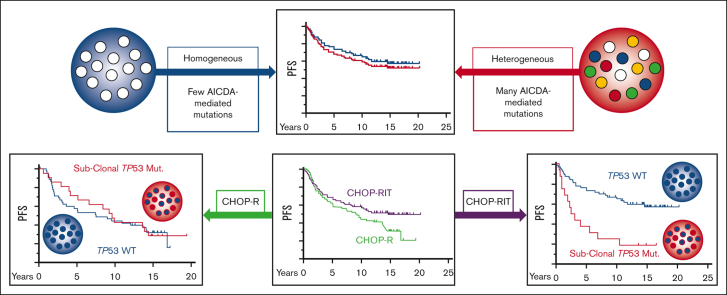
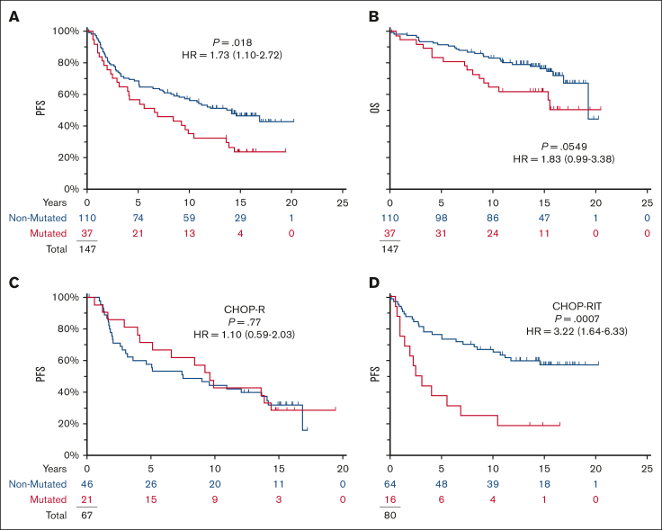
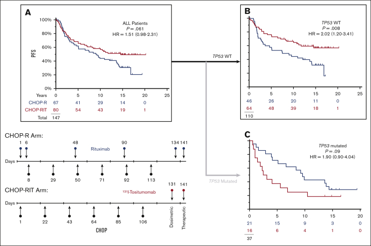
Although TP53 is commonly mutated in transformed follicular lymphoma, mutations are reported in <5% of pretreatment follicular lymphoma (FL) specimens. We assayed archival follicular B-cell non-Hodgkin lymphoma specimens from a completed clinical trial, Southwest Oncology Group S0016, a phase 3 randomized intergroup trial of CHOP (cyclophosphamide, hydroxydaunorubicin, oncovin, and prednisone) chemotherapy plus R-CHOP (rituximab-CHOP) compared with CHOP chemotherapy plus 131-iodine tositumomab (radioimmunotherapy [RIT]-CHOP). Subclonal TP53 mutations (median allele frequency 0.02) were found in 25% of diagnostic FL specimens and in 27% of a separate validation cohort. In the R-CHOP arm, pathogenic TP53 mutations were not associated with progression-free survival (PFS) (10-year PFS 43% vs 44%). In contrast, among patients with no detectable pathogenic TP53 mutation, RIT-CHOP was associated with a longer PFS than with R-CHOP (10-year PFS 67% vs 44%; hazard ratio = 0.49; P = .008). No relationship was detected between PFS and the extent of activation-induced cytidine deaminase (AICDA)-mediated heterogeneity. In summary, subclonal TP53 mutations are common in FL and are a distinct phenomenon from AICDA-mediated genetic heterogeneity. The absence of a detectable subclonal mutation in TP53 defined a population that particularly benefited from RIT.


Conflict of interest statement

Conflict-of-interest disclosure: The authors declare no competing financial interests.

Figures

None
Graphical abstract
Figure 1.
Figure 1.
TP53 mutations are common in S0016 FL specimens and have a low VAF. (A) The pathogenic mutations detected in S0016 include canonical hotspots for TP53 mutations. The location and numbers of mutations at each site are indicated as missense (orange) or nonsense mutations (cyan). The colored regions indicate assigned functionality of each protein domain (from left, transactivation 1, transactivation 2, proline-rich domain, DNA binding, hinge domain, oligomerization domain, and alpha domain). (B) The median VAF of pathogenic mutations in FL is low. In the 37 of 147 FL specimens with a pathogenic TP53 mutation, the median VAF for these mutations is 0.02 (FL). In a validation set, 28 mutations were detected in 19 specimens (FL validation) with a median VAF of 0.03. Additional control samples of MZL and mantle cell lymphoma MCL (36 and 38 specimens) have a substantially higher median VAF. Boxes contain second and third quartiles, and whiskers 10 to 90 percentiles. Specimens with a single detectable mutation are noted with black squares. For specimens with >1 TP53 mutation, all the mutations in that specimen share a unique symbol.
Figure 2.
Figure 2.
TP53 mutations are prognostic in S0016 and specifically affect the prognosis on the RIT-CHOP arm but not on the R-CHOP arm. Of 147 patients, 72 showed disease progression and 44 died with a median of 15 years follow-up among those last known alive. (A,B) Mutations in TP53 were associated with shortened PFS in S0016 but not OS. (C,D) TP53 mutations affected PFS on RIT-CHOP arm but not on the R-CHOP arms (blue, no mutation detected; red, mutation detected).
Figure 3.
Figure 3.
In patients with NO detectable TP53 mutation, CHOP-RIT is associated with longer PFS. (A) There is a trend to longer PFS with CHOP-RIT similar to that previously published for the entire study. (B) Removing the 37 patients with TP53 mutations, the PFS was significantly longer on the RIT-CHOP arm compared with the R-CHOP arm. (C) In the subset of 37 patients with a TP53 mutation, there is an opposite trend with shortened PFS on the RIT-CHOP arm. (Red, RIT-CHOP; Blue R-CHOP).
Figure 4.
Figure 4.
TP53 mutations are evanescent and their VAF does not overtly increase with relapse. Out of 22 patients with serial specimens, 11 patients had at least 1 specimen with a pathogenic mutation. The VAFs for these mutations are shown in Figure 1B FL validation. To estimate changes in tumor cell content, we determined the sum of VAFs for mutations in a single amplicon of uBCL2 (solid black lines with orange blocks); this provides an estimate of the minimum tumor content. The TP53 mutations are depicted on the thick gray lines (red blocks mark the VAF; blue blocks denote that the mutation was not detected at that time point [for TP53] or only germ line sequence for uBCL2).
Figure 5.
Figure 5.
The extent of AICDA-mediated diversity as marked by uBCL2 and IGVH varies widely and is unrelated to the presence of a TP53 mutation. (A) The number of distinct sequences for uBCL2 is the maximum number seen for a single amplicon within that region (refer to Methods). The number of distinct IGVH sequences is the number of distinct variants detected in the clonal IGVH sequence (refer to Methods). Patients with no variants are scored as 1. No relationship is apparent among the number of distinct uBCL2 sequences, the number of distinct IGVH sequences, and the status of TP53 (filled denotes detectable pathogenic lesion; open denotes no detectable lesion). (B-C) No relationship is apparent between the number of variant uBCL2 or IGVH sequences and the time to progression or progression at any time. The number of variant IGVH and uBCL2 sequences was not different between the patients who showed disease progression (blue) or who did not show disease progression (red). Furthermore, there is no suggestion that the number of sequence variants detected differed between those who showed disease progression within 2 years and those who showed disease progression later.

References

    1. Pasqualucci L. Molecular pathogenesis of germinal center-derived B cell lymphomas. Immunol Rev. 2019;288(1):240–261. - PubMed
    1. Pasqualucci L, Bhagat G, Jankovic M, et al. AID is required for germinal center-derived lymphomagenesis. Nat Genet. 2008;40(1):108–112. - PubMed
    1. Pasqualucci L, Khiabanian H, Fangazio M, et al. Genetics of follicular lymphoma transformation. Cell Rep. 2014;6(1):130–140. - PMC - PubMed
    1. Casulo C, Burack WR, Friedberg JW. Transformed follicular non-Hodgkin lymphoma. Blood. 2015;125(1):40–47. - PubMed
    1. Correia C, Schneider PA, Dai H, et al. BCL2 mutations are associated with increased risk of transformation and shortened survival in follicular lymphoma. Blood. 2015;125(4):658–667. - PMC - PubMed

Publication types

MeSH terms