. 2023 Jun 28;15(702):eabo3826.
doi: 10.1126/scitranslmed.abo3826. Epub 2023 Jun 28.

Targeting CCR7-PI3Kγ overcomes resistance to tyrosine kinase inhibitors in ALK-rearranged lymphoma

Affiliations

Targeting CCR7-PI3Kγ overcomes resistance to tyrosine kinase inhibitors in ALK-rearranged lymphoma

Cristina Mastini et al. Sci Transl Med. .

Abstract

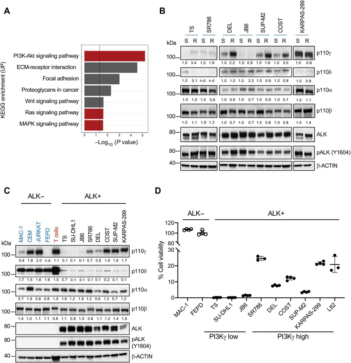
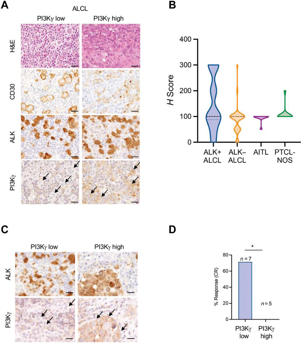
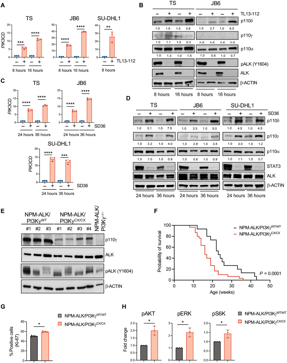
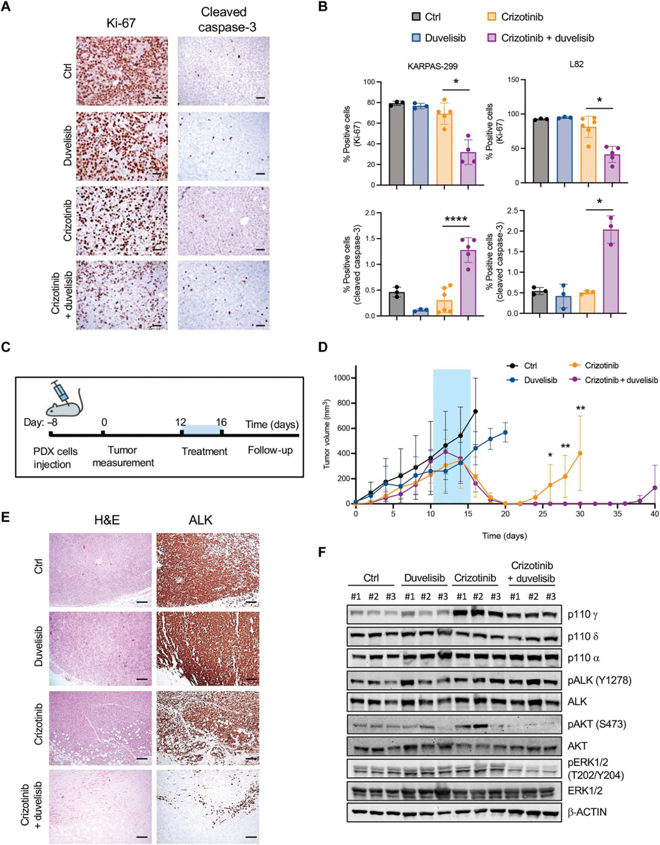
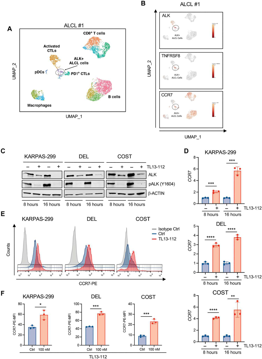
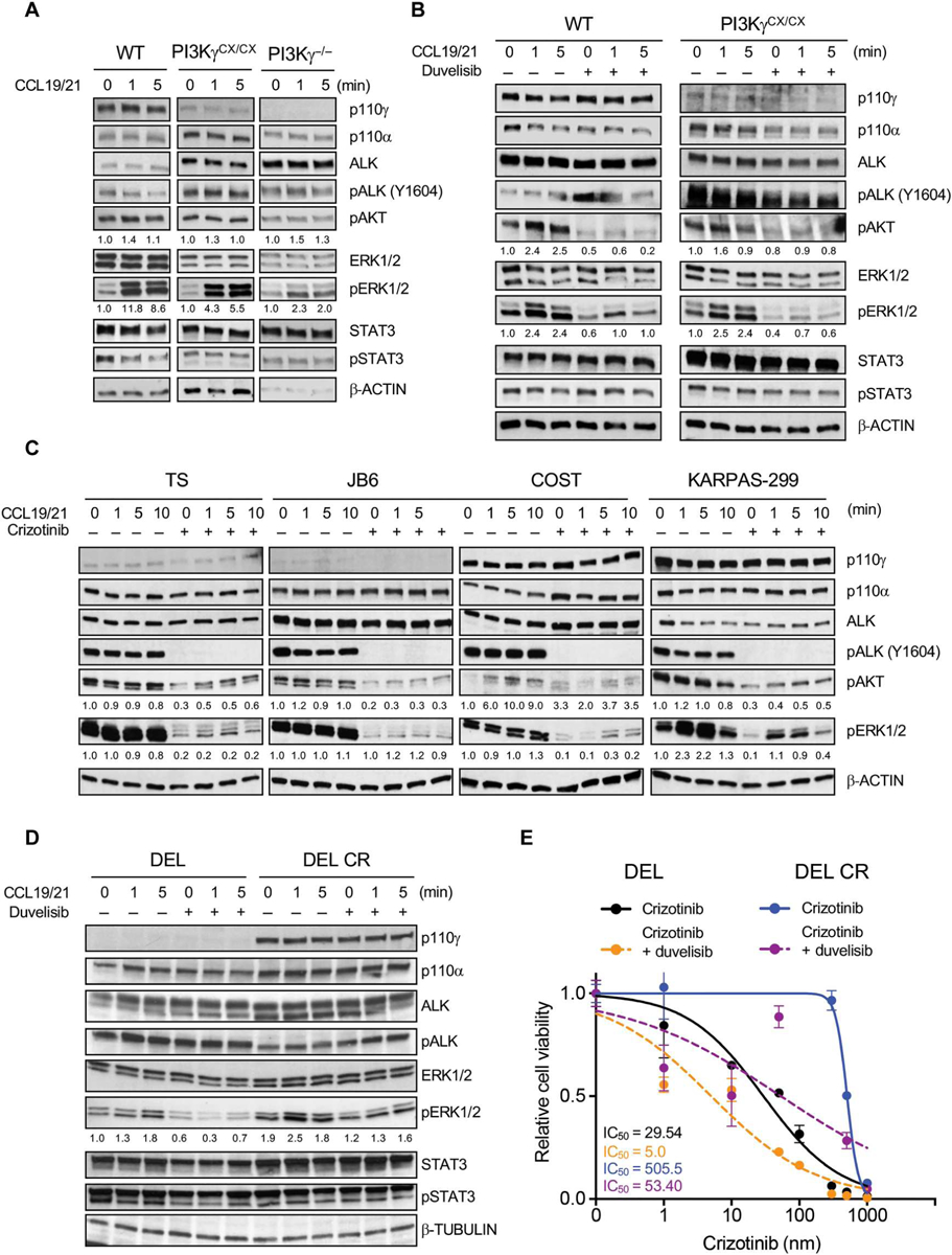
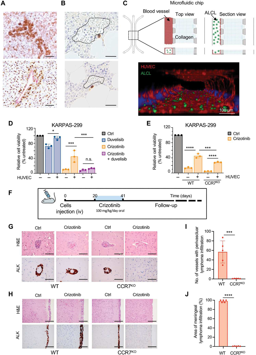
Anaplastic lymphoma kinase (ALK) tyrosine kinase inhibitors (TKIs) show potent efficacy in several ALK-driven tumors, but the development of resistance limits their long-term clinical impact. Although resistance mechanisms have been studied extensively in ALK-driven non-small cell lung cancer, they are poorly understood in ALK-driven anaplastic large cell lymphoma (ALCL). Here, we identify a survival pathway supported by the tumor microenvironment that activates phosphatidylinositol 3-kinase γ (PI3K-γ) signaling through the C-C motif chemokine receptor 7 (CCR7). We found increased PI3K signaling in patients and ALCL cell lines resistant to ALK TKIs. PI3Kγ expression was predictive of a lack of response to ALK TKI in patients with ALCL. Expression of CCR7, PI3Kγ, and PI3Kδ were up-regulated during ALK or STAT3 inhibition or degradation and a constitutively active PI3Kγ isoform cooperated with oncogenic ALK to accelerate lymphomagenesis in mice. In a three-dimensional microfluidic chip, endothelial cells that produce the CCR7 ligands CCL19/CCL21 protected ALCL cells from apoptosis induced by crizotinib. The PI3Kγ/δ inhibitor duvelisib potentiated crizotinib activity against ALCL lines and patient-derived xenografts. Furthermore, genetic deletion of CCR7 blocked the central nervous system dissemination and perivascular growth of ALCL in mice treated with crizotinib. Thus, blockade of PI3Kγ or CCR7 signaling together with ALK TKI treatment reduces primary resistance and the survival of persister lymphoma cells in ALCL.


Conflict of interest statement

Competing interests: C.C.−M. works for Catapult Therapeutics that has developed the anti-CCR7 CAP100 antibody and is an employee and a shareholder of Immunological and Medical Products (IMMED S.L.).

Figures

Fig. 1.
Fig. 1.. Resistance to crizotinib is associated with PI3Kγ up-regulation in ALK+ ALCL.
(A) Kyoto Encyclopedia of Genes and Genomes (KEGG) pathway enrichment analysis for up-regulated genes identified by RNA-seq in samples from patients with ALK+ ALCL that relapsed on ALK inhibitor treatment (n = 2) versus sensitive to ALK inhibitor treatment (n = 2). The PI3K-Akt, Ras, and MAPK signaling pathways are highlighted in red. (B) Western blot analysis performed on paired crizotinib-sensitive (S) and resistant (R) ALK+ ALCL cell lines. (C) Western blot analysis performed on human ALK+ ALCL cell lines, ALK− T lymphoma lines, or normal T cells. Western blot bands were normalized on β-actin. (D) Cell viability measured using CellTiter-Glo of ALK+ and ALK− ALCL cell lines treated with crizotinib (100 nM) for 72 hours. n = 3 or 4 technical replicates. Data are shown as means ± SD. For Western blots, β-actin was used as a loading control, and two independent experiments with similar results were performed.
Fig. 2.
Fig. 2.. High PI3Kγ expression induces spontaneous resistance to crizotinib in patients with ALK+ ALCL.
(A) Photomicrographs of representative hematoxylin and eosin (H&E) staining and immunohistochemical staining performed with the indicated antibodies in primary ALK+ lymphoma samples with low or high expression of PI3Kγ. PI3Kγ antibody was validated in formalin-fixed samples of cells transduced with a PI3Kγ-encoding retrovirus (fig. S5A). Black arrows indicate PI3Kγ expression in lymphoma cells. Scale bars, 100 μm. (B) PI3Kγ staining in human T cell lymphoma subtypes. Expression across the violin plot is shown. ALK+ ALCL, n = 41; ALK− ALCL, n = 29; angioimmunoblastic T cell lymphoma (AITL), n = 10; PTCL-NOS n = 11. The number of patient samples is indicated for each lymphoma subtype. PI3Kγ expression was quantified by immunostaining, and an H score was assigned. (C) Immunohistochemical staining of ALK and PI3Kγ in primary samples of ALK+ ALCL. Black arrows indicate PI3Kγ expression in lymphoma cells. Scale bars, 100 μm. (D) Percentage of response to crizotinib treatment of patients with ALK+ ALCL expressing low (H score < 100; n = 7) or high (H score > 100; n = 5) PI3Kγ. C Scale bars, 50 μm. *P < 0.05. Significance was determined by unpaired, two-tailed Student’s t test.
Fig. 3.
Fig. 3.. PI3Kδ is repressed by ALK in ALK+ ALCL cell lines and constitutive activation of PI3Kγ accelerates ALK-dependent lymphomagenesis.
(A) qRT-PCR analysis of PIK3CD mRNA expression performed on TS, JB6, and SU-DHL1 cell lines treated with TL134–112 (100 nM). n = 3 technical replicates. (B) Western blot analysis on TS and JB6 cell lines treated with TL13–112 (100 nM). (C) qRT-PCR analysis of PIK3CD mRNA expression performed on ALK+ ALCL cell lines treated with SD36 (1 μM). n = 3 technical replicates. (D) Western blot analysis on ALK+ ALCL human cell lines treated with SD36 (1 μM). (E) Western blot analysis of lymphomas obtained from C57BL/6 mice with the indicated genotypes. (F) Kaplan-Meier survival analysis of NPM-ALK transgenic mice crossed with mice expressing an active form of PI3Kγ (PI3KγCX/CX) (black, NPM-ALK/PI3KγWT/WT, n = 30 mice; red, NPM-ALK/PI3KγCX/CX, n = 30 mice). ****P < 0.0001. Significance was determined by log-rank (Mantel-Cox) test. (G) Quantification of Ki-67–positive cells in sections of primary lymphomas (n = 4) with the indicated genotypes. (H) Amount of phosphorylated Akt, ERK, and S6K measured in murine primary tumors with the indicated genotypes by Bio-Rad Bio-Plex. *P < 0.05, **P < 0.01, ***P < 0.001, and ****P < 0.0001. Significance was determined by unpaired, two-tailed Student’s t test. Data are shown as means ± SD. For Western blots, β-actin was used as a loading control, and two independent experiments with similar results were performed.
Fig. 4.
Fig. 4.. Duvelisib potentiates crizotinib treatment in ALK+ ALCL xenografts.
(A) Representative immunohistochemistry (IHC) for Ki-67 and cleaved caspase-3 of xenografted tumors collected from NSG mice injected subcutis with ALK+ ALCL cells and treated when tumors reached 300 to 500 mm3. Mice were treated with a vehicle, duvelisib (10 mg/kg), crizotinib (30 mg/kg), or duvelisib and crizotinib for 6 days. Scale bars, 50 μm. (B) Quantification of Ki-67 (top) and cleaved caspase-3–positive (bottom) cells in ALK+ ALCL xenograft lymphoma as in (A) (n = 3 to 6 tumors). (C) Schematic representation of the in vivo experiment with ALK+ ALCL PDXs. PDXs were injected subcutis. Treatment started when tumors reached 300 to 500 mm3. Mice were treated for 5 days and monitored for follow-up until day 40 after treatment. (D) Tumor growth of PDXs treated with a vehicle (n = 9 tumors), duvelisib (10 mg/kg; n = 6 tumors), crizotinib (30 mg/kg; n = 8 tumors), or duvelisib and crizotinib (n = 8 tumors) for 5 days. (E) Representative H&E staining and IHC for ALK of PDX tumors treated as indicated in (C) and (D). Tumors were collected as follows: control mice, day 16; duvelisib, day 20; crizotinib, day 30; crizotinib and duvelisib, day 40. Scale bars, 100 μm. (F) Western blot analysis on ALK+ ALCL PDX tumor samples treated and collected as indicated in (E). *P < 0.05, ** P < 0.01, and ****P < 0.0001. Significance was determined by unpaired, two-tailed Student’s t test. Data are shown as means ± SD. For Western blots, β-actin was used as a loading control, and two independent experiments with similar results were performed.
Fig. 5.
Fig. 5.. CCR7 is specifically expressed in ALK+ cells in primary ALCL and derepressed by ALK degradation.
(A) Uniform Manifold Approximation and Projection (UMAP) plot of scRNA-seq data for CD45+ cells from a primary lymph node of a patient with ALK+ ALCL, color-coded by the main group of cell type. (B) UMAP plots [CD45+ cell positioning as shown in (A)] normalized for expression of selected genes. (C) Western blot analysis of high-PI3Kγ–expressing ALK+ ALCL cells treated with TL13–112 (100 nM). (D) qRT-PCR analysis of CCR7 mRNA expression performed on ALK+ ALCL cell lines treated with TL13–112 (100 nM). n = 3 technical replicates. (E) CCR7 cell surface expression intensity measured by flow cytometry in ALK+ ALCL cell lines treated with dimethyl sulfoxide or TL13–112 (100 nM). KARPAS-299 cells were treated for 24 hours and DEL and COST cells for 16 hours. (F) Histograms show mean fluorescence intensity (MFI) of CCR7 cell surface expression in ALK+ ALCL cells treated as in (E). *P < 0.05, **P < 0.01, ***P < 0.001, and ****P < 0.0001. Significance was determined by unpaired, two-tailed Student’s t test. Data are shown as means ± SD. For Western blots, β-actin was used as a loading control. Blots are representative of two independent experiments with similar results.
Fig. 6.
Fig. 6.. PI3Kγ signaling activates MAPK pathway through CCR7.
(A) Western blot analysis performed on of NPM-ALK+ lymphoma cells derived from primary tumors collected in mice with the indicated genotypes and stimulated with CCL19/21 (100 ng/ml) ex vivo. (B) Western blot analysis of NPM-ALK+ lymphoma cells derived from primary tumors collected in mice with the indicated genotypes. Cells were stimulated with CCL19/21 (100 ng/ml) and treated with duvelisib (1 μM) for 3 hours ex vivo. (C) Western blot analysis performed on human ALK+ ALCL cell lines. Cells were stimulated with CCL19/21 (100 ng/ml) and left untreated or treated with crizotinib (300 nM) for 3 hours ex vivo. (D) Western blot analysis of a crizotinib-sensitive and -resistant (CR) ALK+ ALCL cell lines stimulated with CCL19/21 (100 ng/ml) in the presence or absence of duvelisib (1 μM). β-Tubulin was used as a loading control. (E) Dose-response curves of crizotinib-sensitive (DEL) and CR (DEL-CR) ALK+ ALCL cells treated with increasing concentrations of crizotinib in single or in combination with duvelisib (10 μM). n = 3 technical replicates. Data are shown as means ± SD. For Western blots, β-actin was used as a loading control. Blots are representative of two independent experiments with similar results.
Fig. 7.
Fig. 7.. The perivascular niche protects ALK+ ALCL cells from crizotinib–induced apoptosis.
(A) Representative IHC for ALK in primary ALK+ ALCL lymph nodes showing perivascular colonization of ALK+ cells. Scale bars, 100 μm. (B) Perivascular distribution of rare persister ALK+ cells (stained in brown) detected by anti-ALK IHC in bone marrow biopsies obtained from patients with ALK+ ALCL in clinical remission. Dotted line: bone marrow vascular space. Scale bars, 100 μm. (C) Top: Schematic representation of the microfluidic chip (left) and schematic of microphysio-logical model of ALCL-vascular interaction and blood vessel formation in a 3D microfluidic model (right). A commercial microfluidic chip with a central collagen hydrogel channel (collagen), flanked by two fluidic media channels, was used (blood vessels). ALCL cells inside the channel (ALCL). Bottom: Confocal microscope imaging of macrovessels (F-actin, red) and ALK+ ALCL (GFP, green) in the microfluidic model. ALK+ ALCL cells were transduced with a lentivirus-expressing enhanced green fluorescent protein. Scale bar, 100 μm. (D) Cell viability of KARPAS-299CCR7WT and KARPAS-299CCR7KO in coculture with or without blood vessels (HUVEC) treated with crizotinib (300 nM) or duvelisib (10 μM) or in combination. (E) Cell viability KARPAS-299CCR7WT and KARPAS-299CCR7KO in coculture with or without HUVEC treated with crizotinib (300 nM) for 72 hours. n = 3 biological replicates. *P < 0.05, ***P < 0.001, and ****P < 0.0001, n.s., not significant. Significance was determined by one-way ANOVA. (F) Schematic representation of the in vivo experiment. COSTCCR7WT and COSTCCR7KO were injected intravenously (iv) into NSG mice, and mice were treated with crizotinib (100 mg/kg/die). (G and H) Representative H&E staining (top) and IHC for ALK (bottom) of the central nervous system of mice injected with COSTCCR7WT and COSTCCR7KO without or with crizotinib treatment. Perivascular [(G), scale bars, 100 μm] or meningeal [(H), scale bars, 200 μm] infiltration of ALK+ lymphoma cells. (I and J) Histograms of lymphoma cell infiltration in the brain perivascular vessels (I) or meninges (J) of mice inoculated with COSTCCR7WT (n = 5) and COSTCCR7KO (n = 5) and treated with crizotinib as in (F). ***P < 0.001 and ****P < 0.0001. Significance was determined by unpaired, two-tailed Student’s t test. Data are shown as means ± SD.

References

    1. Holla VR, Elamin YY, Bailey AM, Johnson AM, Litzenburger BC, Khotskaya YB, Sanchez NS, Zeng J, Shufean MA, Shaw KR, Mendelsohn J, Mills GB, Meric-Bernstam F, Simon GR, ALK: A tyrosine kinase target for cancer therapy. Cold Spring Harb. Mol. Case Stud 3, a001115 (2017). - PMC - PubMed
    1. Gambacorti-Passerini C, Orlov S, Zhang L, Braiteh F, Huang H, Esaki T, Horibe K, Ahn J-S, Beck JT, Edenfield WJ, Shi Y, Taylor M, Tamura K, Van Tine BA, Wu S-J, Paolini J, Selaru P, Kim TM, Long-term effects of crizotinib in ALK-positive tumors (excluding NSCLC): A phase 1b open-label study. Am. J. Hematol 93, 607–614 (2018). - PMC - PubMed
    1. Mossé YP, Voss SD, Lim MS, Rolland D, Minard CG, Fox E, Adamson P, Wilner K, Blaney SM, Weigel BJ, Targeting ALK with crizotinib in pediatric anaplastic large cell lymphoma and inflammatory myofibroblastic tumor: A children’s oncology group study. J. Clin. Oncol 35, 3215–3221 (2017). - PMC - PubMed
    1. Rindone G, Aroldi A, Bossi E, Verga L, Zambrotta G, Tarantino S, Piazza R, Mussolin L, Chiarle R, Gambacorti-Passerini C, A monocentric analysis of the long-term safety and efficacy of crizotinib in relapsed/refractory ALK+ lymphomas. Blood Adv. 7, 314–316 (2023). - PMC - PubMed
    1. Gambacorti Passerini C, Farina F, Stasia A, Redaelli S, Ceccon M, Mologni L, Messa C, Guerra L, Giudici G, Sala E, Mussolin L, Deeren D, King MH, Steurer M, Ordemann R, Cohen AM, Grube M, Bernard L, Chiriano G, Antolini L, Piazza R, Crizotinib in advanced, chemoresistant anaplastic lymphoma kinase-positive lymphoma patients. J. Natl. Cancer Inst 106, djt378 (2014). - PubMed

Publication types

MeSH terms