. 2023 Jun 28;15(702):eadd1175.
doi: 10.1126/scitranslmed.add1175. Epub 2023 Jun 28.

Notch signaling drives intestinal graft-versus-host disease in mice and nonhuman primates

Affiliations

Notch signaling drives intestinal graft-versus-host disease in mice and nonhuman primates

Victor Tkachev et al. Sci Transl Med. .

Abstract

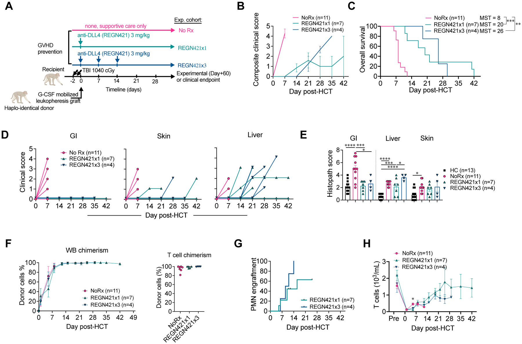
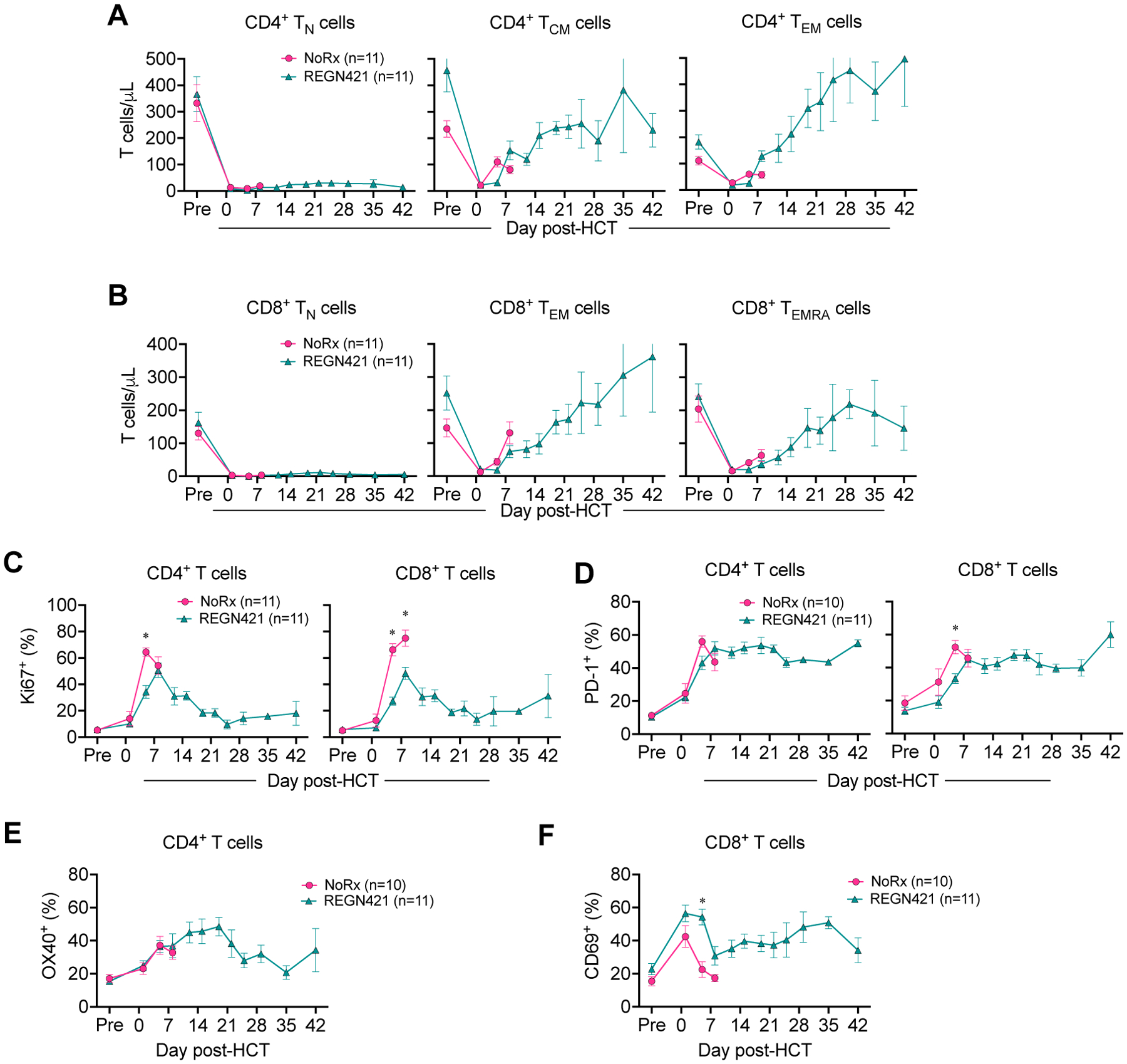
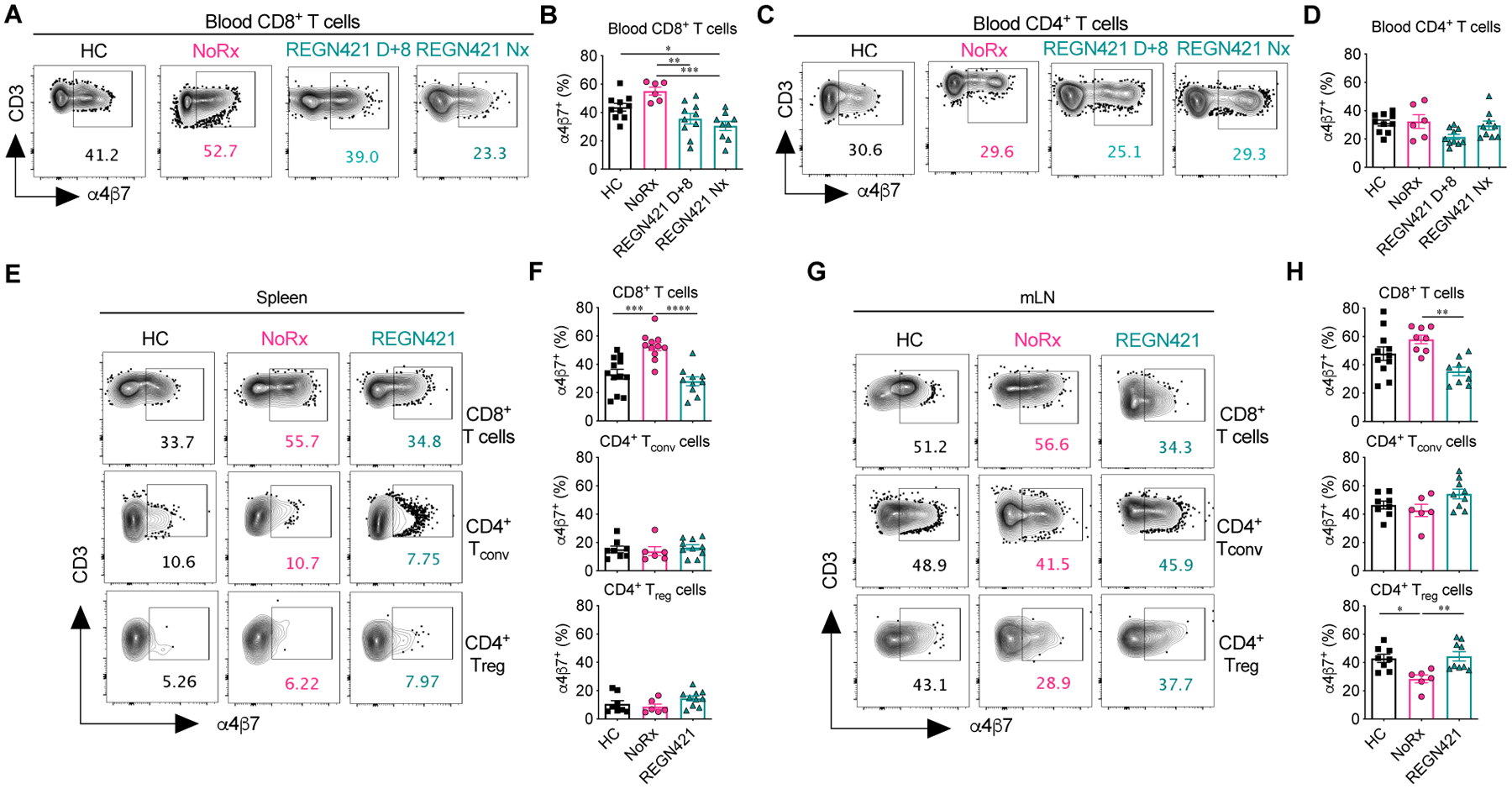
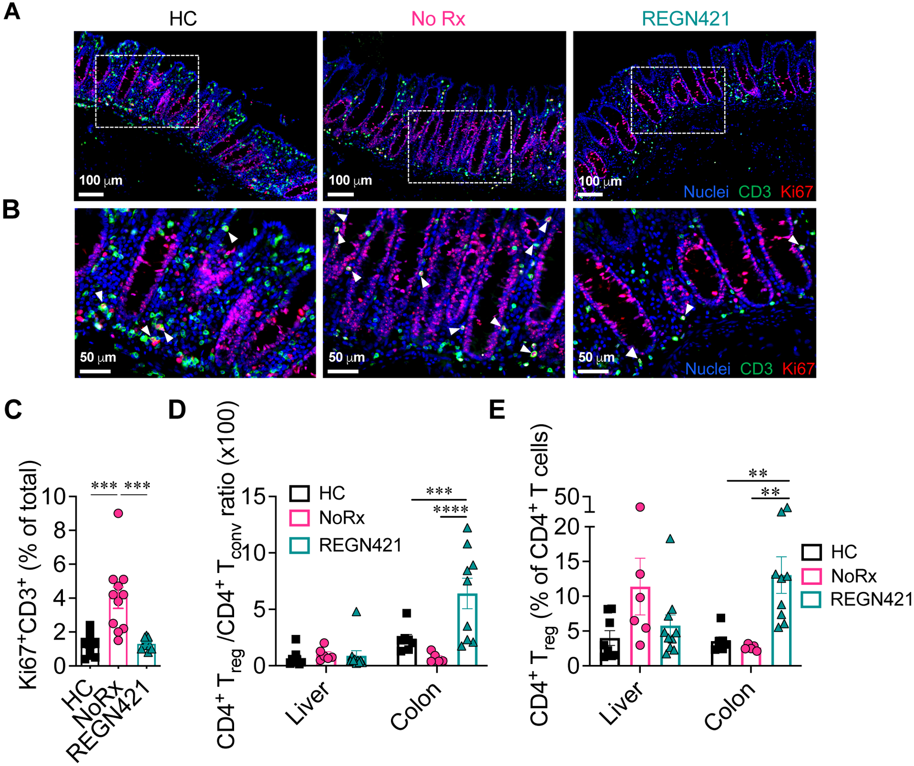
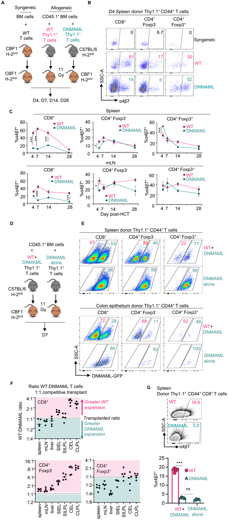
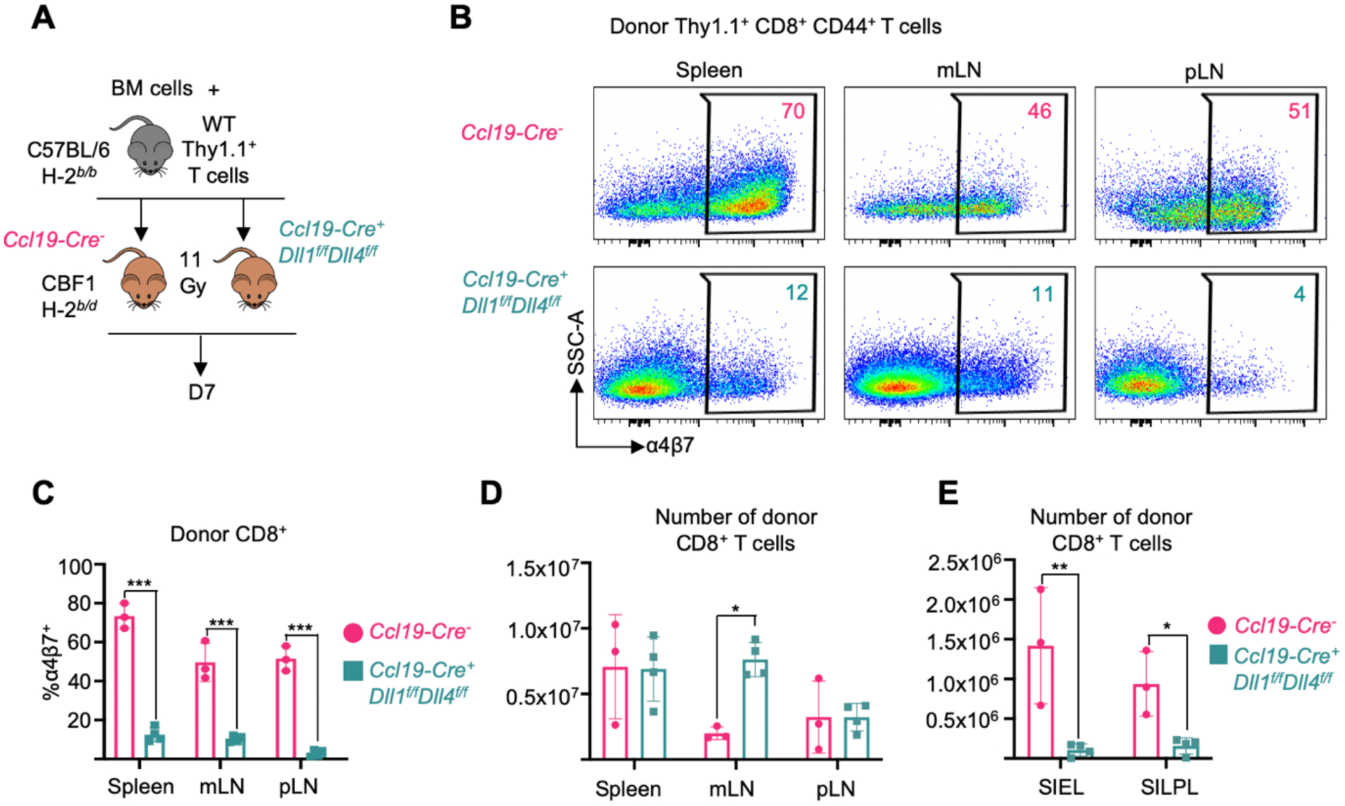
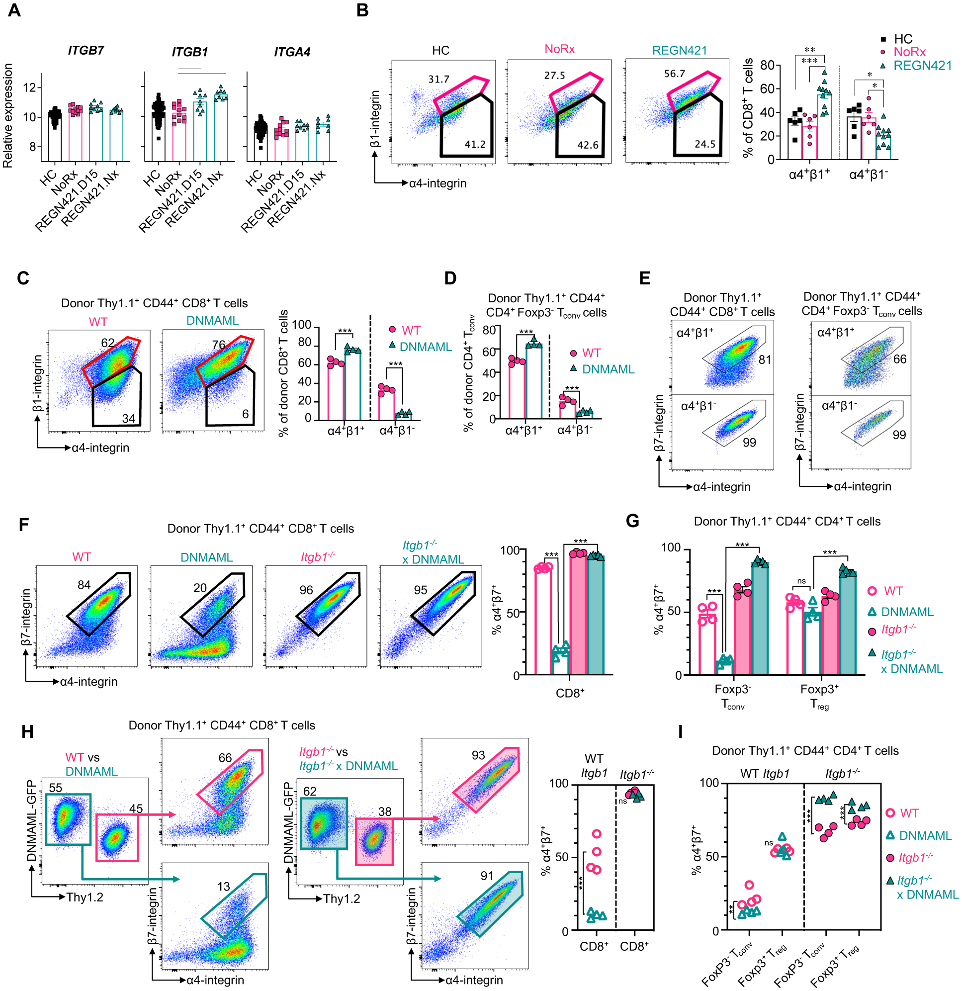
Notch signaling promotes T cell pathogenicity and graft-versus-host disease (GVHD) after allogeneic hematopoietic cell transplantation (allo-HCT) in mice, with a dominant role for the Delta-like Notch ligand DLL4. To assess whether Notch's effects are evolutionarily conserved and to identify the mechanisms of Notch signaling inhibition, we studied antibody-mediated DLL4 blockade in a nonhuman primate (NHP) model similar to human allo-HCT. Short-term DLL4 blockade improved posttransplant survival with durable protection from gastrointestinal GVHD in particular. Unlike prior immunosuppressive strategies tested in the NHP GVHD model, anti-DLL4 interfered with a T cell transcriptional program associated with intestinal infiltration. In cross-species investigations, Notch inhibition decreased surface abundance of the gut-homing integrin α4β7 in conventional T cells while preserving α4β7 in regulatory T cells, with findings suggesting increased β1 competition for α4 binding in conventional T cells. Secondary lymphoid organ fibroblastic reticular cells emerged as the critical cellular source of Delta-like Notch ligands for Notch-mediated up-regulation of α4β7 integrin in T cells after allo-HCT. Together, DLL4-Notch blockade decreased effector T cell infiltration into the gut, with increased regulatory to conventional T cell ratios early after allo-HCT. Our results identify a conserved, biologically unique, and targetable role of DLL4-Notch signaling in intestinal GVHD.


Conflict of interest statement

D.G.A. is currently employed by GlaxoSmithKline. G.C., O.H., F.K., and G.T. are employed by Regeneron, and S.C. is a former employee. B.R.B. has received remuneration as an advisor to Magenta Therapeutics and BlueRock Therapeutics; research funding from BlueRock Therapeutics, Rheos Medicines, Carisma Therapeutics, Inc., and is a co-founder of Tmunity Therapeutics. L.S.K. is on the scientific advisory board for Mammoth Biosciences and HiFiBio. She reports research funding from Magenta Therapeutics, Tessera Therapeutics, Novartis, EMD-Serono, Gilead Pharmaceuticals, and Regeneron Pharmaceuticals. She reports consulting fees from Vertex. L.S.K. reports grants and personal fees from Bristol Myers Squibb. L.S.K.’s conflict-of-interest with Bristol Myers Squibb is managed under an agreement with Harvard Medical School. In addition, L.S.K. has a patent “WO2020172391A1: Methods and compositions relating to the treatment of GVHD” with royalties paid. I.M. has received research funding from Regeneron and Genentech, and he is a member of Garuda Therapeutics’s scientific advisory board.

Figures

Figure 1.
Figure 1.. DLL4 blockade early after allo-HCT protects from GI-aGVHD in the NHP model.
(A) Experimental design, depicting major components of the NHP aGVHD model and dosing regimens with a single or three weekly doses of REGN421 (anti-DLL4). (B) Composite clinical score of allo-HCT recipients in NoRx, REGN421x1, and REGN421x3 cohorts. (C) Overall survival of allo-HCT recipients in the NoRx aGVHD cohort, REGN421x1, and REGN421x3 cohorts. Recipients euthanized based on pre-determined experimental endpoints were censored at terminal analysis. **p<0.01, ***p<0.001, log-rank (Mantel-Cox) test. (D) Clinical scores for GI, skin, and liver aGVHD in allo-HCT recipients, based on established criteria (diarrhea, skin rash and serum bilirubin) (14). (E) Histopathological aGVHD scores for skin, liver, and GI tract (terminal ileum and colon). *p<0.05, ***p<0.001, ****p<0.0001, ANOVA with Tukey post-hoc test. (F) Donor chimerism in whole blood (WB) and CD3+CD20 T cells sorted at terminal analysis. (G) Polymorphonuclear neutrophil (PMN) engraftment in REGN421x1 and REGN421x3 cohorts. (H) Absolute number of T cells in peripheral blood.
Figure 2.
Figure 2.. DLL4 blockade does not interfere with T cell differentiation and reconstitution following allo-HCT.
(A and B) Absolute number of CD4+ (A) and CD8+ (B) T cells with CD45RA+CCR7+CD95 naïve (TN), CD45RACCR7+ central memory (TCM), CD45RACCR7 effector memory (TEM) and CD45RA+CCR7 terminal effector (TEMRA) phenotypes in the blood of allo-HCT recipients. *p<0.05, Tukey-corrected t-test. (C and D) Relative number of Ki67+ (C) or PD-1+ (D) CD4+ and CD8+ T cells in the blood of allo-HCT recipients. *p<0.05, Tukey-corrected t-test. (E and F) Frequency of OX40+CD4+ (E) and CD69+CD8+ (F) T cells in the blood of allo-HCT recipients.
Figure 3.
Figure 3.. DLL4 blockade prevents T cell expression of the gut-homing α4β7 integrin during GVHD.
(A to D) Representative flow cytometry plots (A and C) and data (B and D) depicting the frequency of α4β7+CD8+ (A and B) and α4β7+CD4+ T cells (C and D) in the blood of healthy controls (HC; n=10) and allo-HCT recipients from the NoRx aGVHD (n=6) and REGN421 (n=10) cohorts. Data from the REGN421 cohort was collected at terminal analysis and day +8 (time-matched with the NoRx cohort). (E to H) Representative flow cytometry plots (E and G) and data (F and H) depicting the frequency of α4β7+CD8+, α4β7+CD4+ Tconv and α4β7+CD4+ Treg cells in spleen and mesenteric lymph nodes (mLN) of healthy controls (n=8 to 12 depending on the organ) versus allo-HCT recipients from the NoRx (n=8 to 11 depending on the organ) or REGN421 (n=10) experimental cohorts. For all plots: *p<0.05, **p<0.01, ***p<0.001, ***p<0.001, ****p<0.0001, one-way ANOVA with Tukey post-hoc test.
Figure 4.
Figure 4.. DLL4 blockade inhibits the accumulation of activated tissue-infiltrating T cells in the intestine during aGVHD.
(A to C) Immunofluorescence microcopy of paraffin-embedded colon collected at terminal analysis. Samples from healthy controls (HC) were compared to terminal analysis of allo-HCT recipients from untreated aGVHD (NoRx) or REGN421-treated experimental cohorts. (A) Representative staining for CD3 (green), Ki67 (red) and nuclei visualized by Hoechst (blue). (B) Cropped areas in (A) are enlarged with white arrowheads pointing to CD3+Ki67+ T cells. (C) Quantification of Ki67+CD3+ T cells among total nucleated cells (n=11 images from 5 animals per group). ***p<0.001, Kruskal-Wallis multiple comparison test. (D and E) Treg:Tconv ratio (D) and %Foxp3+ Treg cells (E) were quantified among CD4+ T cells in the liver and colon from healthy controls (n=8) versus at terminal analysis in allo-HCT recipients from untreated aGVHD (NoRx, n=7) or REGN421-treated (n=10) cohorts. **p<0.01, ***p<0.001, ****p<0.0001, one-way ANOVA with Tukey post-hoc test.
Figure 5.
Figure 5.. Cell-intrinsic canonical Notch signals control cell surface α4β7 and gut-homing potential in alloreactive T cells.
(A) 1×10⁶ CBF1 syngeneic T cells (H-2b/d), allogeneic wild-type B6-Thy1.1+ T cells (H-2b/b), or allogeneic Notch-deprived B6-DNMAML-Thy1.1+ T cells (H-2b/b) were transplanted into lethally irradiated CBF1 recipients (H-2b/d) with CD45.1+Thy1.2+ T cell-depleted bone marrow (BM) to distinguish BM-derived cells from the T cell inoculum. (B and C) Representative flow cytometric analysis of α4β7 in donor-derived CD44+CD8+, Foxp3CD4+ and Foxp3+CD4+ T cells in the spleen at day 4 (B) and summary data at indicated times post-transplant in spleen and mLN (C). n=3 mice per group and time point. (D) CBF1 recipients were transplanted as in (A), but with 1:1 wild type:DNMAML donor T cells or DNMAML T cells alone. (E) Representative flow cytometric plots showing relative accumulation of wild-type compared to DNMAML-GFP+ T cells in spleen (top) versus colon epithelium (bottom) at day 7. (F) Organ-specific accumulation of wild-type to DNMAML T cells from the 1:1 competitive transplant was calculated for each T cell subset and expressed as greater (pink) or less than (teal) the initial ratio. mLN, mesenteric lymph node; SIEL, small intestine epithelial lymphocytes; SILPL, small intestine lamina propria lymphocytes; CEL, colon epithelial lymphocytes; CLPL, colon lamina propria lymphocytes. (G) Representative flow cytometric analysis of α4β7 in donor-derived CD8+ T cells from inocula of wild-type and DNMAML T cells (1:1, left) versus DNMAML T cells alone. ns, not significant and ***p<0.001, one-way ANOVA with Tukey’s post-hoc-test.
Figure 6.
Figure 6.. Secondary lymphoid organ Ccl19-Cre+ fibroblastic reticular cells are critical sources of Delta-like Notch ligands.
(A) 1×10⁶ alloreactive T cells plus 1×10⁶ BM cells from C57BL/6 mice were transplanted into Ccl19-Cre+ Dll1f/fDll4f/f or Ccl19-Creneg littermate control CBF1 mice after irradiation (11 Gy). Secondary lymphoid organs and small intestinal lymphocyte fractions were isolated at day 7 post-transplant. (B and C) Representative flow cytometry plots (B) and quantification (C) of α4β7 expression in donor Thy1.1+CD44+CD8+ T cells. SSC-A, side scatter area. (D and E) Absolute number of donor-derived CD8+ T cells in secondary lymphoid organs (D) and small intestinal lymphocyte fractions (E). n=3 or 4 mice per group. *p<0.05, **p<0.01, ***p<0.001, one-way ANOVA with Tukey’s post-hoc tests. mLN, mesenteric lymph node; pLN, peripheral (cervical, axial, brachial, inguinal) lymph nodes; SIEL, small intestine epithelial lymphocytes; SILPL, small intestine lamina propria lymphocytes.
Figure 7.
Figure 7.. Notch signaling regulates α4β7 expression in alloreactive T cells through β1-integrin-dependent mechanisms.
(A) Relative abundance of ITGA4, ITGB7 and ITGB1 mRNA in T cells from healthy control NHP (HC), untreated allo-HCT recipients (NoRx) and allo-HCT recipients receiving anti-DLL4 antibodies (REGN421.D15: blood T cells at day 15; REGN421.Nx: blood T cells at necropsy). Lines indicate significant differences between cohorts (p<0.05, Benjamini-Hochberg correction). (B) Representative flow cytometry plots and summary data depicting the proportion of spleen CD8+ T cells with cell surface α4 and β1 integrins, versus α4 without β1 in HC (n=6), NoRx aGVHD (n=6), and REGN421 (n=10) NHP cohorts. (C to G) 20×106 B6-Thy1.1+ or B6-Thy1.1/1.2+ splenocytes and lymph node cells from mice with wild-type (WT), Notch-deprived (DNMAML), integrin β1-deficient (Itgb1−/−) or Notch-deprived and β1-deficient (Itgb1−/− xDNMAML) T cells were transplanted into lethally irradiated CBF1 recipients (Thy1.2+, H-2b/d) with 1×10⁶ T cell-depleted B6-Thy1.2+ BM cells. (C and D) Representative flow cytometry plots and summary data for α4β1 (spleen, day 4.5) in donor-derived CD44+CD8+ T cells (C) and CD44+Foxp3CD4+ Tconv cells (D). (E) Flow cytometry plots for α4β7 in α4+β1+ (top) and α4+β1 (bottom) wild-type donor-derived spleen CD8+ (left) and CD4+ Tconv cells (right). (F and G) Flow cytometry plots and summary data for α4β7 in donor-derived spleen CD8+ T cells (F), and summary data in donor-derived spleen CD4+ Tconv cells and Foxp3+CD4+ Treg cells (G). (H and I) CBF1 mice were transplanted as in (C to G), but with 15×106:15×106 wild type:DNMAML T cells, or 15×106:15×106 Itgb1−/−:Itgb1−/− DNMAML T cells. (H) Flow cytometry plots showing α4β7 in wild-type versus DNMAML donor-derived spleen CD8+ T cells (left) and Itgb1−/− versus Itgb1−/− × DNMAML CD8+ T cells (right) with summary data (far right). (I) Cell surface α4β7 in donor-derived spleen CD4+ Tconv and CD4+ Treg cells. n=4 mice per group. *p<0.05, **p<0.01, ***p<0.001; ns, not significant. Data were analyzed by a two-way ANOVA with Tukey’s post-hoc-test.

References

    1. Hill GR, Betts BC, Tkachev V, Kean LS, Blazar BR, Current Concepts and Advances in Graft-Versus-Host Disease Immunology. Annu Rev Immunol, (2021). - PMC - PubMed
    1. Ara T et al., Intestinal goblet cells protect against GVHD after allogeneic stem cell transplantation via Lypd8. Sci Transl Med 12, (2020). - PubMed
    1. Piper C et al., Pathogenic Bhlhe40+ GM-CSF+ CD4+ T cells promote indirect alloantigen presentation in the GI tract during GVHD. Blood 135, 568–581 (2020). - PMC - PubMed
    1. Koyama M et al., MHC Class II Antigen Presentation by the Intestinal Epithelium Initiates Graft-versus-Host Disease and Is Influenced by the Microbiota. Immunity 51, 885–898 e887 (2019). - PMC - PubMed
    1. Fu YY et al., T Cell Recruitment to the Intestinal Stem Cell Compartment Drives Immune-Mediated Intestinal Damage after Allogeneic Transplantation. Immunity 51, 90–103 e103 (2019). - PMC - PubMed

Publication types

Substances