. 2023 Sep:75:101764.
doi: 10.1016/j.molmet.2023.101764. Epub 2023 Jun 26.

The gut-brain axis mediates bacterial driven modulation of reward signaling

Affiliations

The gut-brain axis mediates bacterial driven modulation of reward signaling

Jiyoung S Kim et al. Mol Metab. 2023 Sep.

Abstract

Objective: Our goal is to investigate if microbiota composition modulates reward signaling and assess the role of the vagus in mediating microbiota to brain communication.

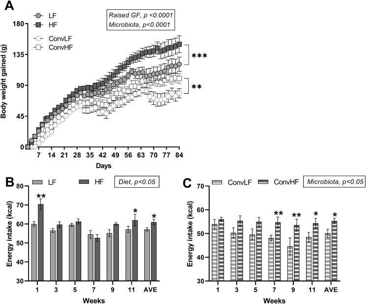
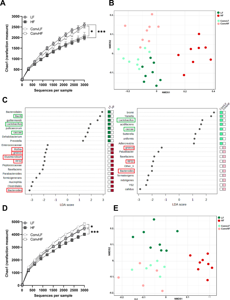
Methods: Male germ-free Fisher rats were colonized with gastrointestinal contents from chow (low fat (LF) ConvLF) or HF (ConvHF) fed rats.

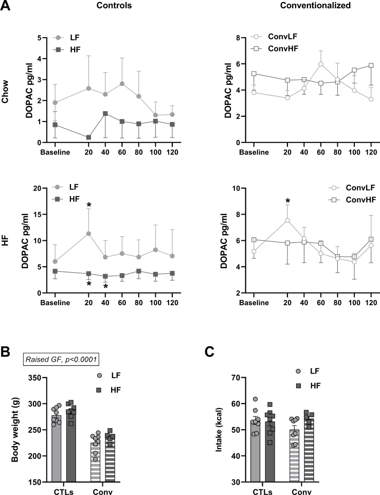
Results: Following colonization, ConvHF rats consumed significantly more food than ConvLF animals. ConvHF rats displayed lower feeding-induced extracellular DOPAC levels (a metabolite of dopamine) in the Nucleus Accumbens (NAc) as well as reduced motivation for HF foods compared to ConvLF rats. Dopamine receptor 2 (DDR2) expression levels in the NAc were also significantly lower in ConvHF animals. Similar deficits were observed in conventionally raised HF fed rats, showing that diet-driven alteration in reward can be initiated via microbiota. Selective gut to brain deafferentation restored DOPAC levels, DRD2 expression, and motivational drive in ConvHF rats.

Conclusions: We concluded from these data that a HF-type microbiota is sufficient to alter appetitive feeding behavior and that bacteria to reward communication is mediated by the vagus nerve.

Keywords: Appetitive behavior; Dopamine; Germ-free rats; Microbiota; Vagus afferent neuron.


Figures

Figure 1
Figure 1
Experimental design, groups, and timeline.
Figure 2
Figure 2
A HF microbiota transfer is sufficient to increase food intake and body weight gain. Body weight gain (A) of control and conventionalized animals. HF fed rats gained significantly more weight than LF (chow) fed animals (p < 0.001) and colonization with a HF-type microbiota (ConvHF) led to excessive weight gained compared to colonization with a LF-type microbiota (ConvLF) (p < 0.01). Rats born germ free (GF) were significantly smaller than control animals (three-way mixed-effects model, GF effect, p < 0.0001; Microbiota effect, p < 0.0001, Time effect, p < 0.0001). Weekly food intake of control (B) and conventionalized (C) rats. HF and ConvHF rats consumed more kcals than LF and ConvLF animals (p < 0.05). Data is presented as mean ± SEM. LF: low fat (chow), HF: high fat, ConvLF: conventionalized low fat, ConvHF: conventionalized high fat, GF: germ free. Main effects p-values are displayed on the graphs while symbols are used for post hoc analysis. ∗p < 0.05, ∗∗p < 0.01, ∗∗∗p < 0.001. Group sizes are detailed in Figure 1.
Figure 3
Figure 3
A HF type microbiota induces dopamine signaling deficits. Intake during microdialysis is presented in (A); there were no differences in 20min intake between groups with animals consuming more calories when HF pellets were presented compared to chow pellets (p < 0.0001). Extracellular DOPAC levels (normalized to baseline) in the NAc increased in response to both chow and HF stimuli in LF and ConvLF rats, however this response was reduced or absent in HF and ConvHF animals (B). Consumption of chow pellets induced a gradual increase in DOPAC levels in LF fed rats (peak at 60 min after food introduction, LF DOPAC60min vs. LF DOPACBaseline, p < 0.01) which was absent in HF fed rats. HF pellets led to a more rapid increase in extracellular DOPAC (peak at 20 min, LF DOPAC20min vs. LF DOPACBaseline, p < 0.001), again this response was absent in HF fed rats. GF rats colonized with a chow or HF-type microbiota recapitulated their respective donor phenotype. Consumption of chow pellets induced a gradual increase in DOPAC levels in ConvLF fed (ConvLF DOPAC60min vs. ConvLF DOPACBaseline, p < 0.01). This response was absent in ConvHF rats. Similarly, to LF rats, in ConvLF animals, consumption of HF pellets led to a rapid increase in NAc extracellular DOPAC (ConvLF DOPAC20min vs. ConvLF DOPACBaseline, p < 0.05) which was absent in ConvHF rats. HF feeding and colonization with a HF type microbiota led to a decreased number of DRD2 expressing cells in the ventral striatum, where the NAc is located, and decreased DRD1 positive cells in the dorsal striatum (D). Pictures of DRD2 stained coronal sections of ventral striatum and pictures of DRD1 stained coronal sections of the dorsal striatum are presented in (D), AC: anterior commissure. Microbiota associated differences were not related to differences in total DA (E) or DA synthesis (F) in the ventral striatum. However, being born GF was associated with an increase in TH levels in the ventral striatum (F) and elevated DOPAC levels (C) indicating that the absence of a microbiota early in life influences the mesolimbic DA system and DA synthesis. Raw DOPAC levels can be found in Supp Fig 3. Total area fraction quantification for DRDs can be found in Supp Fig 7. Data is presented as mean ± SEM. LF: low fat (chow), HF: high fat, CTLs: control (LF and HF) rats, ConvLF: conventionalized low fat, ConvHF: conventionalized high fat, DOPAC: 3,4-Dihydroxyphenylacetic acid, a DA (dopamine) metabolite, DRD1/2: dopamine receptor 1/2, GF: germ free, TH: tyrosine hydroxylase. Main effects p-values are displayed on the graphs while symbols are used for post hoc analysis. (B) Time point vs. Baseline: ∗p < 0.05, ∗∗p < 0.01, ∗∗∗p < 0.001; LF vs. HF or ConvLF vs. ConvHF: #p < 0.05, ##p < 0.01. (A,C,D,E,F) ∗p < 0.05, ∗∗p < 0.01, ∗∗∗∗p < 0.0001. Group sizes are detailed in Figure 1.
Figure 4
Figure 4
Microbiota modulates task acquisition and motivational drive. A HF-type microbiota in adulthood was associated with a reduction in motivational drive characterized by decreases in breakpoint (A, two-way ANOVA, microbiota effect, p < 0.05) and total presses (B, two-way ANOVA, microbiota effect, p < 0.05), this effect was especially evident in recipient animals. A HF-type microbiota also resulted in deficits in task acquisition (C, two-way ANOVA, microbiota effect, p < 0.05). These data showed that microbiota composition is sufficient to alter task acquisition and motivational drive. Animals born GF displayed an overall increase in motivated behavior characterized by increases in breakpoint (A, two-way ANOVA, GF effect, p < 0.0001) and total presses (B, two-way ANOVA, GF effect, p < 0.0001) during a PR schedule of reinforcement compared to control rats (reward was a HF pellet). GF rats also took longer than control animals.to learn the pressing task in a FR paradigm (C, two-way ANOVA, GF effect, p < 0.0001). The lack of microbial exposure in early life had long-term consequences on behavior showing that microbiota is necessary for proper brain development. Data is presented as mean ± SEM. CTLs: control (LF and HF) rats, Conv: conventionalized (ConvLF and ConvHF) rats, HF: high fat, GF: germ free, PR: progressive ration, FR: fixed ratio. Main effects p-values are displayed on the graphs while symbols are used for post hoc analysis. ∗p < 0.05, ∗∗p < 0.01, ∗∗∗∗p < 0.0001. Group sizes are detailed in Figure 1.
Figure 5
Figure 5
Deafferentation leads to a reduction in food intake and body weight gain in dysbiotic rats. Deafferentation led to sustained reduction in body weight gain and decreased food intake in ConvHF (A, B, C) rats compared to ConvLF animals. ConvHF rats gained more weight than ConvLF animals following colonization (A, three-way mixed-effects model, Microbiota x Time effect, p < 0.05). Deafferentation led to a reduction in body weight in both ConvLF and ConvHF rats (B, three-way mixed-effects model, CCK-SAP effect, p < 0.001). Deafferented ConvLF rats (ConvLF-CCK-SAP) regained weight, and by the end of the experiment, there were no differences in body weight gain between the ConvLF-CCK-SAP rats and the sham-operated group, ConvLF-SAP. In dysbiotic rats, deafferentation led to a sustained reduction in body weight gain (B, ConvHF-SAP vs. ConvHF-CCK-SAP, p < 0.01). Changes in weight were likely related to changes in intake as deafferentation led to a decrease in food intake in the ConvHF rats (C, week 11 and 13, ConvHF-SAP vs. ConvHF-CCK-SAP, p < 0.05). Data is presented as mean ± SEM. LF: low fat (chow), HF: high fat, ConvLF: conventionalized low fat, ConvHF: conventionalized high fat. SAP: nodose saporin injections, CCK-SAP: nodose injections of saporin conjugated to CCK to delete gut to brain vagal afferent innervation. Main effects p-values are displayed on the graphs while symbols are used for post hoc analysis. ∗p < 0.05, ∗∗∗p < 0.001. Group sizes are detailed in Figure 1.
Figure 6
Figure 6
The vagus nerve is required for microbiota-driven deficit in dopamine signaling. Intake during microdialysis is presented in (A); there were no differences in 20min intake between groups with animals consuming more calories when HF pellets were presented compared to chow pellets (p < 0.0001). Extracellular DOPAC levels (normalized to baseline) increased in response to chow and HF (B) consumption in intact ConvLF rats (ConvLF-SAP) (Chow, ConvLF-SAP DOPAC20min vs. ConvLF-SAP DOPACBaseline, p < 0.01; HF, ConvLF-SAP DOPAC60min vs. ConvLF-SAP DOPACBaseline, p < 0.05), however, this response was absent in ConvHF-SAP animals. Deafferentation restored DOPAC levels in ConvHF rats (Chow, ConvHF-CCK-SAP DOPAC40min vs. ConvHF-CCK-SAP DOPACBaseline, p < 0.05; HF, ConvHF-CCK-SAP DOPAC60min vs. ConvHF-CCK-SAP DOPACBaseline, p < 0.05). Loss of gut to brain innervation blunted food-induced increase in NAc DOPAC levels in ConvLF animals (ConvLF-CCK-SAP). Intact dysbiotic animals (ConvHF-SAP) displayed decreases in the number of cells expressing DRD2 in the ventral striatum and DRD1 in the dorsal striatum (C). Deafferentation in dysbiotic rats (ConvHF-CCK-SAP) restored both DRD2 and DRD1 levels in the ventral and dorsal striatum (ConvHF-CCK-SAP vs. ConvHF-SAP, p < 0.05) and did not affect DRDs expression in ConvLF animals. Pictures of DRD2 stained coronal sections of ventral striatum and pictures of DRD1 stained coronal sections of the dorsal striatum are presented in (B), AC: anterior commissure. Raw DOPAC levels can be found in Supp Fig 6. Total area fraction quantification for DRDs can be found in Supp Fig 7. Data is presented as mean ± SEM. LF: low fat (chow), HF: high fat, ConvLF: conventionalized low fat, ConvHF: conventionalized high fat, SAP: nodose saporin injections, CCK-SAP: nodose injections of saporin conjugated to CCK to delete gut to brain vagal afferent innervation, DOPAC: 3,4-Dihydroxyphenylacetic acid, a DA (dopamine) metabolite, NAc: nucleus accumbens, DRD1/2: dopamine receptor ½(B). Time point vs. Baseline: ∗p < 0.05, ∗∗p < 0.01; within groups comparisons: #p < 0.05. (A,C) ∗p < 0.05, ∗∗∗∗p < 0.0001. Group sizes are detailed in Figure 1.
Figure 7
Figure 7
The vagus nerve is required for microbiota-driven deficit in motivational drive. Prior to deafferentation, ConvHF rats displayed deficits in motivational drive characterized by reduced breakpoint (A) and pressing behavior (B) during a PR schedule of reinforcement compared to ConvLF rats (reward was a 35% fat, HF pellet). Surgery had no effects on these parameters. Deafferentation restored motivational drive in dysbiotic animals. Breakpoint and total presses during PR were significantly increased in ConvHF-CCK-SAP rats compared to sham operated ConvHF-SAP animals and baseline ConvHF rats (A, B) but had no effects in ConvLF rats (A, B). Deafferentation also improved all GF rats' performance on task acquisition (C). Data is presented as mean ± SEM. LF: low fat (chow), HF: high fat, ConvLF: conventionalized low fat, ConvHF: conventionalized high fat, SAP: nodose saporin injections, CCK-SAP: nodose injections of saporin conjugated to CCK to delete gut to brain vagal afferent innervation. GF: germ free, PR: progressive ration, FR: fixed ratio. Main effects p-values are displayed on the graphs while symbols are used for post hoc analysis. ∗p < 0.05, ∗∗p < 0.01, ∗∗∗<p < 0.001. Group sizes are detailed in Figure 1.
Supplementary Figure 1
Control and colonized animals share similar microbiota profiles. (A) α-diversity (measured by Chao 1 index) was significantly lower in HF fed rats compared to LF fed rats. Similarly, ConvHF displayed reduced α-diversity compared to ConvLF rats two weeks post-colonization. (B) β-diversity was assessed by non-metric multi-dimensional scaling (NMDS) with distances determined using the Bray–Curtis index. Significant dissimilarities between groups were determined via permutational multivariate ANOVA. All groups were significantly different from one another (in particular LF vs. HF, p < 0.001 and ConvLF vs. ConvHF, p < 0.05). Colonized animals cluster in proximity with their respective controls two weeks post-colonization. (C) Linear discriminant analysis (LDA) effect size analysis (LefSe) positively identified specific taxa characteristic of LF (green) or HF (red) diets. Taxa identified by a thick rectangle were also identified as characteristics of ConvLF or ConvHF conditions two weeks post-colonization. Analysis of feces collected at sacrifice revealed that some differences between ConvLF and ConvHF were maintained over the course of the experiment. (D) At sacrifice, both HF and ConvHF rats displayed a reduction in α-diversity when compared to their respective controls. (E) Overall microbiota profile of the ConvLF and ConvHF rats had converged (ConvLF vs. ConvHF, p < 0.1) but differences in specific abundances were still present (See Supp Fig 2). LF: low fat (chow), HF: high fat, ConvLF: conventionalized low fat, ConvHF: conventionalized high fat. Group sizes are detailed in Figure 1.
Supplementary Figure 2
Control and colonized animals share similar microbiota profiles. (A) Example of taxa displaying similar patterns of abundances between LF vs. HF rats and ConvLF vs. ConvHF animals (Relative abundances are presented. For very low abundances, taxa total reads are plotted). (B) Example of differences in abundances still present at sacrifice. (C) Peak extracellular DOPAC levels was correlated with several bacterial taxa abundances (two-week post-colonization abundances). LF: low fat (chow), HF: high fat, ConvLF: conventionalized low fat, ConvHF: conventionalized high fat. Group sizes are detailed in Figure 1.
Supplementary Figure 3
HF microbiota induces deficit in food-driven DOPAC levels in the NAc (raw DOPAC values). (A) Extracellular DOPAC levels increased in response to both chow and HF stimuli in LF and ConvLF rats, however, this response was reduced or absent in HF and ConvHF animals. Unlike the data presented in Figure 3A, the values presented here are the raw DOPAC values (pg/ml). The resulting increase in variability reduced statistical significance. Changes in extracellular DOPAC were not related to differences in body weight (B) or food intake (C). (B) and (C) represent average values during the weeks microdialysis was conducted. Data is presented as mean ± SEM. LF: low fat (chow), HF: high fat, CTLs: control (LF and HF) rats, ConvLF: conventionalized low fat, ConvHF: conventionalized high fat, DOPAC: 3,4-Dihydroxyphenylacetic acid, a DA (dopamine) metabolite, NAc: nucleus accumbens, (A) Time point vs. Baseline: ∗p < 0.05. (B) Main effects p-values are displayed on the graphs. Group sizes are detailed in Figure 1.
Supplementary Figure 4
Injection of CCK-SAP into the NG led to a significant reduction in vagal innervation in the NTS compared to control SAP NG injections. Isolectin B4 (IB4) selectively binds to unmyelinated c fibers, which at the level of the medial NTS are almost exclusively of vagal origin. Bilateral injection of the toxin saporin conjugated to CCK (CCK-SAP) into the NG significantly reduces IB4+ staining in the medial NTS (by about 40%, p < 0.0001) compared to SAP NG injections (A, 20X) in all groups. Quantification is presented (B). Data is presented as mean ± SEM. LF: low fat (chow), HF: high fat, ConvLF: conventionalized low fat, ConvHF: conventionalized high fat, NTS: nucleus of solitary tract, AP: area postrema, NG: nodose ganglion. Main effects p-values are displayed on the graphs while symbols are used for post hoc analysis. ∗p < 0.05, ∗∗p < 0.01, ∗∗∗p < 0.001, ∗∗∗∗p < 0.0001. Group sizes are detailed in Figure 1.
Supplementary Figure 5
Loss of gut to brain innervation had little effect on microbiota composition. (A) α-diversity (Chao 1) was significantly lower in ConvHF rats compared ConvLF animals (p < 0.05), with no significant effect of deafferentation. (B) β-diversity was assessed by non-metric multi-dimensional scaling (NMDS) with distances determined using the Bray–Curtis index. Significant dissimilarities between groups were determined via permutational multivariate ANOVA. ConvLF and ConvHF displayed different microbiota profile (p < 0.001) with no significant differences associated with deafferentation status. (C) Linear discriminant analysis (LDA) Effect Size was used to identify taxa characteristic of a condition. ConvLF-SAP and ConvLF-CCK-SAP animals shared common characteristic taxa, including the previously identified uniformis. ConvHF-SAP and ConvHF-CCK-SAP also shared signature taxa, with increased presence of members of the Lachnospiraceae family, in particular Blautia. (D) When analyzed separately, we found very few difference between deafferented ConvLF rats (ConvLF-CCK-SAP) compared to ConvLF-SAP rats and between ConvHF-SAP and ConvHF-CCK-SAP. The few identified taxa did not translate into differences in abundances. LF: low fat (chow), HF: high fat, ConvLF: conventionalized low fat, ConvHF: conventionalized high fat. SAP: nodose saporin injections, CCK-SAP: nodose injections of saporin conjugated to CCK to delete gut to brain vagal afferent innervation. Group sizes are detailed in Figure 1.
Supplementary Figure 6
The vagus nerve is required for microbiota-driven deficit in food-driven NAc DOPAC levels (raw DOPAC values). Extracellular DOPAC levels increased in response to chow (A) and HF (B) consumption in intact ConvLF rats (ConvLF-SAP), however this response was absent in ConvHF-SAP animals. Deafferentation restored DOPAC levels in ConvHF rats. Conversely, loss of gut to brain innervation blunted food-induced increase in extracellular DOPAC in the NAc in ConvLF rats (A, B). Values presented here are the raw DOPAC values (pg/ml), resulting in an increased variability and reduced statistical significance. Differences in DOPAC levels were not due to differences in baseline DOPAC levels (C), total DA levels (D) or DA synthesis (E). Data is presented as mean ± SEM. LF: low fat (chow), HF: high fat, ConvLF: conventionalized low fat, ConvHF: conventionalized high fat, SAP: nodose saporin injections, CCK-SAP: nodose injections of saporin conjugated to CCK to delete gut to brain vagal afferent innervation, DOPAC: 3,4-Dihydroxyphenylacetic acid, a DA (dopamine) metabolite, NAc: nucleus accumbens, TH: tyrosine hydroxylase, ∗p < 0.05. Group sizes are detailed in Figure 1.
Supplementary Figure 7
The vagus nerve is required for microbiota induced DRDs deficits. HF feeding and colonization with a HF type microbiota led to a decreased DRD2 expression level in the ventral striatum, where the NAc is located, and decreased DRD1 levels in the dorsal striatum (A). Pictures of DRD2 stained coronal sections of ventral striatum and pictures of DRD1 stained coronal sections of the dorsal striatum are presented in (A), scale bar = 400 μm, AC: anterior commissure. Intact dysbiotic animals (ConvHF-SAP) displayed decreases in expression levels of DRD2 in the ventral striatum and DRD1 in the dorsal striatum (B). DRD2 reduction in ConvHF-SAP rats was not significant compared to control ConvLF-SAP animals (p < 0.1), while DRD1 expression levels was significantly reduced in in ConvHF-SAP rats compared to ConvLF-SAP animals (p < 0.05). Deafferentation in dysbiotic rats (ConvHF-CCK-SAP) restored both DRD2 and DRD1 levels in the ventral and dorsal striatum (ConvHF-CCK-SAP vs. ConvHF-SAP, p < 0.05) and did not affect DRDs expression in ConvLF animals. Pictures of DRD2 stained coronal sections of ventral striatum and pictures of DRD1 stained coronal sections of the dorsal striatum are presented in (B), scale bar = 200 μm, AC: anterior commissure. Data is presented as mean ± SEM. LF: low fat (chow), HF: high fat, CTLs: control (LF and HF) rats, ConvLF: conventionalized low fat, ConvHF: conventionalized high fat, SAP: nodose saporin injections, CCK-SAP: nodose injections of saporin conjugated to CCK to delete gut to brain vagal afferent innervation, DRD1/2: dopamine receptor 1/2, GF: germ free. Main effects p-values are displayed on the graphs while symbols are used for post hoc analysis. ∗p < 0.05, ∗∗p < 0.01. Group sizes are detailed in Figure 1.

Similar articles

Cited by

References

    1. Hernandez L., Hoebel B.G. Feeding and hypothalamic stimulation increase dopamine turnover in the accumbens. Physiol Behav. 1988;44:599–606. doi: 10.1016/0031-9384(88)90324-1. - DOI - PubMed
    1. Valdivia S., Patrone A., Reynaldo M., Perello M. Acute high fat diet consumption activates the mesolimbic circuit and requires orexin signaling in a mouse model. PLoS One. 2014;9 doi: 10.1371/journal.pone.0087478. - DOI - PMC - PubMed
    1. Berridge K.C., Robinson T.E., Aldridge J.W. Dissecting components of reward: 'liking', 'wanting', and learning. Curr Opin Pharmacol. 2009;9:65–73. doi: 10.1016/j.coph.2008.12.014. - DOI - PMC - PubMed
    1. Han W., Tellez L.A., Perkins M.H., Perez I.O., Qu T., Ferreira J., et al. A neural circuit for gut-induced reward. Cell. 2018;175:887–888. doi: 10.1016/j.cell.2018.10.018. - DOI - PubMed
    1. Tellez L.A., Medina S., Han W., Ferreira J.G., Licona-Limon P., Ren X., et al. A gut lipid messenger links excess dietary fat to dopamine deficiency. Science. 2013;341:800–802. doi: 10.1126/science.1239275. - DOI - PubMed

Publication types

Substances