Review
. 2023 Aug;299(8):104966.
doi: 10.1016/j.jbc.2023.104966. Epub 2023 Jun 26.

The diverse structural modes of tRNA binding and recognition

Affiliations
Review

The diverse structural modes of tRNA binding and recognition

Anna Biela et al. J Biol Chem. 2023 Aug.

Abstract

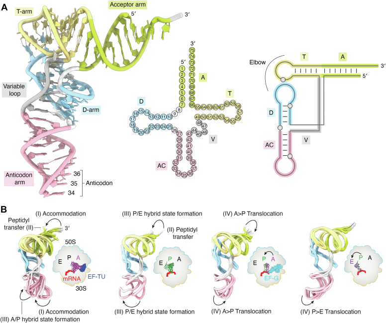
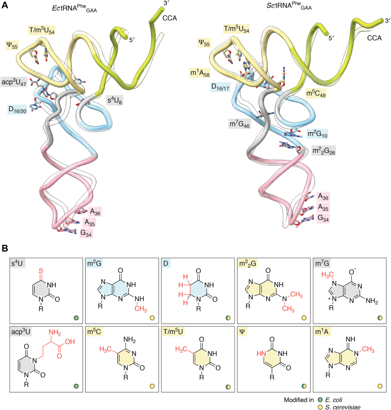
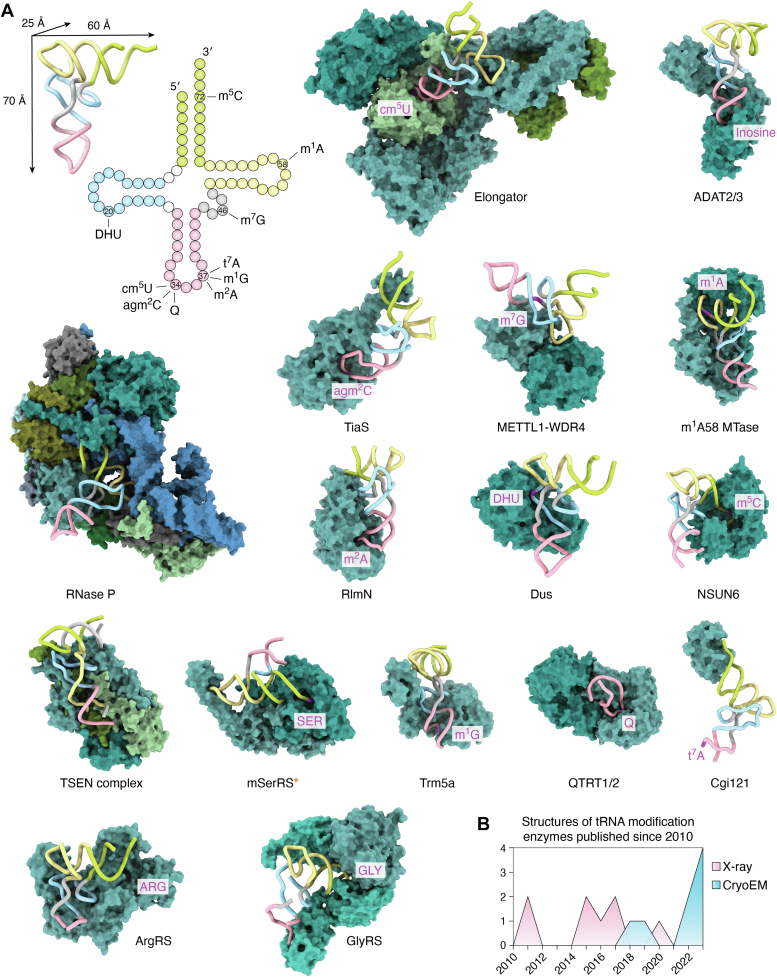
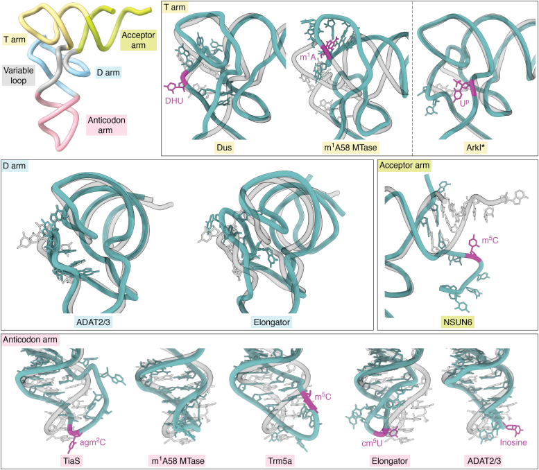
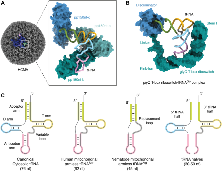
tRNAs are short noncoding RNAs responsible for decoding mRNA codon triplets, delivering correct amino acids to the ribosome, and mediating polypeptide chain formation. Due to their key roles during translation, tRNAs have a highly conserved shape and large sets of tRNAs are present in all living organisms. Regardless of sequence variability, all tRNAs fold into a relatively rigid three-dimensional L-shaped structure. The conserved tertiary organization of canonical tRNA arises through the formation of two orthogonal helices, consisting of the acceptor and anticodon domains. Both elements fold independently to stabilize the overall structure of tRNAs through intramolecular interactions between the D- and T-arm. During tRNA maturation, different modifying enzymes posttranscriptionally attach chemical groups to specific nucleotides, which not only affect translation elongation rates but also restrict local folding processes and confer local flexibility when required. The characteristic structural features of tRNAs are also employed by various maturation factors and modification enzymes to assure the selection, recognition, and positioning of specific sites within the substrate tRNAs. The cellular functional repertoire of tRNAs continues to extend well beyond their role in translation, partly, due to the expanding pool of tRNA-derived fragments. Here, we aim to summarize the most recent developments in the field to understand how three-dimensional structure affects the canonical and noncanonical functions of tRNA.

Keywords: NMR; RNA modifications; cryo-EM; crystallography; tRNA.


Conflict of interest statement

Conflict of interest The authors declare that they have no conflicts of interest with the contents of this article.

Figures

Figure 1
Figure 1
The structure of tRNA at different levels and during translation.A, left: specific regions are indicated on yeast tRNAPheGAA (PDB ID 1EHZ) as follows: 3ʹ and 5ʹ end; acceptor arm (light green); T-arm (yellow); D-arm (light blue); variable loop (gray); anticodon arm (pink); and 34, 35, 36 anticodon nt. The dimensions of folded tRNAs are approximately 60 × 70 × 25 Å. Middle: clover-leaf secondary structure representation of tRNA numbered according to the standard convention. Right: tertiary nucleotide interactions responsible for the L-shape tRNA structure. B, multiple structures of tRNA extracted from the context of elongating bacterial 70S ribosome determined by time-resolved cryo-EM. The schematic demonstrates local flexibility of the A-site (magenta), P-site (dark green), and E-site (purple) tRNA associated with the cognate mRNA (red) bound to the 70S ribosome [large 50S (blue) and small 30S (orange)]. The arrows highlight the bending of the 3ʹ end (silver) from the acceptor arm (light green) and the swinging of the anticodon arm (pink). The major sequential steps of the elongation cycle associated with the L-shape distortion are indicated with Roman numerals (I–IV). The two superimpositions on the left (A-site phenylalanine- and P-site methionine-tRNA) follow the steps and changes after cognate aminoacyl-tRNA delivery by elongation factor EF-TU (dark blue). The two superimpositions on the right depict AP proline- and PE methionine-tRNA translocation facilitated by elongation factor EF-G (cyan), which resets the system for the next elongation cycle. EF-Tu, Elongator factor-Tu.
Figure 2
Figure 2
Structure of unmodified and modified tRNAPhe.A, comparison of prokaryotic and eukaryotic tRNAPheGAA. Superimpositions of in vitro transcribed (transparent) and endogenous (opaque) tRNAPheGAA from Escherichia coli (Ec) and Saccharomyces cerevisiae (Sc) obtained by crystallography. These tRNAs are depicted as cartoons using the same color code for each region as in Figure 1. Modifications that are important for the tRNA core structure are represented in sticks and colored according to their position in the tRNA. The PDB IDs and resolution limits of each tRNA are as follows: unmodified EctRNA (PDB ID 3L0U at 3.0 Å) and SctRNA (PDB ID 5AXM at 2.2 Å) and modified EctRNA (PDB ID 6Y3G at 3.1 Å) and SctRNA (PDB ID 1EHZ at 1.9 Å). The m7G47 modification in EctRNA (PDB ID 6Y3G) is present in tRNAPheGAA but is not visible in the electron density map. B, chemical structures of modified nucleosides that are shown in (A). The modifications are indicated in red and the ribose moiety is annotated with R. Base colors correspond to the modified target positions in (A), while the color (green or yellow) of the small circle indicates the presence of modification in E. coli or S. cerevisiae. RMSD values for E. coli tRNAPhe and S. cerevisiae tRNAPhe were calculated with ChimeraX using the backbone atoms of nt 3 to 74. E. coli: overall RMSD 2.669 Å2, acceptor stem 4.426 Å2, D-arm 1.595 Å2, anticodon-loop 2.716 Å2, variable loop 2.362 Å2, T-arm 1.349 Å2; S. cerevisiae: overall RMSD 1.662 Å2, acceptor stem 2.132 Å2, D-arm 2.194 Å2, anticodon-loop 1.628 Å2, variable loop 0.801 Å2, T-arm 0.665 Å2.
Figure 3
Figure 3
tRNA modification enzymes bind and recognize tRNAs using diverse strategies.A, gallery of available structures of tRNA modifying/processing enzymes solved with full-length tRNA (published since 2010). Precise modification sites are marked on the clover-leaf map of tRNA (left upper corner) and in the 3D structures (magenta). Elongator (PDB ID 8ASW), ADAT2/3 (PDB ID 8AW3), METTL1-WDR4 (PDB ID 8CTH), m1A58 MTase (PDB ID 5CD1), mitochondrial seryl-tRNA synthetase (PDB ID 7U2B), Dus (PDB ID 3B0V), Ribonuclease P (PDB ID 6AHU), TiaS (PDB ID 3AMT), Trm5a (PDB ID 5WT3), NSUN6 (PDB ID 5WWT), TSEN complex (PDB ID 7UXA/7ZRZ), RlmN (PDB ID 5HR6), QTRT1/2 (PDB ID 7NQ4), Cgi121 subunit of KEOPS (PDB ID 7KJT). All presented structures are cytoplasmic enzymes except for mitochondrial seryl-tRNA synthetase, which is marked by an orange asterisk. DusC from Escherichia coli (PDB ID 4YCP, 4YCO) is not shown as it is highly similar to TthDus (PDB ID 3B0V). The class I and class II aminoacyl tRNA synthetases are also included: arginyl-tRNA synthetase (ArgRS, PDB ID 5YYN) and glycyl-tRNA synthetase (GlyRS, PDB ID 7YSE). B, summary of the number of structures solved by different structural biology techniques together with full-length tRNA (2010–2022). KEOPS, Kinase, endopeptidase and other protein of small size complex.
Figure 4
Figure 4
Modification enzymes influence tRNA structure in various regions. Yeast tRNAPheGAA (PDB ID 1EHZ) is aligned on each structure by the matchmaker tool in ChimeraX and shown transparently. Structures are sorted by the region affected structurally, and the names of the modifying enzymes are colored, accordingly. Modification sites are marked in magenta. Structures of full length-tRNAs are shown without their respective protein partners - Dus (PDB ID 3B0V), m1A58 MTase (PDB ID 5CD1), Elongator (PDB ID 8ASW), ADAT2/3 (PDB ID 8AW3), TiaS (PDB ID 3AMT), Trm5a (PDB ID 5WT3), NSUN6 (PDB ID 5WWT). ArkI (PDB ID 7VNV) is a crystal structure of tRNA alone, modified by TkArkI.
Figure 5
Figure 5
Examples of tRNAs with unexpected functions and atypical appearances.A, a cartoon representation of a cryo-EM structure of HCMV virion (PDB ID 5VKU) with tRNA bound to pp150 nt (inlet, PDB ID 7LJ3). The asymmetrical unit in the virion is colored in blue and the selected tRNAs are highlighted in pink, purple, and green. B, a cartoon representation of a cryo-EM structure of a full-length of Bacillus subtilis glyQ T-box-tRNAGlyGCC complex (PDB ID 6POM). The tRNA is illustrated as spaghetti representation, while the binding partners are shown with surface representation. C, two-dimensional representation of various tRNA folds, including the canonical and truncated forms. HCMV, human cytomegalovirus.

References

    1. Crick F.H. On protein synthesis. Symp. Soc. Exp. Biol. 1958;12:138–163. - PubMed
    1. Roeder R.G., Rutter W.J. Multiple forms of DNA-dependent RNA polymerase in eukaryotic organisms. Nature. 1969;224:234–237. - PubMed
    1. Weinmann R., Raskas H.J., Roeder R.G. Role of DNA dependent RNA polymerases II and III in transcription of the adenovirus genome late in productive infection. Proc. Natl. Acad. Sci. U. S. A. 1974;71:3426–3430. - PMC - PubMed
    1. Chan P.P., Lowe T.M. GtRNAdb 2.0: an expanded database of transfer RNA genes identified in complete and draft genomes. Nucleic Acids Res. 2016;44:D184–D189. - PMC - PubMed
    1. Baranov P.V., Gesteland R.F., Atkins J.F. Recoding: translational bifurcations in gene expression. Gene. 2002;286:187–201. - PubMed

Publication types

LinkOut - more resources