. 2025 May;35(2):138-151.
doi: 10.1016/j.zemedi.2023.05.007. Epub 2023 Jun 26.

Silicone phantoms fabricated with multi-material extrusion 3D printing technology mimicking imaging properties of soft tissues in CT

Affiliations

Silicone phantoms fabricated with multi-material extrusion 3D printing technology mimicking imaging properties of soft tissues in CT

Sepideh Hatamikia et al. Z Med Phys. 2025 May.

Abstract

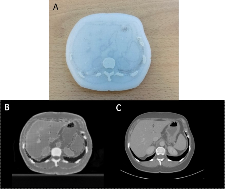
Recently, 3D printing has been widely used to fabricate medical imaging phantoms. So far, various rigid 3D printable materials have been investigated for their radiological properties and efficiency in imaging phantom fabrication. However, flexible, soft tissue materials are also needed for imaging phantoms for simulating several clinical scenarios where anatomical deformations is important. Recently, various additive manufacturing technologies have been used to produce anatomical models based on extrusion techniques that allow the fabrication of soft tissue materials. To date, there is no systematic study in the literature investigating the radiological properties of silicone rubber materials/fluids for imaging phantoms fabricated directly by extrusion using 3D printing techniques. The aim of this study was to investigate the radiological properties of 3D printed phantoms made of silicone in CT imaging. To achieve this goal, the radiodensity as described as Hounsfield Units (HUs) of several samples composed of three different silicone printing materials were evaluated by changing the infill density to adjust their radiological properties. A comparison of HU values with a Gammex Tissue Characterization Phantom was performed. In addition, a reproducibility analysis was performed by creating several replicas for specific infill densities. A scaled down anatomical model derived from an abdominal CT was also fabricated and the resulting HU values were evaluated. For the three different silicone materials, a spectrum ranging from -639 to +780 HU was obtained on CT at a scan setting of 120 kVp. In addition, using different infill densities, the printed materials were able to achieve a similar radiodensity range as obtained in different tissue-equivalent inserts in the Gammex phantom (238 HU to -673 HU). The reproducibility results showed good agreement between the HU values of the replicas compared to the original samples, confirming the reproducibility of the printed materials. A good agreement was observed between the HU target values in abdominal CT and the HU values of the 3D-printed anatomical phantom in all tissues.

Keywords: 3D printing; CT imaging; Radiological properties; Silicone materials.


Conflict of interest statement

Declaration of Competing Interest The authors declare that they have no known competing financial interests or personal relationships that could have appeared to influence the work reported in this paper.

Figures

Figure 1
Figure 1
(Left image) The multi-material printer used in this study. (Right image) Both independently controllable fluid extruders are shown.
Figure 2
Figure 2
The gyroid infill structures generated in PrusaSlicer software for different infills including S1-S15 corresponding to 100% to 30% material volume fraction, with 10% increments between 30% and 60%, 5% increments between 65% and 75%, and 3% increments between 79% and 100%.
Figure 3
Figure 3
(A), (B): Segmented abdominal slice and one example of the selected line profile for HU analysis, separating the (1) bone (yellow), (2) kidney and vessels (pink), (3) liver and spleen (orange), (4) connective tissue (purple), (5) muscle, skin and abdomen (green) as well as the (6) air in the lungs and abdomen (brown). The segments were reassembled in Prusa Slicer (C) and their corresponding gyroid infill percent was assigned to them (D). The red and blue structures in (C) and (D) are printed with Material 1 and Material 2, respectively. Note: several line profiles within each region were selected and the standard deviation over all points for the selections related to those line profiles were calculated for each region, but we only visualize one line profile per region in (B) for simplicity).
Figure 4
Figure 4
The 3D printed samples related to the three printed materials (down: Material 1, middle: Material 2, up: Material 3) at different infill densities (S1-S15) and the corresponding cross sections at S4, S10 and S14.
Figure 5
Figure 5
(Left): The axial view of the CT scan at 120 kV from all samples related to Material 1, Material 2 and Material 3 at different infill densities S1-S15. (Right): Axial slice of the CT scan of the Gammex phantom including different tissue equivalent inserts. The display window shows linear attenuation coefficient and is set to the range [-270–1900].
Figure 6
Figure 6
Resulting HU values for three materials for the three different scan settings including 120 kVp, 100 kVp and 80 kVp.
Figure 7
Figure 7
The relation between the mass density and HU of the samples (at 120 kV) with respect to different infill densities used for material 2.
Figure 8
Figure 8
A) The 3D printed anatomical phantom, B) axial slice of the CT scan from the 3D printed anatomical phantom, C) axial slice of the abdominal CT scan. The phantom was printed with a 45% layer-plane scaling compared to real patient scan. The display window shows linear attenuation coefficient and is set to the range [-270–1600].
Figure 9
Figure 9
The HU values related to line profiles as indicated in Fig. 3B for the patient scan in comparison to the fabricated anatomical phantom. Blue and red plots represent patient and phantom line profiles, respectively.

References

    1. Ligon S.C., Liska R., Stampfl J., Gurr M., Mülhaupt R. Polymers for 3D printing and customized additive manufacturing. Chem Rev. 2017;117(15):10212–10290. doi: 10.1021/acs.chemrev.7b00074. - DOI - PMC - PubMed
    1. Hazelaar C., van Eijnatten M., Dahele M., Wolff J., Forouzanfar T., Slotman B., Verbakel W.F.A.R. Using 3D printing techniques to create an anthropomorphic thorax phantom for medical imaging purposes. Med Phys. 2018;45(1):92–100. doi: 10.1002/mp.12644. - DOI - PubMed
    1. Tino R., Yeo A., Leary M., Brandt M., Kron T. A systematic review on 3d-printed imaging and dosimetry phantoms in radiation therapy. Technol Cancer Res Treat. 2019;18 doi: 10.1177/1533033819870208. 1533033819870208. - DOI - PMC - PubMed
    1. Holmes R.B., Negus I.S., Wiltshire S.J., Thorne G.C., Young P. Alzheimer’s disease neuroimaging initiative. Creation of an anthropomorphic CT head phantom for verification of image segmentation. Med Phys. 2020;47(6):2380–2391. doi: 10.1002/mp.14127. - DOI - PMC - PubMed
    1. Kissick L.A.D., et al. The phantoms of medical and health physics. Med Phys. 2016;43(9):5264. doi: 10.1118/1.4960370. PMID: 27587060. - DOI - PubMed

MeSH terms