. 2023 Jul;619(7970):555-562.
doi: 10.1038/s41586-023-06268-1. Epub 2023 Jun 28.

Continuous synthesis of E. coli genome sections and Mb-scale human DNA assembly

Affiliations

Continuous synthesis of E. coli genome sections and Mb-scale human DNA assembly

Jérôme F Zürcher et al. Nature. 2023 Jul.

Abstract

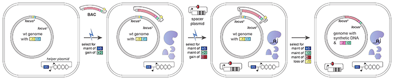
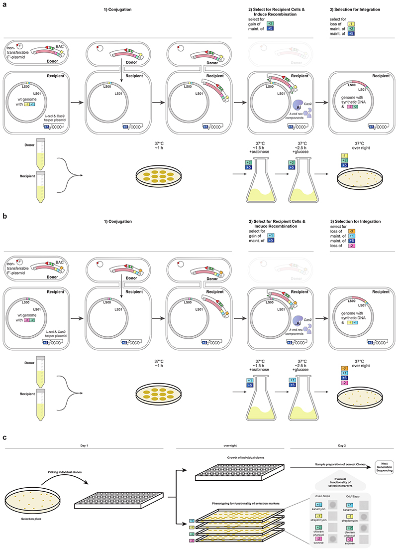
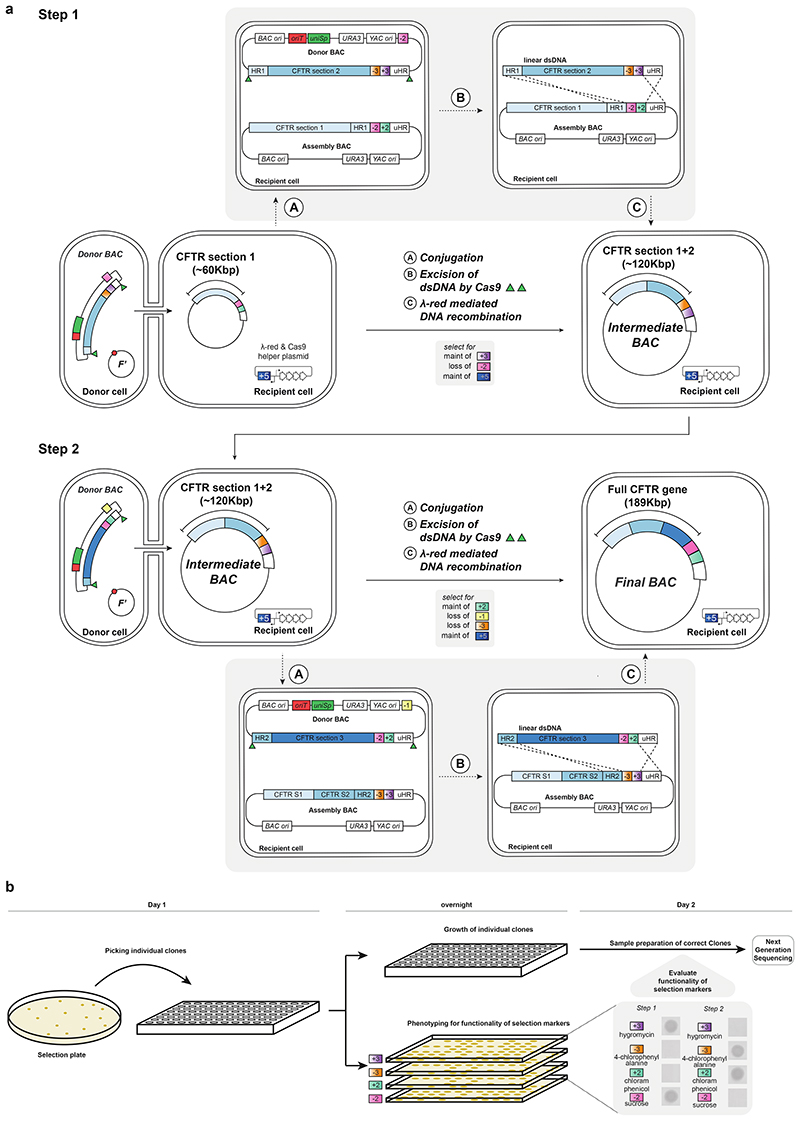
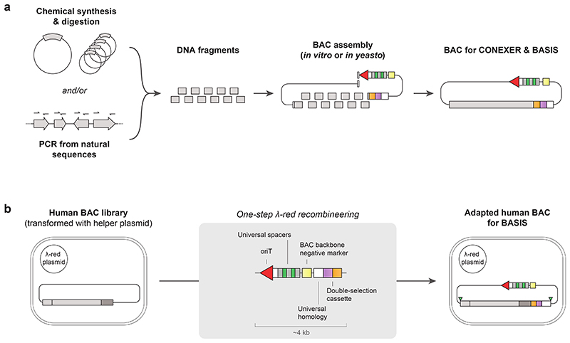
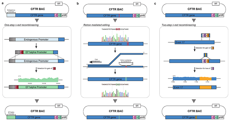
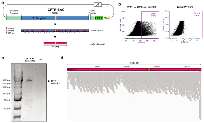
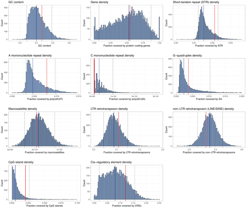
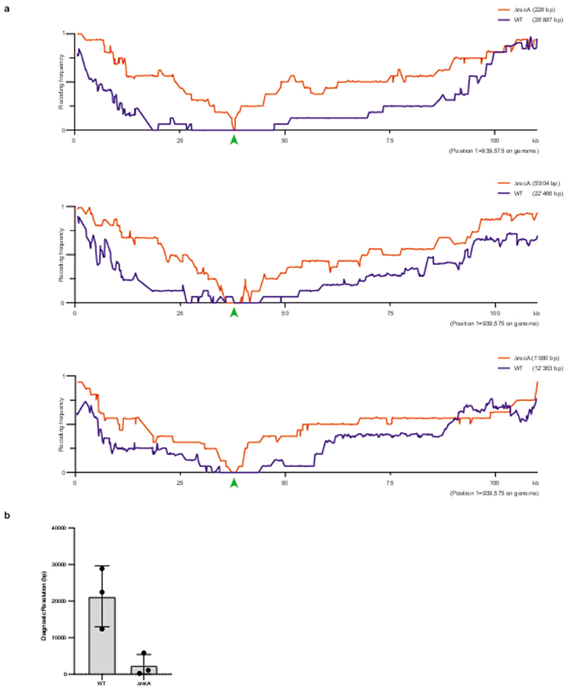
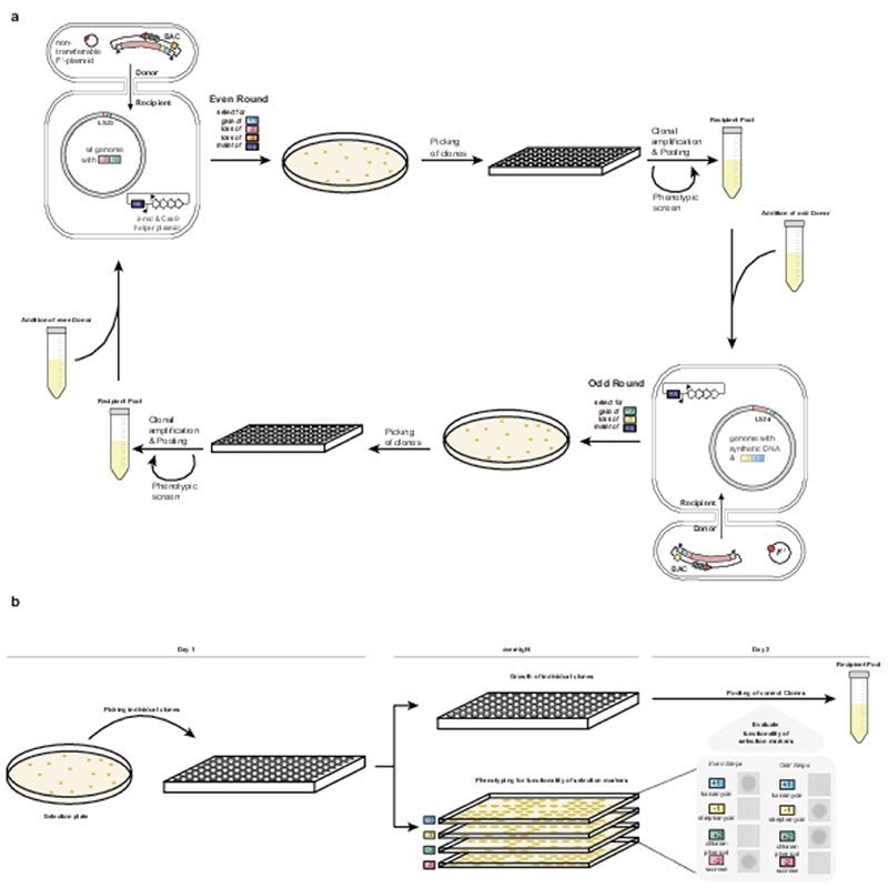
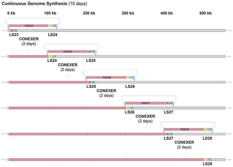
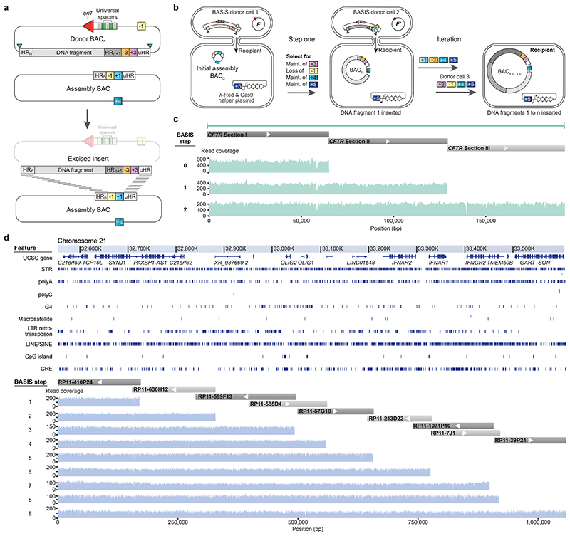
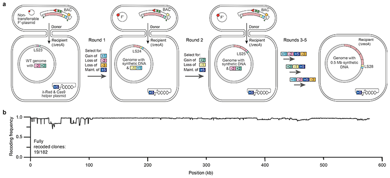
Whole-genome synthesis provides a powerful approach for understanding and expanding organism function1-3. To build large genomes rapidly, scalably and in parallel, we need (1) methods for assembling megabases of DNA from shorter precursors and (2) strategies for rapidly and scalably replacing the genomic DNA of organisms with synthetic DNA. Here we develop bacterial artificial chromosome (BAC) stepwise insertion synthesis (BASIS)-a method for megabase-scale assembly of DNA in Escherichia coli episomes. We used BASIS to assemble 1.1 Mb of human DNA containing numerous exons, introns, repetitive sequences, G-quadruplexes, and long and short interspersed nuclear elements (LINEs and SINEs). BASIS provides a powerful platform for building synthetic genomes for diverse organisms. We also developed continuous genome synthesis (CGS)-a method for continuously replacing sequential 100 kb stretches of the E. coli genome with synthetic DNA; CGS minimizes crossovers1,4 between the synthetic DNA and the genome such that the output for each 100 kb replacement provides, without sequencing, the input for the next 100 kb replacement. Using CGS, we synthesized a 0.5 Mb section of the E. coli genome-a key intermediate in its total synthesis1-from five episomes in 10 days. By parallelizing CGS and combining it with rapid oligonucleotide synthesis and episome assembly5,6, along with rapid methods for compiling a single genome from strains bearing distinct synthetic genome sections1,7,8, we anticipate that it will be possible to synthesize entire E. coli genomes from functional designs in less than 2 months.


Conflict of interest statement

Competing interests

J.W.C. is the founder of Constructive Bio. The MRC has filed a patent application covering the work described.

Figures

Extended Data Fig. 1
Extended Data Fig. 1. Steps in REXER-mediated integration of ~100 kb of synthetic DNA into the E. coli genome using homology region (HR)-specific spacers.
REXER allows integration of more than 100 kb of synthetic DNA (pink) into the genome, through replacement of the corresponding genomic DNA. A bacterial artificial chromosome (BAC) containing the synthetic DNA of interest is electroporated into competent cells with a suitably marked genome, the cells also contain a helper plasmid encoding the Cas9 protein and the lambda red recombination components. Selection for the helper plasmid (+5) and the BAC (+2) is applied. A clonal cell is then expanded and induced with arabinose to express the helper plasmid genes and made electrocompetent again. HR-specific spacer arrays (either plasmid-based as shown, or as linear DNA) are then electroporated into the cell; this leads to CRISPR/Cas9 mediated in vivo excision of the synthetic DNA, flanked by a double selection cassette (+2/-2) and HRs to the genome, from the BAC. The lambda red recombination machinery then uses the HRs to direct the integration of the excised DNA into the genome. Triangles denote the Cas9 cleavage sites at the HRs (grey boxes) flanking the synthetic DNA. Selection on tetracycline (maintenance of +5), ampicillin (maintenance of +6), chloramphenicol (maintenance of +2), and streptomycin (loss of -1) ensures only cells where the recombination took place over the whole section survive. The selectable markers are +1, blue, kanR (selected for with kanamycin); -1, yellow, rpsL (selected against with streptomycin); +2, green, cat (selected for with chloramphenicol); -2, pink, sacB (selected against with sucrose); +5, dark blue tetR (selected for with tetracycline); +6, red ampR (selected for with ampicillin).
Extended Data Fig. 2
Extended Data Fig. 2. Step-wise depiction of CONEXER procedure.
a, In an odd step of CONEXER, recipient cells with a +1/-1 double selection cassette in their genome (+1, kanR (confers growth on kanamycin); -1, rpsL (confers sensitivity to streptomycin)) and a tetracycline resistance (+5, tetR (confers resistance to tetracycline)) conferring plasmid encoding arabinose inducible lambda red components and Cas9 are mixed with donor cells and spotted on an agar plate. Donor cells contain an odd BAC and a non-transferable F’ plasmid. During incubation on the plate (1h, 37 °C) the BAC is conjugated from donor to recipient cells. Subsequently, cells are washed off the plate and inoculated in selective media containing tetracycline (selection for maintenance of +5) and chloramphenicol (selection for gain of +2), this selects for recipient cells that have received the BAC. Arabinose is also added to induce lambda red components and Cas9; excision of the linear DNA and recombination with the genome is induced at this step. Cells are recovered in selective media containing tetracycline (selection for maintenance of +5) and chloramphenicol (selection for maintenance of +2) but no arabinose; this selects for recipient cells that have received the BAC. Finally, cells are plated on selective agar plates containing tetracycline (selection for maintenance of +5) – to select for recipient cells, chloramphenicol (selection for maintenance of +2) – to select for genomic integration of the +2/-2 double selection cassette from the BAC, and streptomycin (selection for loss of -1) – to select for loss of the +1/-1 double selection cassette from the genome and loss of the BAC backbone. b, In an even step of CONEXER, recipient cells with a +2/-2 double selection cassette in their genome (+2, cat (confers growth on chloramphenicol); -2, sacB (confers sensitivity to sucrose) and a tetracycline resistance (+5 tetR (confers resistance to tetracycline)) conferring plasmid encoding arabinose inducible lambda red components and Cas9 are mixed with donor cells and spotted on an agar plate. Donor cells contain an odd BAC and a non-transferable F’ plasmid. During incubation on the plate (1h, 37 °C) the BAC is conjugated from donor to recipient cells. Subsequently, cells are washed off the plate and inoculated in selective media containing tetracycline (selection for maintenance of +5) and kanamycin (selection for gain of +1); this selects for recipient cells that have received the BAC. Arabinose is also added to induce lambda red components and Cas9; excision of the linear DNA and recombination with the genome is induced at this step. Cells are recovered in selective media containing tetracycline (selection for maintenance of +5) and kanamycin (selection for maintenance of +1) but no arabinose; this selects for recipient cells that have received the BAC. Finally, cells are plated on selective agar plates containing tetracycline (selection for maintenance of +5) – to select for recipient cells, kanamycin (selection for maintenance of +1) – to select for genomic integration of the +1/-1 double selection cassette from the BAC, sucrose (selection for loss of -2) – to select for loss of the +2/-2 double selection cassette from the genome, and 4-CP (selection for loss of -3) – to select for loss of the BAC backbone. c, Clones from CONEXER experiments are picked from the selection plate. They are grown up individually in a 96-well plate and phenotyped, for the functionality of selection markers, on agar plates. Subsequently, clones that show the correct growth phenotype (even steps: growth on +1, -2; no growth on -1, +2; odd steps: growth on -1, +2; no growth on +1, -2) are sequenced by NGS. The selectable markers are +1, blue, kanR (selected for with kanamycin); -1, yellow, rpsL (selected against with streptomycin); +2, green, cat (selected for with chloramphenicol); -2, pink, sacB (selected against with sucrose); -3, orange, pheS* (selected against with 4-chlorophenylalanine); +5, dark blue, tetR (selected for with tetracycline).
Extended Data Fig. 3
Extended Data Fig. 3. CFTR assembly via BASIS.
a, Recipient cells containing a BASIS assembly BAC with the first section of the CFTR gene and a plasmid encoding Cas9 and lambda red components were mixed with donor cells. Donor cells contained a BASIS BAC encoding the second section of the CFTR gene and a non-transferable F’ plasmid. The donor BAC was conjugated to the recipient cell (A) and recipient cells selected for on tetracycline (+5, tetR (confers resistance to tetracycline)). Upon induction of protein expression from the helper plasmid, linear dsDNA was excised from the donor BAC (B). The excised DNA inserts into the assembly BAC between HR1 and uHR. Selection on hygromycin (selection for gain of +3) – to select for gain of the selection cassette from the donor BAC, sucrose (selection for loss of -2) – to select for loss of the selection cassette from the assembly BAC and loss of donor BAC backbone, tetracycline (selection for maintenance of +5) – to select for maintenance of the helper plasmid, ensured that only cells with the correctly assembled BAC survive. In step 2, recipient cells containing a BAC with the first and second section of the CFTR gene and a plasmid encoding Cas9, and lambda red components were mixed with donor cells. Donor cells contained a BASIS BAC encoding the third section of the CFTR gene and the non-transferable F’ plasmid. The donor BAC was conjugated to the recipient cell (A) and recipient cells selected for on tetracycline (+5, tetR (confers resistance to tetracycline)). Upon induction of protein expression from the helper plasmid, linear dsDNA was excised from the donor BAC (B). The excised DNA inserts into the assembly BAC between HR2 and uHR. Selection on chloramphenicol (selection for gain of +2) – to select for gain of the selection cassette from the donor BAC), 4-CP (loss of -3) – to select for loss of the selection cassette from the assembly BAC, streptomycin (loss of -1) – to select for loss of the donor BAC backbone, and tetracycline (maintenance of +5) – to select for maintenance of the helper plasmid, ensured that only cells with the correctly assembled BAC survive. b, Clones from BASIS experiments were picked from the selection plate. They were grown up individually in a 96-well plate and phenotyped for the functionality of selection markers on agar plates. Subsequently, clones that showed the correct growth phenotype and in some cases genotype for the assembly junctions by PCR (step 1: growth on hygromycin, growth on sucrose; no growth on 4-CP, no growth on chloramphenicol, and genotyping for insertion of the second section of CFTR (for primers see Supplementary Data 2); step 2: growth on 4-CP, growth on chlopramphenicol; no growth on hygromycin, no growth on sucrose) were sequenced by NGS. The selectable markers are +3, purple, hygR (selected for with hygromycin); -3, orange pheS* (selected against with 4-chlorophenylalanine); +2, green, cat (selected for with chloramphenicol); -2, pink, sacB (selected against with sucrose); -1, yellow rpsL (selected against with streptomycin); +5, dark blue, tetR (selected for with tetracycline).
Extended Data Fig. 4
Extended Data Fig. 4. Assembly of BACs to be used for CONEXER and BASIS, and adaptation of existing BACs derived from human BAC libraries.
a, BAC assembly from DNA fragments of 3-10 kb; these fragments may be derived from chemically synthesized oligonucleotides and/or may be amplified from natural sequences. 10 kb DNA fragments are assembled with established methods, either in vitro or in vivo by yeast assembly. All fragments are assembled with the BAC backbone, which contains components required for subsequent CONEXER or BASIS steps (universal spacers, origin of transfer, marker cassettes). b, BACs used for assembly of a megabase-scale human genomic section are derived from human BAC libraries. The sequence of human DNA in these BACs overlap with each other, and these overlaps constitute the homology regions exploited for assembly. The universal spacer cassette, the origin of transfer, the universal homology region, and appropriate selection markers were introduced into BACs from the human BAC library, by one-step λ-red recombineering, to generate BASIS BACs.
Extended Data Fig. 5
Extended Data Fig. 5. BACs produced by BASIS can be extensively modified by lambda red recombineering and retron-mediated editing – to generate insertions, replacements, and edits.
a, For expression in human tissue culture, the endogenous CFTR promoter was replaced with an EF1alpha constitutive promoter using λ-red recombineering. To this end the EF1alpha promoter was coupled to an ampicillin resistance gene (+6, red ampR (confers resistance to ampicillin)). Following recombineering cells were selected on ampicillin (selection for gain of +6) – to select for replacement of the promoter. Sequence coverage of the EF1alpha prompter is shown (maximum coverage indicated in brackets). b, BACs produced by BASIS can be precisely edited using retron-mediated editing. A single strand binding protein and a retron containing the desired base pair substitutions were expressed in target cells containing the BAC. During replication annealing of the retron to the lagging strand led to the desired edits. We corrected two point mutations in exon 15 of the CFTR gene (Methods). Sanger sequencing traces of the region containing the point mutations are shown before (top - red) and after (bottom - green) editing. c, To distinguish BAC encoded CFTR from the endogenous gene an HA-tag was inserted into exon 17 of the CFTR gene on the BAC. This tag is known to be tolerated in the cDNA of CFTR. First, a double selection cassette (+3, orange hygR (confers resistance to hygromycin); -3, purple pheS* (confers sensitivity to 4-CP)) was inserted into the locus of interest. Following recombineering cells were selected on hygromycin (selection for gain of +3) – to select for insertion of the double selection cassette. Subsequently, λ-red recombineering was used to replace the double selection cassette with an HA-tag. Following recombineering cells were selected on 4-CP (selection for loss of -3) – to select for replacement of the double selection cassette. Sequence coverage of exon 17 containing the HA-tag is shown (maximum coverage indicated in brackets).
Extended Data Fig. 6
Extended Data Fig. 6. Expression of CFTR from a modified BASIS BAC in mammalian cells.
a, Schematic of a 208 kb CFTR BAC: the entire human CFTR gene with an HA-tag (yellow-bar) was expressed under a constitutive EF1alpha promoter. The native transcript, containing exons and introns is spliced to form the mature transcript of approximately 4.5 kb in length. Additionally, eGFP and a Neomycin (Neo) resistance are expressed from a constitutive CAG promoter. b, FACS plots from human embryonic kidney (HEK293) cells transfected with the CFTR BAC (CFTR-HA_GFP) and a control (WT). On the x-axis the fluorescence intensity from GFP protein is displayed. On the y-axis side-scattering in displayed. Approximately, 1.19% of cells transfected with the CFTR BAC are positive for GFP. c, PCR was performed on cDNA from cells with and without the transfected CFTR BAC. The band (around 4.5 kb) corresponding to an amplicon of CFTR transcript is specific to cells transfected with the CFTR BAC. Band sizes of the ladder are indicated to the left (grey arrows). The experiment in c was performed in one biological replicate. d, NGS of the PCR product shown in (c) demonstrates that CFTR has been transcribed and processed properly in cells after transfection. The presence of an HA-tag in the transcript shows that the transcript is derived from the BASIS BAC encoded CFTR. Increased coverage at the flanks likely stems from sequencing of the primers.
Extended Data Fig. 7
Extended Data Fig. 7. Analysis of genomic features in the 1.1 Mb target region on chromosome 21.
Analysis of distribution of genomic features in the 1.1 Mb target region of the human genome assembled by BASIS. The genomic features in this region are compared to the distribution of the genomic feature throughout the whole genome, computed in 1 Mb windows. The red line indicates the fraction of each genomic feature for the 1.1 Mb target region. The blue line indicates the median for each genomic feature for the whole genome. The dotted lines represent the 5th and 95th percentile of the distribution.
Extended Data Fig. 8
Extended Data Fig. 8. CONEXER experiments on sequences that cannot be replaced by recoded synthetic DNA.
a, Codons in yceQ at positions 37’213, 37’227, 37’251 (indicated by green arrow), within 100k09, cannot be replaced by recoded synthetic DNA in which TCG codons are replaced with AGC, TCA codons are replaced with AGT, and TAG codons are replaced with TAA. Each graph shows a compiled recoding landscape in WT genetic background (blue) and ∆recA (red). Landscapes were generated by sequencing 13-16 clones from an independent CONEXER experiment with a CONEXER BAC bearing recoded sequence for 100k09. The resolution of the diagnostic landscapes (distance between the positions of the first and last event with recoding frequency 0) is indicated in parentheses. Three independent replicates of each experiment are shown. b, Bar graph of the diagnostic resolution of CONEXER experiments in section 100k09 in WT and ∆recA conditions; data are from n=3 independent biological replicates shown in panel a. Data are represented as mean +/- standard deviation. The ∆recA condition is significantly better than the WT at localizing disallowed synthetic sequences (two-sided unpaired t test: p-value = 0.021). We note that previous experiments by REXER in WT background yielded a resolution of >20’000 bp.
Extended Data Fig. 9
Extended Data Fig. 9. Continuous genome synthesis method.
Alternating even and odd rounds of CONEXER for continuous replacement of genomic DNA with synthetic DNA. a, Following an even round of CONEXER, clones are picked from an appropriate selection plate containing kanamycin (selection for gain of +1), sucrose (selection for loss of -2), 4-chlorophenylalanine (selection for loss of -3), and tetracycline (selection for maintenance of +5). Clones are amplified overnight and in parallel undergo a phenotypic screen. Amplified clones with all the correct phenotypes are pooled. This pool serves directly as the recipient for an odd round of CONEXER. After an odd round of CONEXER clones are picked from an appropriate selection plate containing chloramphenicol (selection for gain of +2), streptomycin (selection for loss of -1), and tetracycline (selection for maintenance of +5). Clones are amplified overnight and in parallel undergo a phenotypic screen. Amplified clones with all the correct phenotypes are pooled. This pool serves directly as the recipient for an even round of CONEXER. Cycling through even and odd rounds of CONEXER lead to continuous synthesis of a synthetic genome from the corresponding BACs. b, Colonies obtained from a step of CONEXER are picked from the selection plate. They are grown up individually in a 96-well plate and phenotyped for the functionality of selection markers on agar plates. Subsequently, clones that show the correct growth phenotype (even steps: growth on kanamycin (selection for +1), growth on sucrose (selection against -2); no growth on streptomycin (selection against -1), no growth on chloramphenicol (selection for +2); odd steps: growth on streptomycin (selection against -1), growth on chloramphenicol (selection for +2); no growth on kanamycin (selection for +1), no growth on sucrose (selection against -2)) are pooled into one culture. This culture immediately serves as the recipient strain for the next step of CONEXER. The selectable markers are +1, blue, kanR (selected for with kanamycin); -1, yellow, rpsL (selected against with streptomycin); +2, green, cat (selected for with chloramphenicol); -2, pink, sacB (selected against with sucrose); -3, orange, pheS (selected against with 4-chlorophenylalanine); +5, dark blue, tetR (selected for with tetracycline).
Extended Data Fig. 10
Extended Data Fig. 10. Continuous synthesis of 500 kb of a recoded E. coli genome from CONEXER BACs.
Continuous genome synthesis via rounds of CONEXER in ∆recA recipients. Genomic DNA is depicted in grey and synthetic, recoded DNA in pink. The selectable markers are +1, blue, kanR (selected for with kanamycin); -1, yellow, rpsL (selected against with streptomycin); +2, green, cat (selected for with chloramphenicol); -2, pink, sacB (selected against with sucrose). Each round of CONEXER replaces approximately 100 kb of the E. coli genome with synthetic DNA, and takes two days. Continuous synthesis of a 500 kb synthetic section in the E.coli genome was achieved in 10 days.
Fig. 1
Fig. 1. Universal spacers enable scarless replacement of genomic DNA with synthetic DNA.
a, Spacer RNAs (grey boxes) specific for each homology region (HR), as used in REXER; ‘N’ indicates sequences that are complementary to HR sequences. b, Universal spacer RNAs direct Cas9 to the constant sequence of the BAC backbone. In panels a and b: the selectable markers are -1 (yellow, rpsL), +2 (green, cat), and -2 (pink, sacB). PAM sequences are indicated by a black box. The triangles denote Cas9 cleavage sites. c, Verification by PCR of the 5’ and 3’ genomic integration sites after REXER using universal spacers at two genomic loci. A total of 11 post-REXER clones were genotyped for each experiment. The triangles indicate the size of the expected PCR product at each locus before (white) and after (black) REXER. d, Sequence verification of the junctions between the insert and the rest of the genome after REXER using universal spacer RNA. The insert and 6 bp non-homology sequences (flap, tilted) are shown above the sequence that is expected for scarless integration (The full dataset is provided in Supplementary Fig. 3). The triangles indicate the Cas9 cut sites. e, f, Compiled recoding landscapes of REXER with HR-specific (e) and universal (f) spacers. We performed REXER, replacing 95.6 kb of E. coli genomic DNA with synthetic DNA (100k24 from our recoded whole genome synthesis). A total of 20 post-REXER clones were fully sequenced for each experiment. The compiled recoding landscape graphs show the average frequency at which each recoded codon was integrated across the genomic locus,. The experiment in c was performed in one biological replicate. Gel source data are provided in Supplementary Fig. 1.
Fig. 2
Fig. 2. CONEXER is a rapid, simplified, and standardized method for genome synthesis from synthetic DNA in episomes.
a, REXER and CONEXER BACs. The CONEXER BACs contain a universal spacer array (green bars), origin of transfer (oriT, red arrow). b, The CONEXER BAC is conjugatively transferred from donor cells to recipient cells with the aid of a non-transferable F’ plasmid (F’). Recipient cells, with an appropriately marked genome (+2/-2 at LS23 is shown), express Cas9 and the λ-red components. Selection for +5 selects recipient cells. Replacement of genomic DNA with synthetic DNA in the recipient cells is then selected for by selecting for gain of +1 and loss of -2; selection for loss of -3 selects for loss of the BAC backbone. In the examples shown in panels a and b, the selectable markers are +1 (blue, kanR), -1 (yellow, rpsL), +2 (green, cat), -2 (pink, sacB), -3 (orange, pheS*), and +5 (dark blue, tetR). Maint., maintenance. WT, wild type. c, The compiled recoding landscape of 84 clones from CONEXER with 100k24.
Fig. 3
Fig. 3. Megabase-scale assembly of DNA using BASIS.
a, Donor and assembly BACs for BASIS. uHR is the universal homology region. HRn is an HR specific for the nth step of insertion. The donor BAC insert contains HRn+1, which serves as HRn for the (n+1)th step. The green triangles indicate the cut sites for Cas9 excision. b, The BASIS workflow. The donor BAC is delivered by conjugative transfer to the recipient cell. The insert is excised from the donor BAC and inserted into the assembly BAC, as shown in (a). Iteration, using alternating sets of markers, directs the insertion of n DNA fragments into the assembly BAC. In panels a and b the selectable markers are +1 (blue, kanR), -1 (yellow, rpsL), +3 (purple, hygroR), -3 (orange, pheS*), +4 (petrol, gentR), and +5 (dark blue, tetR). c, Assembly of the full-length CFTR gene through BASIS using three BACs and verification using next generation sequencing (NGS). The sequencing coverage tracks for each step of BASIS are shown in a vertical stack; read coverage (y-axis) is plotted against the position in base pairs (bp) from the start of the first insert (x-axis). d, BASIS assembly of the 1.1 Mb target region of chromosome 21. The top blue bar indicates the region targeted for assembly. UCSC database genes are indicated. The vertical blue lines indicate the positions of other features, including: short tandem repeats, guanosine quadruplexes (G4), long terminal repeats (LTR) retrotransposons, LINEs, SINEs, and cis regulatory elements (CRE). The RP number and the grey bars indicate the BAC from which the insert was derived. The sequencing coverage track for each assembly step is shown in a vertical stack, as in panel c.
Fig. 4
Fig. 4. Deletion of recA from the host genome increases the fraction of fully recoded clones in CONEXER mediated genomic replacement.
a, Screening of gene deletion strains in CONEXER mediated replacement of genomic fragment 100k24 for increased frequency of fully recoded clones. We performed a one-way analysis of variance comparing each condition with the WT and corrected for multiple hypotheses using the conservative Bonferroni-correction. Deletions of recA (P < 0.0001) and recO (P = 0.04) significantly increased the fraction of clones with fully synthetic sequences. Data are represented as the mean of n = 3 independent biological replicates. b, Deletion of recA increases the fraction of fully recoded clones across several 100 kb fragments (100k24 - 100k28) after CONEXER.
Fig. 5
Fig. 5. CGS from episomes.
a, In the first round of CGS we performed CONEXER with a donor cell containing a CONEXER BAC bearing synthetic DNA covering 100k24 and a +1/-1 double selection cassette. The recipient cell contained an appropriately marked genome (-2/+2 at LS23), and had a recA deletion. Recipient clones from the selection with the correct set of phenotypes were pooled and acted as the recipient for the next round of CONEXER mediated genome replacement, by virtue of containing +1/-1 at LS24 of their genome. In the next round we performed CONEXER with a donor cell containing a CONEXER BAC bearing synthetic DNA covering 100k25 and a +2/-2 double selection cassette. Recipient clones from the selection with the correct set of phenotypes were pooled and acted as the recipient for the next round of CONEXER mediated genome replacement, by virtue of containing +2/-2 at LS25 of their genome. This process was repeated three more times. After five rounds of CGS, a subset of the resulting cells were expected to have integrated synthetic DNA over the entire 500 kb region. The selectable markers are +1 (blue, kanR), -1 (yellow, rpsL), +2 (green, cat), -2 (pink, sacB), -3 (orange, pheS*), and +5 (dark blue, tetR). b, The compiled recoding landscape of 182 clones from CGS of 100k24 - 100k28. In total, 19 out of the 182 sequenced clones were fully recoded over the whole 500 kb section of the genome.

References

    1. Fredens J, et al. Total synthesis of Escherichia coli with a recoded genome. Nature. 2019;569:514–518. doi: 10.1038/s41586-019-1192-5. - DOI - PMC - PubMed
    1. Gibson DG, et al. Complete chemical synthesis, assembly, and cloning of a Mycoplasma genitalium genome. Science. 2008;319:1215–1220. doi: 10.1126/science.1151721. - DOI - PubMed
    1. Gibson DG, et al. Creation of a bacterial cell controlled by a chemically synthesized genome. Science. 2010;329:52–56. doi: 10.1126/science.1190719. - DOI - PubMed
    1. Wang KH, et al. Defining synonymous codon compression schemes by genome recoding. Nature. 2016;539:59. doi: 10.1038/nature20124. - DOI - PMC - PubMed
    1. Gibson DG, et al. Enzymatic assembly of DNA molecules up to several hundred kilobases. Nat Methods. 2009;6:343–345. doi: 10.1038/nmeth.1318. - DOI - PubMed

Publication types

MeSH terms