. 2023 Jul;619(7971):749-754.
doi: 10.1038/s41586-023-06182-6. Epub 2023 Jun 28.

Femtosecond proton transfer in urea solutions probed by X-ray spectroscopy

Affiliations

Femtosecond proton transfer in urea solutions probed by X-ray spectroscopy

Zhong Yin et al. Nature. 2023 Jul.

Abstract

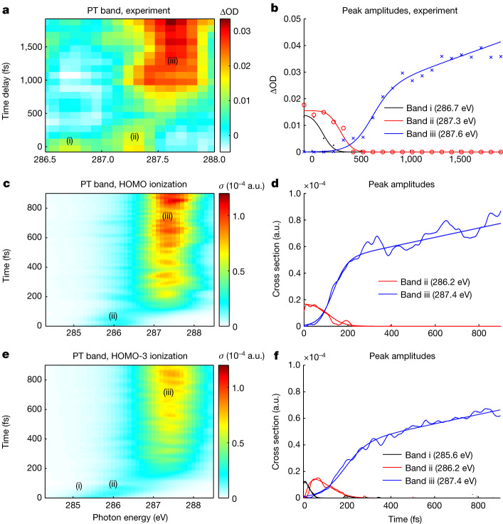
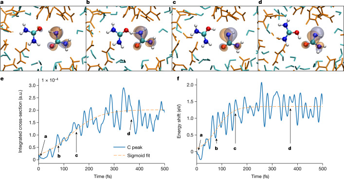
Proton transfer is one of the most fundamental events in aqueous-phase chemistry and an emblematic case of coupled ultrafast electronic and structural dynamics1,2. Disentangling electronic and nuclear dynamics on the femtosecond timescales remains a formidable challenge, especially in the liquid phase, the natural environment of biochemical processes. Here we exploit the unique features of table-top water-window X-ray absorption spectroscopy3-6 to reveal femtosecond proton-transfer dynamics in ionized urea dimers in aqueous solution. Harnessing the element specificity and the site selectivity of X-ray absorption spectroscopy with the aid of ab initio quantum-mechanical and molecular-mechanics calculations, we show how, in addition to the proton transfer, the subsequent rearrangement of the urea dimer and the associated change of the electronic structure can be identified with site selectivity. These results establish the considerable potential of flat-jet, table-top X-ray absorption spectroscopy7,8 in elucidating solution-phase ultrafast dynamics in biomolecular systems.


Conflict of interest statement

The authors declare no competing interests.

Figures

Fig. 1
Fig. 1. Overview of the experimental setup and the pumped and unpumped spectra.
a, Schematic depiction of the experimental setup. b, Schematic molecular-orbital diagram illustrating ionization by a pump pulse, followed by probing of the system with a SXR pulse. c, XAS spectrum of a 10 M urea solution covering the carbon and nitrogen K edges with and without pump, and the time-averaged ∆OD signal.
Fig. 2
Fig. 2. Overview of experimental and theoretical results.
ah, The transient carbon K-edge XAS results of 10 M or 5 M urea solutions (ad) and a direct comparison with the QM/MM calculations after HOMO ionization (eh) (showing cross sections in atomic units (a.u.)). Panels a,c,e,g show an overview of the results, and panels b,d,f,h show XAS spectra at selected time delays (specified in the legends). The 10 M aqueous urea solution (a,b) reveals the signature of proton transfer in the urea dimer, which is comparable with calculations (e,f). At the lower 5 M concentration (c,d), proton transfer is not observed in the signal fluctuations, which is supported by the lack of proton transfer in QM/MM calculations of ionized urea:water (1:1) complexes embedded in the 10 M solution (g,h).
Fig. 3
Fig. 3. Detailed analysis of the proton-transfer band.
a,b, Experimental data. cf, QM/MM calculations. The proton-transfer (PT) bands from the experiment (a) and calculations for ionization of HOMO (c) and HOMO-3 (e) are analysed by fitting three Gaussians corresponding to the two pre- and one post-proton-transfer bands (i–iii) as shown in Extended Data Fig. 4. Their corresponding amplitudes are shown in b, d and f.
Fig. 4
Fig. 4. Separation of electronic and structural rearrangements in transient XAS.
ad, Snapshots from an exemplary QM/MM trajectory displaying proton transfer in the HOMO-ionized urea dimer along with the SOMO (or hole orbital). Time t = 6 fs (a), t = 78 fs (b), t = 150 fs (c) and t = 372 fs (d). e, C 1s → SOMO absorption cross-section as a function of time. f, Energy shift of the corresponding transition. The blue lines in e and f show the calculated position and the dashed orange lines are sigmoidal fits with time constants given in the text.
Extended Data Fig. 1
Extended Data Fig. 1. Broadband SXR probe spectrum.
SXR probe pulse spectrum transmitted through a 100-nm Ti filter. The spectral intensity has been corrected using the Jacobian transformation from wavelengths to photon energies. The carbon-edge absorption originates from carbon-containing contaminations on the optical components.
Extended Data Fig. 2
Extended Data Fig. 2. Rise time of the transient absorption band of the 2.5M urea solution.
Spectrally integrated differential absorbance (285–295 eV) at the carbon K-edge of a 2.5M aqueous urea solution, yielding a rise time of 27 fs (10% to 90%).
Extended Data Fig. 3
Extended Data Fig. 3. Potential energy surfaces from Koopmann’s theorem in comparision with the EOM-CC method.
Scan through the potential-energy surfaces of the lowest ionized states along the proton transfer coordinate for a urea water complex and a urea dimer (cyclic conformation) in vacuum. The solid lines show results obtained using Koopmanns’ theorem, the dashed lines show results obtained using the EOM-CC method. The x-axis indicates displacement of the proton from the neutral ground state equilibrium geometry towards the acceptor oxygen.
Extended Data Fig. 4
Extended Data Fig. 4. Fitting of the proton transfer band.
Fits of Gaussian profiles of the proton transfer band at four different pump-probe time delays.
Extended Data Fig. 5
Extended Data Fig. 5. Proton transfer dynamics of ionised cyclic urea dimer in solution in comparison with gas phase.
(a) Ensemble-averaged C 1s→valence resonance integrated absorption cross section and (b) energy shift of the corresponding transition as a function of time along with error bars for the QM dimer trajectories in 10 M solution that undergo proton transfer following HOMO ionization. For comparison, subfigures (c) and (d) show the same quantities for a cyclic dimer in vacuum from ref. (please note the different time scale here). The blue line shows the calculated values and the dashed orange line is a sigmoidal fit with time constants given in the text.
Extended Data Fig. 6
Extended Data Fig. 6. Theoretical binding energies of urea dimers.
Histogram of the calculated molecular-orbital binding energies from HOMO to HOMO-6 for the initial QM/MM ensemble of the neutral QM urea dimer in 10 M aqueous solution.
Extended Data Fig. 7
Extended Data Fig. 7. Populations dynamics of electronic states accessed following ionization of deeper-lying HOMOs.
Population of electronic states in solvated urea dimer cation states from QM/MM calculations after initial ionization in (a) HOMO-3 and (b) HOMO-5. For initial HOMO-3 ionization (a), the half decay time obtained by an exponential fit to state 3 (hole in HOMO-3) is (6.68 ± 0.02) fs and the half rise time obtained by a sigmoidal fit to state 0 population (hole in HOMO) is (34.19 ± 0.10) fs.
Extended Data Fig. 8
Extended Data Fig. 8. Proton transfer fractions of urea dimers after ionization.
Percentage of completed proton transfer within urea dimer following ionization of (a) HOMO, (b) HOMO-3 and (c) HOMO without MM environment in a 10 M aqueous urea solution, and (d) HOMO ionization of gas-phase cyclic dimer in vacuum from ref. (please note the different time scale here).
Extended Data Fig. 9
Extended Data Fig. 9. Concentration dependence measurement of urea solutions.
Time-resolved XAS at the carbon K-edge of 10M, 8M, 5M and 2.5M urea aqueous solutions, recorded under conditions otherwise identical to Fig. 2 of the main text.
Extended Data Fig. 10
Extended Data Fig. 10. Pump-intensity dependence.
Time-resolved XAS at the carbon K-edge of 10M aqueous urea solutions performed with ∼30 fs pump pulses centered at 400 nm and variable peak intensities. The rise times of the raw (no background subtraction) proton transfer bands obtained from each measurement are given in panel (b),(d) and (f) respectively.

References

    1. Hammes-Schiffer S, Tully JC. Proton transfer in solution: molecular dynamics with quantum transitions. J. Chem. Phys. 1994;101:4657–4667. doi: 10.1063/1.467455. - DOI
    1. Elsässer, T. & Becker, H. Ultrafast Hydrogen Bonding Dynamics and Proton Transfer Processes in the Condensed Phase Vol. 23 (Springer Science & Business Media, 2013).
    1. Pertot Y, et al. Time-resolved x-ray absorption spectroscopy with a water window high-harmonic source. Science. 2017;355:264–267. doi: 10.1126/science.aah6114. - DOI - PubMed
    1. Attar AR, et al. Femtosecond x-ray spectroscopy of an electrocyclic ring-opening reaction. Science. 2017;356:54–59. doi: 10.1126/science.aaj2198. - DOI - PubMed
    1. Saito N, et al. Real-time observation of electronic, vibrational, and rotational dynamics in nitric oxide with attosecond soft x-ray pulses at 400 ev. Optica. 2019;6:1542–1546. doi: 10.1364/OPTICA.6.001542. - DOI

Publication types