. 2023 Jun 28;30(1):49.
doi: 10.1186/s12929-023-00941-3.

The homodimer interfaces of costimulatory receptors B7 and CD28 control their engagement and pro-inflammatory signaling

Affiliations

The homodimer interfaces of costimulatory receptors B7 and CD28 control their engagement and pro-inflammatory signaling

Andrey Popugailo et al. J Biomed Sci. .

Abstract

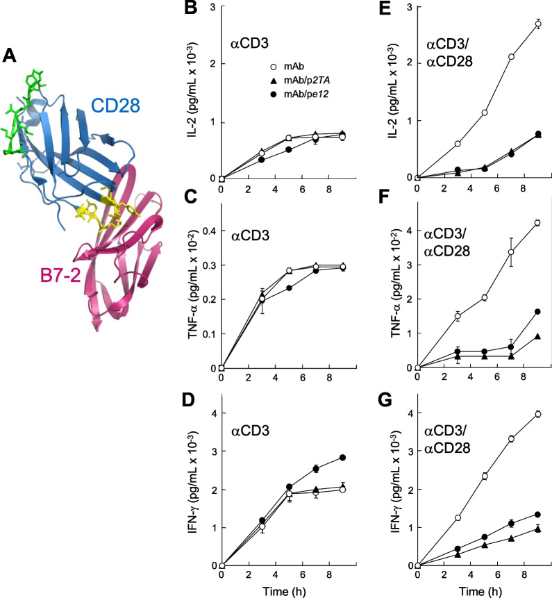
Background: The inflammatory response is indispensable for protective immunity, yet microbial pathogens often trigger an excessive response, 'cytokine storm', harmful to the host. Full T-cell activation requires interaction of costimulatory receptors B7-1(CD80) and B7-2(CD86) expressed on antigen-presenting cells with CD28 expressed on the T cells. We created short peptide mimetics of the homodimer interfaces of the B7 and CD28 receptors and examined their ability to attenuate B7/CD28 coligand engagement and signaling through CD28 for inflammatory cytokine induction in human immune cells, and to protect from lethal toxic shock in vivo.

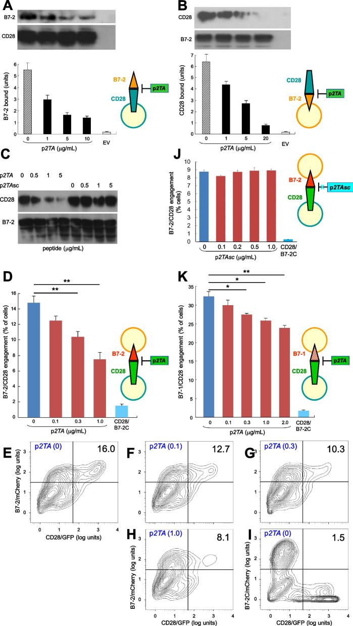
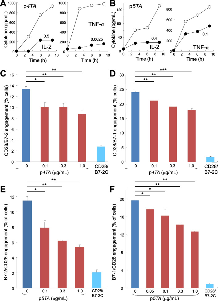
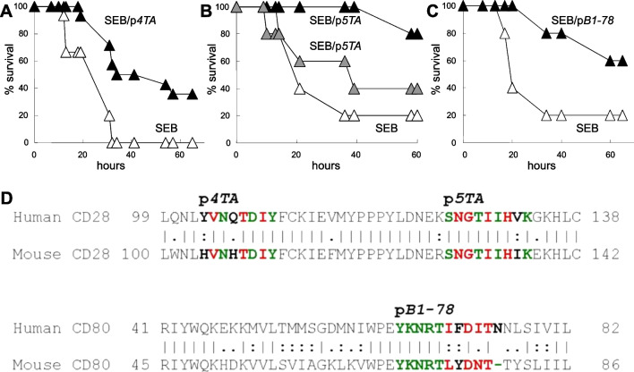
Methods: Short B7 and CD28 receptor dimer interface mimetic peptides were synthesized and tested for their ability to attenuate the inflammatory cytokine response of human peripheral blood mononuclear cells, as well as for their ability to attenuate B7/CD28 intercellular receptor engagement. Mice were used to test the ability of such peptides to protect from lethal superantigen toxin challenge when administered in molar doses far below the toxin dose.

Results: B7 and CD28 homodimer interfaces are remote from the coligand binding sites, yet our finding is that by binding back into the receptor dimer interfaces, short dimer interface mimetic peptides inhibit intercellular B7-2/CD28 as well as the tighter B7-1/CD28 engagement, attenuating thereby pro-inflammatory signaling. B7 mimetic peptides exhibit tight selectivity for the cognate receptor in inhibiting intercellular receptor engagement with CD28, yet each diminishes signaling through CD28. In a prominent example of inflammatory cytokine storm, by attenuating formation of the B7/CD28 costimulatory axis, B7-1 and CD28 dimer interface mimetic peptides protect mice from lethal toxic shock induced by a bacterial superantigen even when administered in doses far submolar to the superantigen.

Conclusions: Our results reveal that the B7 and CD28 homodimer interfaces each control B7/CD28 costimulatory receptor engagement and highlight the protective potential against cytokine storm of attenuating, yet not ablating, pro-inflammatory signaling via these receptor domains.

Keywords: Control of B7/CD28 receptor engagement; Costimulatory receptors B7 and CD28; Inflammatory cytokine storm; Pro-inflammatory signaling; Receptor homodimer interface mimetic peptides; Regulation of B7/CD28 signaling.


Conflict of interest statement

RK, GA and RL are inventors on patents and patent applications for peptides described.

Figures

Fig. 1
Fig. 1
CD28 dimer interface mimetic peptide p2TA inhibits signaling through CD28. A The complex between CD28 (blue) and B7-2 (pink). The extracellular domain of CD28 is oriented such that it enters the T cell at the top and that of B7-2 is oriented such that it enters the antigen-presenting cell at the bottom. In CD28, p2TA sequence within the dimer interface is shown in green and the B7 binding site (MYPPPY) in yellow. Because the structure of the CD28/B7-2 complex remains unresolved, CD28 (1YJD.pdb [7]) was superimposed on CTLA-4 in the CTLA-4/B7-2 complex (1I85.pdb [14]). BG PBMC from a single human donor were induced with αCD3 (BD) or αCD3/αCD28 monoclonal antibodies (mAb) (EG) alone (○) or in the presence of 10 µg/ml of p2TA (▲) or pe12 (●). At times shown, IL-2, TNF-α and IFN-γ in culture medium were quantitated in triplicate. Data are mean and SEM. Representative data of 3 experiments are shown
Fig. 2
Fig. 2
p2TA binds directly to CD28. A, B Representative surface plasmon resonance responses for binding of CD28-IgG-Fc to immobilized peptides p2TA (700 resonance units) (A) and pe12 (1260 resonance units) (B) (top panels); KD, 4 and 3 µM, respectively. Analyte concentrations increased in twofold increments from 0.2 µM. Representative surface plasmon resonance responses for binding of IgG-Fc to immobilized p2TA and pe12 are shown in bottom panels; analyte concentrations increased in twofold increments from 0.125 µM. C Representative surface plasmon resonance responses for binding of disulfide-linked homodimeric CD28 extracellular domain protein without Fc (CD28) to immobilized p2TA (733 resonance units). Analyte concentrations increased in twofold increments from 0.125 µM; KD, 3.4 µM. D The p2TA, p5TA and p4TA regions at the homodimer interface of CD28. In the sequence of the extracellular domain of CD28, dimer interface contact residues are shown in red color, peptide sequences p2TA, p4TA and p5TA are highlighted in yellow, and the αCD28 epitope [13] is underlined. In structure model of the extracellular domain of costimulatory receptor CD28 (green; 1yjd.pdb), a single beta-barrel, region of p2TA is shown in sticks in dark blue with 2 dimer interface contacts in orange, region of p5TA is in red with 4 dimer interface contacts in yellow and on the right the HVK sequence shared with the epitope, and region of p4TA is in cyan with 3 dimer interface contacts in orange
Fig. 3
Fig. 3
CD28 dimer interface mimetic peptide p2TA inhibits intercellular engagement of costimulatory receptor CD28 with B7-2 or B7-1. A p2TA inhibits binding of B7-2 to cell-surface CD28. HEK293T cells were transfected to express cell-surface CD28 or with empty vector (EV). Cells were incubated with soluble B7-2 in the absence or presence of p2TA at concentrations shown. Western blots show binding of B7-2 and equal expression of CD28 by the cells. Bound B7-2 is quantitated in the bar graphs; data are mean and SEM of three independent experiments. B p2TA inhibits binding of CD28 to cell-surface B7-2. HEK293T cells were transfected to express cell-surface B7-2 or with empty vector. Cells were incubated with soluble CD28 in the absence or presence of p2TA at concentrations shown. Western blots show binding of CD28 and equal expression of B7-2 by the cells. Bound CD28 is quantitated in the bar graphs; data are mean and SEM of three independent experiments. C HEK293T cells were transfected to express cell-surface B7-2. Cells were incubated with soluble CD28 in the absence or presence of p2TA or its randomly scrambled form, p2TAsc [13], at concentrations shown. Western blots show binding of CD28 and expression of B7-2 by the cells. D p2TA attenuates intercellular B7-2/CD28 receptor engagement. HEK293T cells transfected to express CD28/GFP fusion protein (green label) were incubated with HEK293T cells transfected to express B7-2/mCherry fusion protein (red label), in absence or presence of p2TA at concentrations shown. As negative control served B7-2C/mCherry, which lacks the ability to bind CD28. Intercellular B7-2/CD28 receptor engagement was scored using flow cytometry to quantitate per cent doubly labeled cells. Data are mean and SEM of three independent experiments. EI Contour plots for a representative experiment in D, upon incubation of cells expressing CD28/GFP with cells expressing B7-2/mCherry (EH) or B7-2C/mCherry (I) in the absence or presence of p2TA (µg/mL). Per cent doubly labeled cells is denoted in upper right quadrant. J p2TAsc fails to attenuate intercellular B7-2/CD28 engagement, assayed as in D. Data are mean and SEM of three independent experiments (contour plots: Additional file 1: Fig. S2). K p2TA attenuates intercellular B7-1/CD28 engagement. Synapse formation was assayed as in D, using B7-1/mCherry fusion protein instead of B7-2/mCherry (contour plots: Additional file 1: Fig. S3). Intercellular receptor engagement was compared using the one-tailed unpaired student’s t-test; *p < 0.05, **p < 0.005
Fig. 4
Fig. 4
CD28 dimer interface mimetic peptides p4TA and p5TA attenuate B7/CD28 engagement, signaling through CD28, and inflammatory cytokine production. A, B Human PBMC were induced with αCD3/αCD28 monoclonal antibodies alone (open symbols) or in the presence of p4TA (A) or p5TA (B) at concentrations shown (µg/ml) (filled symbols). At times shown, IL-2 and TNF-α in culture medium were quantitated. Data in A and B are for PBMC from distinct human donors. Representative data of 3 experiments are shown. C, D p4TA attenuates intercellular B7/CD28 engagement. Receptor engagement was assayed by flow cytometry as in Fig. 3D for B7-2/CD28 engagement and as in Fig. 3K for B7-1/CD28 engagement. Data are mean and SEM of three independent experiments (contour plots: Additional file 1: Fig. S5A and S5B). E, F p5TA attenuates intercellular B7/CD28 engagement. Receptor engagement was assayed by flow cytometry as in C and D. Data are mean and SEM of three independent experiments (contour plots: Additional file 1: Fig. S5C, D). Intercellular receptor engagement was compared using the one-tailed unpaired student’s t-test; *p < 0.05, **p < 0.005, ***p < 0.001
Fig. 5
Fig. 5
B7-1 and B7-2 dimer interface mimetic peptides attenuate engagement of CD28 by the cognate B7 costimulatory receptor. A In cartoon model of the extracellular domain of costimulatory receptor B7-1 (CD80) (green; 1dr9.pdb), a double beta-barrel, amino acid residues forming pB1-8 are modeled in sticks, with 2 residues making homodimer interface contacts shown in yellow. B, C pB1-8 selectively attenuates intercellular B7-1/CD28 engagement (B) but not B7-2/CD28 engagement (C). Receptor engagement was assayed by flow cytometry as in Fig. 3K for B7-1/CD28 engagement and as in Fig. 3D for B7-2/CD28 engagement. D In the extracellular domain of B7-1, amino acid residues forming pB1-78 are modeled in sticks, with 4 residues making homodimer interface contacts shown in yellow and orange. E, F pB1-78 selectively attenuates intercellular B7-1/CD28 engagement (E) but not B7-2/CD28 engagement (F). G, H pB2-7 selectively attenuates intercellular B7-2/CD28 engagement (H) but not B7-1/CD28 engagement (G). Data are mean and SEM of three independent experiments (contour plots: Additional file 1: Fig. S6). Intercellular receptor engagement was compared using the one-tailed unpaired student’s t-test; *p < 0.05, **p < 0.005, ***p < 0.001, ****p < 0.0001; n.s, not significant
Fig. 6
Fig. 6
B7-1 and B7-2 dimer interface mimetic peptides attenuate signaling through CD28 and inflammatory cytokine production. AF Human PBMC were induced with αCD3/αCD28 monoclonal antibodies alone (open symbols) or in the presence of pB1-8 (A, B), pB1-78 (C, D) or pB2-7 (E, F) at concentrations shown (µg/mL) (filled symbols). At times shown, IL-2 and TNF-α in culture medium were quantitated. PBMC from three distinct human donors were used to generate the data in AF, respectively. Representative data of 3 experiments are shown
Fig. 7
Fig. 7
At doses submolar to toxin, CD28 and B7-1 dimer interface mimetic peptides protect mice from lethal superantigen challenge. A Mice were injected with SEB alone (n = 15 per group) (open symbols) or together with 0.04 µg p4TA (n = 14 per group) (black symbols); p for survival, 0.062. B Mice (n = 5 per group) were injected with SEB alone or together with 0.045 µg p5TA (grey symbols) or 0.09 µg p5TA (black symbols); p for survival, 0.029. C Mice (n = 5 per group) were injected with SEB alone or together with 0.052 µg pB1-78 (black symbols); p for survival, 0.192. D Protein sequences of human and mouse CD28 and CD80, aligned by EMBOSS Needle. Sequences shown are portions of human (P10747) and mouse (P31041) CD28 and of human (P33681) and mouse (Q3U4B5) CD80. Dimer interface contacts in p4TA, p5TA and pB1-78 are shown in red, non-contact residues are shown in green if identical and in black if non-identical

References

    1. Lindsten T, Lee KP, Harris ES, Petryniak B, Craighead N, Reynolds PJ, Lombard DB, Freeman GJ, Nadler LM, Gray GS, Thompson CB, June CH. Characterization of CTLA-4 structure and expression on human T cells. J Immunol. 1993;151:3489–3499. doi: 10.4049/jimmunol.151.7.3489. - DOI - PubMed
    1. Collins AV, Brodie DW, Gilbert RJ, Iaboni A, Manso-Sancho R, Walse B, Stuart DI, van der Merwe PA, Davis SJ. The interaction properties of costimulatory molecules revisited. Immunity. 2002;17:201–210. doi: 10.1016/S1074-7613(02)00362-X. - DOI - PubMed
    1. Sharpe AH, Freeman GJ. The B7-CD28 superfamily. Nat Rev Immunol. 2002;2:116–126. doi: 10.1038/nri727. - DOI - PubMed
    1. Riley JL, June CH. The CD28 family: a T-cell rheostat for therapeutic control of T-cell activation. Blood. 2005;105:13–21. doi: 10.1182/blood-2004-04-1596. - DOI - PubMed
    1. Greenwald RJ, Freeman GJ, Sharpe AH. The B7 family revisited. Annu Rev Immunol. 2005;23:515–548. doi: 10.1146/annurev.immunol.23.021704.115611. - DOI - PubMed