. 2023 Jul 1;16(7):dmm050145.
doi: 10.1242/dmm.050145. Epub 2023 Jul 20.

ARPC5 deficiency leads to severe early-onset systemic inflammation and mortality

Affiliations

ARPC5 deficiency leads to severe early-onset systemic inflammation and mortality

Elena Sindram et al. Dis Model Mech. .

Abstract

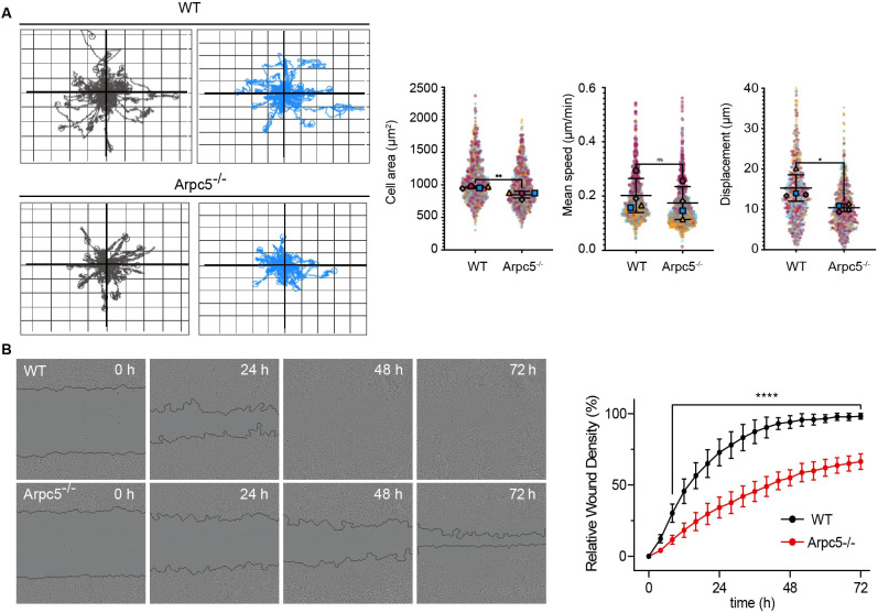
The Arp2/3 complex drives the formation of branched actin networks that are essential for many cellular processes. In humans, the ARPC5 subunit of the Arp2/3 complex is encoded by two paralogous genes (ARPC5 and ARPC5L) with 67% identity. Through whole-exome sequencing, we identified a biallelic ARPC5 frameshift variant in a female child who presented with recurrent infections, multiple congenital anomalies, diarrhea and thrombocytopenia, and suffered early demise from sepsis. Her consanguineous parents also had a previous child who died with similar clinical features. Using CRISPR/Cas9-mediated approaches, we demonstrate that loss of ARPC5 affects actin cytoskeleton organization and function in vitro. Homozygous Arpc5-/- mice do not survive past embryonic day 9 owing to developmental defects, including loss of the second pharyngeal arch, which contributes to craniofacial and heart development. Our results indicate that ARPC5 is important for both prenatal development and postnatal immune signaling, in a non-redundant manner with ARPC5L. Moreover, our observations add ARPC5 to the list of genes that should be considered when patients present with syndromic early-onset immunodeficiency, particularly if recessive inheritance is suspected.

Keywords: Actin; Actinopathy; Arp2/3; CRISPR/Cas9; Immunodeficiency.


Conflict of interest statement

Competing interests The authors declare no competing or financial interests.

Figures

Fig. 1.
Fig. 1.
ARPC5 deficiency manifests as a severe systemic autosomal recessive disorder. (A) Index patients (VI.4 and VI.5) with parents and siblings. Complete family pedigree is available in Fig. S2. Half-filled symbols represent unaffected ARPC5 heterozygous mutation carriers. Filled symbols represent affected homozygous (and presumed homozygous) carriers. (B) Intolerance scores (GDI, gene damage index; RVIS, residual variation intolerance score) for genes in which homozygous variants were identified during WES analysis of VI.5 (WES candidate genes) compared to the scores for genes that are already known to be associated with IEIs (Disease-related AR genes) and related to ARPC5, or encoding other subunits of the Arp2/3 complex (ARP2/3 complex genes). (C) Sanger sequencing electropherograms of the index family showing the single nucleotide deletion.
Fig. 2.
Fig. 2.
ARPC5:p.Ile64Thrfs*8 leads to loss of ARPC5 and upregulation of ARPC5L. (A) Schematic of mRNA and Sanger sequencing of guide DNA from WT THP1 cells, the index patient VI.5 and c.191delT-THP1cells. (B) mRNA quantification of ARPC5 exons 1-2 mRNA (Ex.1-2) and exons 2-4 (Ex.2-4) in WT and c.191delT-THP1 cells. (C) Immunoblot analysis of ARPC5 and ARPC5L in WT, c.191delT-THP1 and c.191delT-THP1 cells reconstituted with WT or c.189delT ARPC5 cDNA. The graph shows the relative quantification of four independent replicates. Data are mean±s.e.m. *P<0.05 (paired two-tailed t-test).
Fig. 3.
Fig. 3.
Arpc5 deficiency affects cell spreading, actin stress fiber morphology and focal adhesions. (A) Immunoblot analysis of ARPC5L in WT and ARPC5−/− HeLa cells (top). Quantification and statistical analysis of independent triplicates (bottom). Data are mean±s.e.m. *P<0.05 (paired two-tailed t-test). (B) Immunofluorescence images of HeLa WT and ARPC5−/− cells labeled with Alexa Fluor 488 Phalloidin (actin, magenta), Vinculin (focal adhesions, green) and DAPI (DNA, blue) (left). Quantification of cell and focal adhesion area of WT and ARPC5 KO HeLa cells (right). Three independent experiments were analyzed. n=20-30 cells per experiment. Data are mean±s.d. *P<0.05 (unpaired two-tailed t-test).
Fig. 4.
Fig. 4.
Arpc5 deficiency affects cells migration. (A) Representative spider graphs illustrating the displacement of WT or Arpc5−/− Hela cells during 24-h random cell migration. Fifty cell tracks are shown in each graph (left). Gray and blue graphs show representative displacement tracks from two independent experiments. Quantification of cell area, speed and displacement of WT and Arpc5−/− Hela cells during random migration (right). Four independent experiments were analyzed. n>200 cells per experiment. Data are mean±s.d. *P<0.05, **P<0.01 (unpaired two-tailed t-test). ns, not significant. (B) Representative phase images of HeLa WT and ARPC5−/− cells are shown for the indicated times in hours after the scratch (left). Relative wound density of four technical replicates for each time point is shown (right). Data are mean±s.d. ****P<0.0001 (two-way ANOVA).
Fig. 5.
Fig. 5.
Arpc5 deficiency impairs cell migration and actin polymerization of Jurkat cells. (A) Immunoblot analysis of ARPC5L in WT and ARPC5−/− Jurkat cells (left). Quantification and statistical analysis of independent triplicates (right). Data are mean±s.e.m. *P<0.05 (paired two-tailed t-test). (B) Mean velocity of WT and ARPC5−/− Jurkat cells with and without CXCL12 treatment. Three independent experiments, >160 cells per experiment were analyzed. Data are mean±s.e.m. **P<0.01 (one-way ANOVA with Tukey's multiple comparison test). (C) F-actin polymerization in Jurkat cells with or without ARPC5 at the indicated time point after stimulation with CXCL12. MFI, mean fluorescence intensity. Three independent experiments were analyzed. Data are mean±s.d. *P<0.05, **P<0.01, ****P<0.0001 (two-way ANOVA).
Fig. 6.
Fig. 6.
Arpc5 deficiency in mice results in defective organogenesis and embryonic lethality. (A) Schematic of the genomic organization of the Arpc5 locus highlighting the four exons (pink rectangles) as well the position of the start (black arrow) and stop (black asterisk) codons. The strategy used to generate the conditional KO mice and the position of two loxP sites (white triangles) flanking exon 2 are indicated. Cre-mediated removal of exon 2 results in a frameshift within exon 3 and a new stop codon (red asterisk). (B) Representative images of control and Arpc5 KO embryos. Most Arpc5 KO embryos are resorbed earlier (right) but a few show growth arrest and die by 9.5 dpf (bottom shows side and dorsal views). Scale bar: 1 mm. (C) Maximum intensity projection images and orthoview (xz, yz) of control and Arpc5 KO embryos stained with α-smooth muscle actin (α-SMA, green) and DAPI (blue) at 9.5 dpf. Red dotted lines indicate partial closure of spinal neuropore (control and Arpc5 KO) and cranial neuropore (Arpc5 KO only). The second pharyngeal arch (PA2) and the cardiac outflow tract (oft) are absent from Arpc5 KO embryos. The first pharyngeal arch (PA1), right (rv) and left (lv) ventricular myocardium, auditory pit (ap), optic pit (op) and forelimb bud (fb) are indicated where present. Scale bar: 0.5 mm.

References

    1. Abella, J. V., Galloni, C., Pernier, J., Barry, D. J., Kjaer, S., Carlier, M. F. and Way, M. (2016). Isoform diversity in the Arp2/3 complex determines actin filament dynamics. Nat. Cell Biol. 18, 76-86. 10.1038/ncb3286 - DOI - PubMed
    1. Albert, M. H., Notarangelo, L. D. and Ochs, H. D. (2011). Clinical spectrum, pathophysiology and treatment of the Wiskott-Aldrich syndrome. Curr. Opin Hematol. 18, 42-48. 10.1097/MOH.0b013e32834114bc - DOI - PubMed
    1. Alekhina, O., Burstein, E. and Billadeau, D. D. (2017). Cellular functions of WASP family proteins at a glance. J. Cell Sci. 130, 2235-2241. 10.1242/jcs.199570 - DOI - PMC - PubMed
    1. Ansar, M., Ebstein, F., Ozkoc, H., Paracha, S. A., Iwaszkiewicz, J., Gesemann, M., Zoete, V., Ranza, E., Santoni, F. A., Sarwar, M. T.et al. (2020). Biallelic variants in PSMB1 encoding the proteasome subunit beta6 cause impairment of proteasome function, microcephaly, intellectual disability, developmental delay and short stature. Hum. Mol. Genet. 29, 1132-1143. 10.1093/hmg/ddaa032 - DOI - PubMed
    1. Balasubramanian, M. K., Feoktistova, A., Mccollum, D. and Gould, K. L. (1996). Fission yeast Sop2p: a novel and evolutionarily conserved protein that interacts with Arp3p and modulates profilin function. EMBO J. 15, 6426-6437. 10.1002/j.1460-2075.1996.tb01034.x - DOI - PMC - PubMed

Publication types