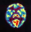
[Functional neuroimaging options for tinnitus]
- PMID: 37382658
- PMCID: PMC10520110
- DOI: 10.1007/s00106-023-01319-5
[Functional neuroimaging options for tinnitus]
Abstract
Background: The pathophysiology behind tinnitus is still not well understood. Different imaging methods help in the understanding of the complex relationships that lead to the perception of tinnitus.
Objective: Herein, different functional imaging methods that can be used in the study of tinnitus are presented.
Materials and methods: Considering the recent literature on the subject, the relevant imaging methods used in tinnitus research are discussed.
Results and conclusion: Functional imaging can reveal correlates of tinnitus. Due to the still limited temporal and spatial resolution of current imaging modalities, a conclusive explanation of tinnitus remains elusive. With increasing use of functional imaging, additional important insights into the explanation of tinnitus will be gained in the future.
Zusammenfassung: HINTERGRUND: Die Pathophysiologie des Tinnitus ist nach wie vor nicht ausreichend verstanden. Verschiedene Bildgebungsmethoden helfen beim besseren Verständnis der komplexen Zusammenhänge, die zur Wahrnehmung von Tinnitus führen.
Ziel der arbeit: Es erfolgt die Vorstellung von verschiedenen funktionellen Bildgebungsmethoden, die in der Erforschung von Tinnitus eingesetzt werden können.
Material und methoden: Unter Einbezug der aktuellen Fachliteratur zum Thema gehen die Autoren auf die relevanten Bildgebungsmethoden der Tinnitusforschung ein.
Ergebnisse und schlussfolgerung: Die funktionelle Bildgebung kann Korrelate von Tinnitus aufzeigen. Aufgrund der noch eingeschränkten zeitlichen und räumlichen Auflösung der aktuellen Bildgebungsmodalitäten lässt eine abschließende Erklärung von Tinnitus auf sich warten. Mit der weiteren Verbreitung der funktionellen Bildgebung lassen sich in Zukunft zusätzliche wichtige Erkenntnisse zur Aufklärung von Tinnitus gewinnen.
Keywords: Diagnostic imaging; Electroencephalography; Functional brain imaging; Magnetic resonance imaging; Positron emission tomography computed tomography.
© 2023. The Author(s).
References
-
- Brozoski T, Odintsov B, Bauer C. Gamma-aminobutyric acid and glutamic acid levels in the auditory pathway of rats with chronic tinnitus: a direct determination using high resolution point-resolved proton magnetic resonance spectroscopy (H-MRS) Front Syst Neurosci. 2012;6:9. doi: 10.3389/fnsys.2012.00009. - DOI - PMC - PubMed
Publication types
MeSH terms
LinkOut - more resources
Full Text Sources
Medical


