. 2023 Jun 13:14:1204543.
doi: 10.3389/fimmu.2023.1204543. eCollection 2023.

A novel ACE2 decoy for both neutralization of SARS-CoV-2 variants and killing of infected cells

Affiliations

A novel ACE2 decoy for both neutralization of SARS-CoV-2 variants and killing of infected cells

Alexandra Kegler et al. Front Immunol. .

Abstract

The coronavirus disease 2019 (COVID-19) pandemic caused by severe acute respiratory syndrome coronavirus 2 (SARS-CoV-2) led to millions of infections and deaths worldwide. As this virus evolves rapidly, there is a high need for treatment options that can win the race against new emerging variants of concern. Here, we describe a novel immunotherapeutic drug based on the SARS-CoV-2 entry receptor ACE2 and provide experimental evidence that it cannot only be used for (i) neutralization of SARS-CoV-2 in vitro and in SARS-CoV-2-infected animal models but also for (ii) clearance of virus-infected cells. For the latter purpose, we equipped the ACE2 decoy with an epitope tag. Thereby, we converted it to an adapter molecule, which we successfully applied in the modular platforms UniMAB and UniCAR for retargeting of either unmodified or universal chimeric antigen receptor-modified immune effector cells. Our results pave the way for a clinical application of this novel ACE2 decoy, which will clearly improve COVID-19 treatment.

Keywords: ACE2 decoy; COVID-19; SARS–CoV–2; T-cell based immunotherapy; adapter CAR platform; bispecific antibody.


Conflict of interest statement

Author DM was employed by CROmed Translational Research Ltd. The remaining authors declare that the research was conducted in the absence of any commercial or financial relationships that could be construed as a potential conflict of interest.

Figures

Figure 1
Figure 1
Mechanism of action of the angiotensin-converting enzyme 2 (ACE2)-minibody (Mb) target module (TM). Our novel TM is based on the extracellular domain of human ACE2 fused to the hinge and CH3 region of human IgG1, as well as the peptide epitope E5B9 (red box). Consequently, it should bind to the receptor-binding domain (RBD) of SARS-CoV-2 and block SARS-CoV-2 infections in human cells (left). In addition, this novel TM should be able to bind to virus-infected cells expressing SARS-CoV-2 RBD on their surface (RBD+ human cells), thereby mediating their elimination via T-cell-based immunotherapy (right). For this purpose, either unmodified T-cells in combination with the previously described effector module (EM) of the UniMAB system or T-cells equipped with the universal artificial receptor of the UniCAR system can be used for targeting virus-infected cells.
Figure 2
Figure 2
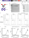
Biochemical and functional characterization of the ACE2-Mb TM. (A) Schematic representation of the ACE2-Mb TM containing a portion of the extracellular domain of the human ACE2 receptor (AA18-615) linked to the hinge and CH3 region of a human IgG1 Ab and the peptide tag E5B9. L, leader peptide. (B) As schematically shown, two ACE2-Mb TM monomers can form a homodimer via disulfide bonds in the hinge region. (C, D) The TM was recombinantly expressed in either (1) 3T3 or (2) a GMP-grade producer cell line derived from CHO cells. The ACE2-Mb TM was purified from cell culture supernatants via its Strep-tag. (C) In order to estimate the concentration and purity of the isolated ACE2-Mb TM, elution fractions were analyzed by SDS-PAGE followed by staining with Quick Coomassie® Stain. (D) In order to identify the ACE2-Mb TM, SDS-PAGE and immunoblotting were performed. The TM was detected via its E5B9-tag. (E, F) Analysis of binding of the ACE2-Mb TM to the RBD domain of SARS-CoV-2 via flow cytometry. For this purpose, cell lines were established permanently expressing the SARS-CoV-2 RBDWT fusion protein tagged with the E7B6 peptide epitope. (E) As a negative control, cells lacking the SARS-CoV-2 RBDWT fusion protein (w/o transgene) were used. The binding of the ACE2-Mb TM was detected using as the primary Ab the anti-La mAb (5B9) and a fluorescently labeled secondary goat anti-mouse IgG (minimal x-reactivity) Ab. For anti-La mAb (7B6) and sACE2 detection, only fluorescently labeled goat anti-mouse IgG (minimal x-reactivity) Ab and anti-His mAb were applied, respectively. Histograms depict stained cells (black) and the respective control (grey). (F) To determine affinity towards SARS-CoV-2 RBDWT, cells were incubated with increasing amounts of sACE2, 3T3-, or CHO-produced ACE2-Mb TM. Relative median fluorescence intensity (rel. MFI) as mean ± SD and K D values are shown (n = 3).
Figure 3
Figure 3
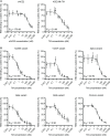
Blocking of ACE2 receptor binding. Increasing concentrations of (A, B) ACE2-Mb TM or (A) soluble ACE2 (sACE2) were added together with His-tagged (A) SARS-CoV-2 RBDWT or (B) RBD from different SARS-CoV-2 variants on ELISA plates coated with human ACE2. The binding of RBD to coated ACE2 was detected by using an HRP-conjugated anti-His mAb. The data represent the mean ± SD and IC50 of three independent experiments.
Figure 4
Figure 4
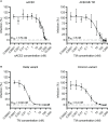
Blocking of infection by SARS-CoV-2 S-pseudo-typed virus particles. Luciferase-encoding virus particles pseudo-typed with (A) SARS-CoV-2 SWT protein or (B) S protein from Delta and Omicron VOCs were added together with increasing concentrations of (A, B) ACE2-Mb TM or (A) soluble ACE2 (sACE2) to HEK 293T ACE2 cells. After 3 days, the infection rate was determined by using a bioluminescence-based assay. Summarized data are shown as mean ± SD and IC50 values are depicted (n ≥ 3).
Figure 5
Figure 5
Blocking of infection of live SARS-CoV-2. Increasing concentrations of the ACE2-Mb TM were incubated with an early isolate, Delta or Omicron VOC, for 1 h at 37°C and subsequently added to Vero E6 cells. After 1 h, Avicel/DMEM was supplemented, and plates were incubated for 2 to 3 days followed by fixation, inactivation, crystal violet staining, and plaque number counting. Plaque-forming units (PFU) per milliliter were plotted against the TM concentration. Summarized data of triplicates as mean ± SD and IC50 values are shown.
Figure 6
Figure 6
Recruitment of T-cells via the UniMAB or UniCAR system results in efficient lysis of SARS-CoV-2 RBD+ cells in the presence of the ACE2-Mb TM. (A, B) Bioluminescence-based cytotoxicity assays were performed with luciferase+ PC3 cells either possessing (SARS-CoV-2 RBD+, left and right diagrams) or lacking (w/o transgene, left diagrams) SARS-CoV-2 RBD expression. These PC3 cells were coincubated at an E:T ratio of 5:1 with (A) unmodified T-cells for 24 h or (B) UniCAR T-cells for 6 h in the absence (w/o) or presence of 25 nM (left diagrams) or decreasing concentrations (right diagrams) of indicated Ab components. Summarized data of three individual donors as mean ± SD and EC50 values are shown. Statistical analysis was performed by using two-way ANOVA with (A) Tukey’s or (B) Šidák’s multiple comparisons tests (*** p < 0.001 with respect to (A, B) w/o and (A) only ACE2-Mb TM as well as only EM controls).
Figure 7
Figure 7
Nasal application of the ACE2-Mb TM protects Syrian hamsters against SARS-CoV-2 infection. (A, B) Five Syrian hamsters were infected by intranasal inoculation of the SARS-CoV-2 Delta VOC (B.1.617.2) in the absence (control) or presence (treated) of the ACE2-Mb TM (see Material and methods). Virus load was estimated by qRT-PCR in (A) oral/buccal samples collected over 5 days after infection or (B) in oral/buccal samples, blood, and depicted organs harvested at day 5 after infection. (A, B) Summarized data of two (control) or three (treated) animals are shown as mean ± SEM (data points on the x-axis represent the value 0). Statistical analysis was performed by using an unpaired two-tailed Student’s t-test (* p < 0.05; ** p < 0.01; *** p < 0.001).

References

    1. WHO . WHO COVID-19 dashboard (2023). Available at: https://covid19.who.int/ (Accessed May 04, 2023).
    1. Garcia-Beltran WF, Lam EC, St. Denis K, Nitido AD, Garcia ZH, Hauser BM, et al. . Multiple SARS-CoV-2 variants escape neutralization by vaccine-induced humoral immunity. Cell (2021) 184(9):2372–83.e9. doi: 10.1016/j.cell.2021.03.013 - DOI - PMC - PubMed
    1. Liu L, Iketani S, Guo Y, Chan JFW, Wang M, Liu L, et al. . Striking antibody evasion manifested by the omicron variant of SARS-CoV-2. Nature (2022) 602(7898):676–81. doi: 10.1038/s41586-021-04388-0 - DOI - PubMed
    1. Wibmer CK, Ayres F, Hermanus T, Madzivhandila M, Kgagudi P, Oosthuysen B, et al. . SARS-CoV-2 501Y.V2 escapes neutralization by south African COVID-19 donor plasma. Nat Med (2021) 27(4):622–5. doi: 10.1038/s41591-021-01285-x - DOI - PubMed
    1. Wang Q, Iketani S, Li Z, Liu L, Guo Y, Huang Y, et al. . Alarming antibody evasion properties of rising SARS-CoV-2 BQ and XBB subvariants. Cell (2023) 186(2):279–86.e8. doi: 10.1016/j.cell.2022.12.018 - DOI - PMC - PubMed

Publication types

Substances

Supplementary concepts