. 2023;43(1):161-167.

Needle Arthroscopy as a Reduction Aid for Lower Extremity Peri-Articular Fractures: Case Series and Technical Tricks

Affiliations

Needle Arthroscopy as a Reduction Aid for Lower Extremity Peri-Articular Fractures: Case Series and Technical Tricks

Luke A Lopas et al. Iowa Orthop J. 2023.

Abstract

Background: Intra-articular fractures represent a challenging group of injuries that can occur in many different locations. In addition to restoring the mechanical alignment and stability of the extremity, accurate reduction of the articular surface is a primary goal for the treatment of peri-articular fractures. A variety of methods have been deployed to assist in the visualization and subsequent reduction of the articular surface, each with a unique set of pros and cons. The ability to visualize the articular reduction must be balanced against the soft tissue trauma required for extensile exposures. Arthroscopic assisted reduction has gained popularity for the treatment of a variety of articular injuries. Recently, needle based arthroscopy has been developed, predominantly as an outpatient tool for the diagnosis of intra-articular pathology. We present an initial experience with and technical tricks for the use of a needle based arthroscopic camera in the treatment of lower extremity peri-articular fractures.

Methods: A retrospective review of all cases where needle arthroscopy was used as a reduction adjunct in lower extremity peri-articular fractures at a single, academic, level one trauma center was performed.

Results: Five patients with six injuries were treated with open reduction internal fixation with adjunctive needle based arthroscopy. Early experience and tips and tricks for successful utilization of this technique are presented.

Conclusion: Needle based arthroscopy may represent a valuable adjunct in the treatment of peri-articular fractures and warrants further investigation. Level of Evidence: IV.

Keywords: arthroscopic assisted reduction; articular fracture; needle arthroscopy.


Figures

Figure 1.
Figure 1.
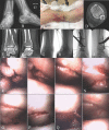
74-year-old female transferred from another facility after spanning external fixation of an intra-articular pilon fracture. Initial x-rays upon presentation demonstrating inadequate reduction in frame, (A) mortise ankle, (B) lateral ankle. (C) Axial computed tomography demonstrating classic pilon articular fragments. Intraoperative fluoroscopy of final reduction and fixation construct (D). Pre clamp placement view with camera looking at articular surface (E), and post clamp placement view using camera (F) to confirm reduction and compression across fracture lines. View at medial shoulder confirming reduction of anterior, medial, and posterior fragments (G). Axial post op CT at joint (H), sagittal post op CT (I) confirming reduction of anterior and posterior articular fragments, and coronal post op CT confirming medial reduction (J).
Figure 2.
Figure 2.
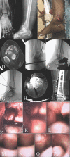
39-year-old male who fell from a ladder sustaining a right hip dislocation with a posterior wall fracture. Injury AP pelvis x-ray (A). Injury axial (B), coronal (C), sagittal (D), and 3D reconstruction (E) computed tomography images demonstrating a comminuted posterior wall fracture with intra-articular debris. Intra-operative fluoroscopy with schantz pin in the femoral neck for distraction and provisional reduction stabilized with k-wires. Intra-operative use of camera to aid in visualization and judgment of articular reduction as well as assure that no further intra-articular debris needs to be removed (G, H, I, J). Postoperative axial (K), coronal (L), and sagittal (M) computed tomography demonstrating adequate reduction and remaining debris confined to the fovea.
Figure 3.
Figure 3.
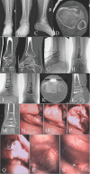
47-year-old male who sustaining a twisting lower extremity injury. Mortise (A) and lateral (B) x-rays demonstrating a spiral, intra-articular distal tibia fracture. Clinical photo of the patient’s limb demonstrating fracture blisters and significant edema (C). Pre-operative axial (D), coronal (E), and sagittal (F) computed tomography slices demonstrating medial and posterior intra-articular fractures, with anterior articular block connecting with anterior-lateral metaphyseal spike. Intra-operative fluoroscopy of mortise (G) and lateral (H) spanning external fixation performed on the day of injury. Intra-operative fluoroscopy two weeks later at time of definitive fixation with provisional reduction and plate balance obtained. Intra-operative images obtained with needle arthroscopic camera of articular surface pre-reduction (K, L, M, N), and images obtained post reduction (O, P, Q, R), confirming articular reduction and obviating the need for post-operative computed tomography to confirm reduction.
Figure 4.
Figure 4.
57-year-old female in a motor vehicle collision who sustained a Gustilo-Anderson type 3A open right pilon fracture. Injury mortise (A) and lateral (B) ankle x-rays. Clinical photo of medial, transverse, tension failure wound (C) in the trauma bay. Pre-operative axial (D) computed tomography scan demonstrating significant anterior and medial comminution. Intra-operative fluoroscopy mortise (E) and lateral (F) images during the initial debridement, irrigation, and spanning external fixation demonstrating overall restoration of length, alignment, and rotation. Lateral (G) intra-operative fluoroscopy at the time of definitive fixation with provisional stabilization and reconstruction of the articular block. Post-operative axial (H) and coronal (I) computed tomography demonstrating reduction of the articular block. Intra-operative view of the lateral joint pre reduction (J) to post reduction (K, L). Intra-operative view of the joint post reduction progressing from the central joint space to the medial joint space (M, N, O, P) confirming accurate medial reduction without needing to re-open the traumatic medial wound.
Figure 5.
Figure 5.
48-year-old male pedestrian struck by an automobile sustaining a left sided tibial plateau fracture and an ipsilateral pilon fracture (see Fig 6). Injury AP (A) and Lateral (B) knee x-rays and axial (C), sagittal (D), coronal (E), and 3D reconstruction (F) computed tomography images demonstrate a predominantly antero-medial injury with an additional large posterior-medial fragment. Intraoperative final AP (G) and lateral (H) fluoroscopic images demonstrate buttress fixation of both the anterior-medial and posterior-medial fragments. Postoperative axial (I), coronal (J), and sagittal (K) computed tomography images demonstrate reduction and fixation of the articular fragments. Intra-operative images obtained with the needle arthroscopic camera confirming reduction of the articular fragments (L, M, N).
Figure 6.
Figure 6.
Pilon fracture sustained by the same patient illustrated in figure 5. Injury AP (A), mortise (B), and lateral (C) x-rays, and axial (D), coronal (E), and sagittal (F) computed tomography slices demonstrate an anterior crush injury to the distal tibia. Intraoperative fluoroscopic lateral (G), and mortise (H) with provisional reduction obtained and stabilized with k-wires. Final lateral (I), and mortise (J) intra-operative fluoroscopy. Postoperative axial (K), sagittal (L), and coronal (M) computed tomography scans confirming reduction of anterior joint. Intra-operative needle arthroscopic camera images pre-reduction (N, O, P), and post reduction (Q, R, S), confirming reduction and hardware safety.

References

    1. Dei Giudici L, Di Muzio F, Bottegoni C, Chillemi C, Gigante A. The role of arthroscopy in articular fracture management: the lower limb. Eur J Orthop Surg Traumatol. 2015;25(5):807–13. - PubMed
    1. Hamilton GA, Doyle MD, Castellucci-Garza FM. Arthroscopic-Assisted Open Reduction Internal Fixation. Clin Podiatr Med Surg. 2018;35(2):199–221. - PubMed
    1. Ono A, Nishikawa S, Nagao A, Irie T, Sasaki M, Kouno T. Arthroscopically assisted treatment of ankle fractures: arthroscopic findings and surgical outcomes. Arthroscopy. 2004;20(6):627–31. - PubMed
    1. Chen XZ, Chen Y, Liu CG, Yang H, Xu XD, Lin P. Arthroscopy-Assisted Surgery for Acute Ankle Fractures: A Systematic Review. Arthroscopy. 2015;31(11):2224–31. - PubMed
    1. Egol KA, Cantlon M, Fisher N, Broder K, Reisgo A. Percutaneous Repair of a Schatzker III Tibial Plateau Fracture Assisted by Arthroscopy. J Orthop Trauma. 2017;3(31 Suppl):S12–S3. - PubMed