

A Rare Type of Hepatocellular Carcinoma Presenting With Cardiac Thrombus
- PMID: 37384080
- PMCID: PMC10299851
- DOI: 10.7759/cureus.39611
A Rare Type of Hepatocellular Carcinoma Presenting With Cardiac Thrombus
Abstract
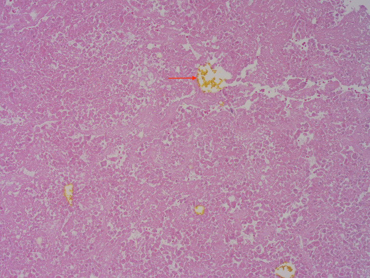
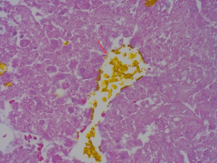
Hepatocellular carcinoma (HCC) is a complication of end stage liver disease. Even rarer is right atrial tumor thrombus burden due to HCC. Common metastatic sites of HCC in descending order are lung, peritoneum, and bone. We present a patient with liver cirrhosis due to nonalcoholic fatty liver disease (NAFLD) admitted due to incidental finding of right atrial thrombus on echocardiography after missing HCC surveillance for four years. Patient received a computed tomography (CT) scan that showed an inconclusive liver lesion despite two liver biopsies, and patient was incidentally found to have clear cell HCC diagnosed after right hepatectomy. Right atrial thrombus was treated with surgical thrombectomy and pathology showed necrotic HCC thrombi in right atrium with bile pigment. Due to the possibility of tumor growth with extrahepatic manifestations, screening in compensated cirrhosis is essential.
Keywords: cirrhosis; hepatocellular carcinoma; hepatology; non alcoholic fatty liver; right atrial thrombus.
Copyright © 2023, Aboona et al.
Conflict of interest statement
The authors have declared that no competing interests exist.
Figures


References
-
- Hepatocellular carcinoma with inferior vena cava and right atrial tumor thrombus: a case report. Chen M, Huang X, Yang Q. Echocardiography. 2019;36:2110–2113. - PubMed
-
- Hepatocellular carcinoma with intracavitary cardiac involvement: a case report and review of the literature. Sung AD, Cheng S, Moslehi J, Scully EP, Prior JM, Loscalzo J. Am J Cardiol. 2008;102:643–645. - PubMed
-
- Is it possible? Invasion of the heart with hepatocellular carcinoma in a short time. Senarslan O, Kantarci UH, Eyuboglu M, Senarslan DA. Int J Cardiovasc Acad. 2016;2:124–126.
Publication types
LinkOut - more resources
Full Text Sources