. 2023 Aug 15;133(16):e138207.
doi: 10.1172/JCI138207.

Reduction of nemo-like kinase increases lysosome biogenesis and ameliorates TDP-43-related neurodegeneration

Affiliations

Reduction of nemo-like kinase increases lysosome biogenesis and ameliorates TDP-43-related neurodegeneration

Leon Tejwani et al. J Clin Invest. .

Abstract

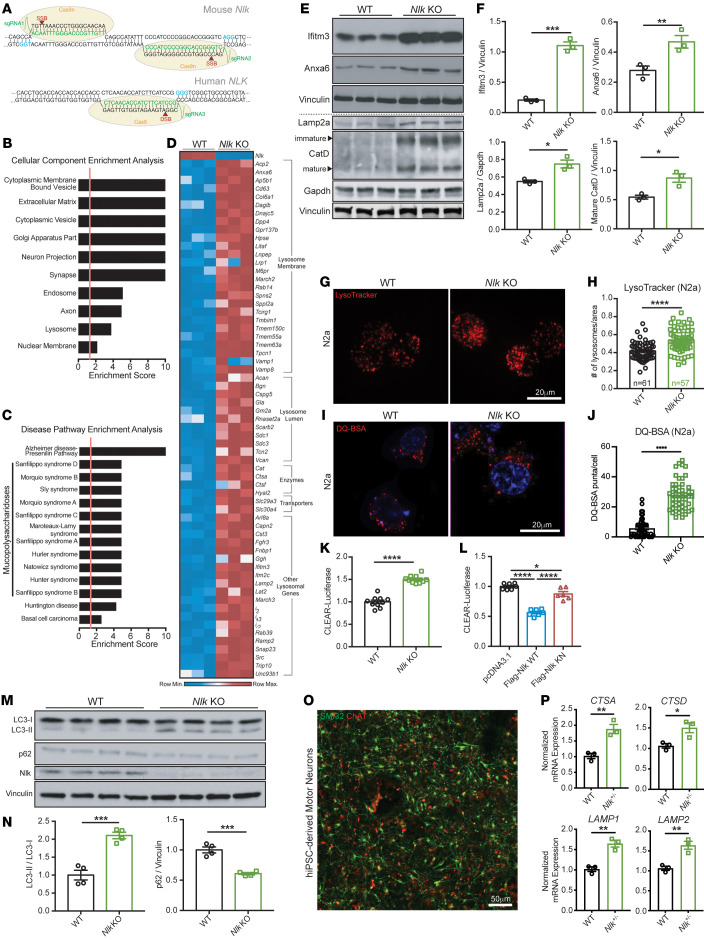
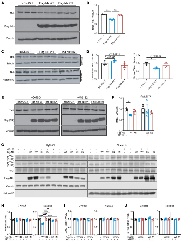
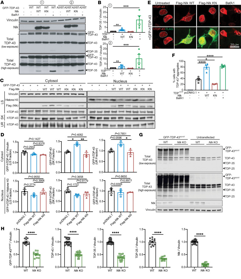
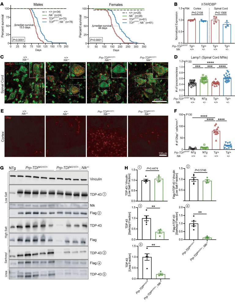
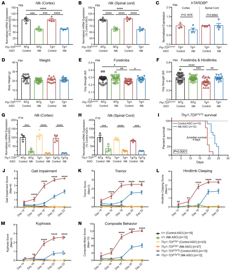
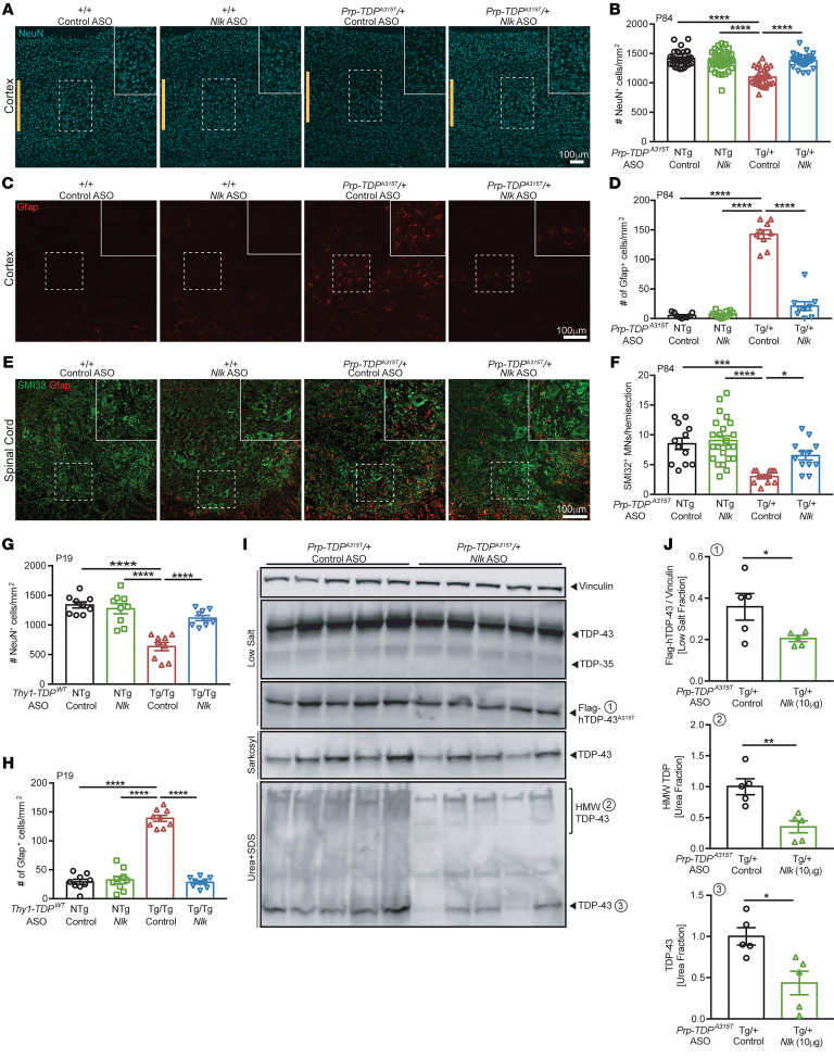
Protein aggregation is a hallmark of many neurodegenerative disorders, including amyotrophic lateral sclerosis (ALS). Although mutations in TARDBP, encoding transactive response DNA-binding protein 43 kDa (TDP-43), account for less than 1% of all ALS cases, TDP-43-positive aggregates are present in nearly all ALS patients, including patients with sporadic ALS (sALS) or carrying other familial ALS-causing (fALS-causing) mutations. Interestingly, TDP-43 inclusions are also present in subsets of patients with frontotemporal dementia, Alzheimer's disease, and Parkinson's disease; therefore, methods of activating intracellular protein quality control machinery capable of clearing toxic cytoplasmic TDP-43 species may alleviate disease-related phenotypes. Here, we identify a function of nemo-like kinase (Nlk) as a negative regulator of lysosome biogenesis. Genetic or pharmacological reduction of Nlk increased lysosome formation and improved clearance of aggregated TDP-43. Furthermore, Nlk reduction ameliorated pathological, behavioral, and life span deficits in 2 distinct mouse models of TDP-43 proteinopathy. Because many toxic proteins can be cleared through the autophagy/lysosome pathway, targeted reduction of Nlk represents a potential approach to therapy development for multiple neurodegenerative disorders.

Keywords: Genetics; Lysosomes; Mouse models; Neurodegeneration; Neuroscience.


Figures

Figure 1
Figure 1. Reduction of Nlk increases lysosome biogenesis in vitro.
(A) Schematic of dual-guide RNA targeting of Cas9 nickase (Cas9n) to Nlk for generation of isogenic Nlk KON 2a cells and NLK+/- hiPSCs. (B and C) Enrichment analyses for all significantly differentially expressed genes from RNA-Seq of isogenic WT and Nlk KO N2a cells. Significant enrichment scores of 1.3 (FDR adjusted P value q < 0.05) are designated by vertical red lines. (D) Heatmap of significantly upregulated lysosomal genes from RNA-Seq (n = 3 biological replicates). (E and F) Western blot validation (E) confirmed RNA-Seq results, quantified in F (n = 3 biological replicates). Blot lanes separated by the dotted line were run on lysates from distinct experimental replicates and normalized to their respective housekeeping proteins. (G and H) Representative LysoTracker images of N2a cells (G), quantified in H, demonstrated increased lysosome number in Nlk KO cells (WT, n = 61 cells; Nlk KO, n = 57 cells). (I and J) Representative images of functional lysosomes in N2a cells incubated with DQ BSA (I), quantified in J (WT, n = 50 cells; Nlk KO, n = 50 cells). (K and L) CLEAR-luciferase assay in Nlk KO N2a cells (K) or WT N2a cells transfected with WT or KN Nlk (L) demonstrated Nlk kinase activity suppresses CLEAR network transcription (K, n = 10 replicates from 2 different Nlk KO clones; L, n = 7 replicates from 2 experimental repeats). (M and N) Western blots (M) showed increased LC3-II/LC3-I and decreased p62 levels in Nlk KO N2a cells, quantified in N (n = 4 biological replicates). (O) Representative immunostaining images of hiPSC-derived motor neurons. (P) qPCR showing NLK+/– iPSC–derived motor neurons expressed higher levels of CTSA, CTSD, LAMP1, and LAMP2 compared with isogenic controls (n = 3 biological replicates from distinct motor neuron differentiations). Two-tailed t tests (F, H, J, K, N, and P) or 1-way ANOVA (L) analyses were performed, and data are represented as mean ± SEM. *P < 0.05; **P < 0.01; ***P < 0.001; ****P < 0.0001.
Figure 2
Figure 2. Nlk destabilizes nuclear Tfeb via proteasomal degradation.
(A and B) Western blots of N2a whole-cell protein lysates following transfection with Flag-Nlk WT or Flag-Nlk KN (A). Tfeb levels decreased upon Nlk overexpression in a kinase activity–dependent manner, quantified in B (n = 3 biological replicates). (C and D) Western blots of WT N2a cells transfected with Flag-Nlk WT or Flag-Nlk KN showed a kinase activity–dependent decrease of nuclear Tfeb levels (C), quantified in D (n = 3 biological replicates). (E and F) Western blots of N2a whole-cell protein lysates following transfection with Flag-Nlk WT or Flag-Nlk KN, with DMSO vehicle or MG132 treatment (E). Tfeb levels decreased upon Nlk overexpression in a kinase activity–dependent manner, but this destabilization was not observed in the presence of the proteasome inhibitor MG132, quantified in F (n = 3 biological replicates). (GJ) Western blots of subcellular fractionated N2a protein lysates following transfection with Flag-Nlk WT or Flag-Nlk KN, with DMSO or MG132 treatment (G). Only nuclear Tfeb levels were reduced upon Nlk overexpression in a kinase activity–dependent manner, and this decrease was dependent on the proteasomal degradation pathway, quantified in H. Phosphorylation of Tfeb at S122 and S142 was not affected by Nlk overexpression, quantified in I and J (n = 4 biological replicates). One-way ANOVA analyses (B, D, F, and HJ) were performed, and data are represented as mean ± SEM. *P < 0.05; **P < 0.01; ***P < 0.001; ****P < 0.0001.
Figure 3
Figure 3. Genetic reduction of Nlk reduces TDP levels in vitro.
(A and B) Western blots showing coexpression of Nlk-WT with GFP-tagged TDPWT or TDPA315T in NSC-34 cells (A) increased levels of TDP-35, while Nlk-KN did not. V-ATPase inhibitor BafA1 treatment similarly increased TDP-35 levels in the absence of Nlk overexpression to an even greater extent. Quantification shows TDP-35 levels in B (n = 4 biological replicates). (C and D) Western blots of sequentially extracted proteins based on detergent solubility from subcellular fractionated NSC-34 cells (C). Quantification of TDP-43 species shows Nlk WT overexpression increased cytosolic insoluble TDP-43 (sarkosyl and urea fractions) (D) (n = 3 biological replicates). LS, low salt; SK, sarkosyl; UR, urea. (E and F) Representative immunostaining of NSC-34 cells cotransfected with GFP-tagged TDP-43 and Nlk showing that WT Nlk increased formation of GFP-positive cytosolic TDP-43 aggregates (E), quantified in F. Nuclei are outlined with dotted lines (n = 4 biological replicates). (G and H) Western blots of cell lysates from WT or Nlk KO N2a cells transfected with GFP–TDP-43A315T (G), quantified in H (n = 12–24 replicates pooled from 4 individual experiments). Levels of exogenous GFP–TDP-43A315T, endogenous TDP-43, and truncated fragments of TDP-43 were significantly reduced. One-way ANOVA analyses (B, D, and F) or 2-tailed t tests (H) were performed to compare all listed genotypes/treatments unless otherwise noted, and data are represented as mean ± SEM. *P < 0.05; **P < 0.01; ***P < 0.001; ****P < 0.0001.
Figure 4
Figure 4. Genetic reduction of Nlk improves survival and pathology in TDP-43 mice.
(A) Kaplan-Meier survival curves showing 50% genetic reduction of Nlk increased Prp-TDPA315T/+ (Prp–TDP-43 Tg) male and female animal survival. Data are combined from 2 independent Nlk gene trap insertion lines. Sample sizes for each genotype are provided in the figure. Curves were compared by log-rank test. (B) A 50% genetic reduction of Nlk did not affect cortex or spinal cord hTARDBP mRNA levels in P84 Prp-TDPA315T/+ mice (Nlk+/+, n = 4 animals; Nlk+/–, n = 3 animals). (CF) Representative immunohistochemical images showing genetic reduction of Nlk increased Lamp1+ lysosomes in the soma (small dotted lines) of lumbar spinal cord motor neurons (C, quantified in D; n = 18–22 motor neurons from 3 animals per genotype) and rescued layer V astrogliosis (E, quantified in F; n = 12 sections from 3 animals per genotype) in P130 Prp-TDPA315T/+ female mice. (G and H) Western blots of various forms of TDP-43 in fractions of female mouse spinal cords at P200 (G). Quantification of TDP-43 species shows levels of insoluble TDP-43 (sarkosyl and urea fractions) were significantly reduced with 50% genetic reduction of Nlk (H) (TDP-Tg, n = 5 animals; TDP-Tg/Nlk, n = 4 animals). Note that TDP-43A315T/+ protein is Flag tagged at the N-terminal end. Two-tailed t tests (B and H) or 1-way ANOVA analyses (D and F) were performed to compare all listed genotypes/treatments unless otherwise noted, and data are represented as mean ± SEM. NTg, nontransgenic; Tg, transgenic. **P < 0.01; ***P < 0.001; ****P < 0.0001.
Figure 5
Figure 5. Pharmacological reduction of Nlk using ASOs improves behavior in 2 TDP-43 mouse models.
(A and B) An i.c.v. injection of 10 μg of Nlk ASO at P1 resulted in a 40% reduction of Nlk mRNA at P84 in the cortex (A) and spinal cord (B) (n = 4 animals per condition). (C) ASO reduction of Nlk did not affect transcript levels of hTARDBP (n = 3–4 animals per condition). (D) Overall body weight was not affected by ASO administration (n = 12–19 animals per condition). (E and F) Nlk ASO administration at P1 rescued forelimb (E) and forelimb with hind limb (F) grip-strength deficits in Prp-TDPA315T/+ mice at P84 (n = 18–27 animals per condition). (G and H) Injection of 10 μg of Nlk ASO at P1 resulted in a 90% and 75% reduction of Nlk mRNA at P19 in the cortex (G) and spinal cord (H), respectively (n = 3–5 animals per condition). (I) Kaplan-Meier survival curves showing administration of 10 μg Nlk ASO increased Thy1-TDPTg/Tg animal survival (n = 10–12 animals per condition). Curves were compared by log-rank test. (JN) Nlk ASO administration reduced gait impairment (J), tremor (K), hind limb clasping (L), kyphosis (M), and composite motor score (N) in Thy1-TDPTg/Tg animals between P14 and P22 (n = 10–28 animals per condition). One-way ANOVA analyses (AH and JN) were performed to compare all listed genotypes/treatments per day unless otherwise noted, and data are represented as mean ± SEM. *P < 0.05; **P < 0.01; ***P < 0.001; ****P < 0.0001.
Figure 6
Figure 6. Pharmacological reduction of Nlk using ASOs decreases TDP-43 levels in vivo and ameliorates pathology in 2 TDP-43 mouse models.
(AF) Administration of 10 μg of Nlk ASO at P1 rescued loss of layer V cortical neurons (A, quantified in B; n = 29–39 sections from 4–5 animals per genotype; marked by yellow bars), layer V astrogliosis (C, quantified in D; n = 9–12 sections from 3–4 animals per genotype), and lumbar spinal cord motor neuron loss (E, quantified in F; n = 12–21 sections from 3–5 animals per genotype) in Prp-TDPA315T/+ male mice at P84. (G and H) 10 μg of Nlk ASO at P1 rescued loss of layer V cortical neurons (G; n = 9 sections from 3 animals per genotype) and layer V astrogliosis (H; n = 9 sections from 3 animals per genotype) in Thy1-TDPTg/Tg mice at P19. (I and J) Western blots of various forms of TDP-43 in fractions of male mouse spinal cords at P84. Quantification of TDP species from (I) shows levels of Flag-hTDP-43 (low-salt fraction), total TDP-43 (urea fraction), and high-molecular weight TDP (urea fraction) were significantly reduced in Nlk ASO–injected animals (J) (n = 5 animals per condition). One-way ANOVA analyses (B, D, F, G, and H) or 2-tailed t tests (J) were performed to compare all listed genotypes/conditions unless otherwise noted, and data are represented as mean ± SEM. *P < 0.05; **P < 0.01; ***P < 0.001; ****P < 0.0001.

References

    1. Arai T, et al. TDP-43 is a component of ubiquitin-positive tau-negative inclusions in frontotemporal lobar degeneration and amyotrophic lateral sclerosis. Biochem Biophys Res Commun. 2006;351(3):602–611. doi: 10.1016/j.bbrc.2006.10.093. - DOI - PubMed
    1. Ling SC, et al. Converging mechanisms in ALS and FTD: disrupted RNA and protein homeostasis. Neuron. 2013;79(3):416–438. doi: 10.1016/j.neuron.2013.07.033. - DOI - PMC - PubMed
    1. Neumann M, et al. Ubiquitinated TDP-43 in frontotemporal lobar degeneration and amyotrophic lateral sclerosis. Science. 2006;314(5796):130–133. doi: 10.1126/science.1134108. - DOI - PubMed
    1. Amador-Ortiz C, et al. TDP-43 immunoreactivity in hippocampal sclerosis and Alzheimer’s disease. Ann Neurol. 2007;61(5):435–445. doi: 10.1002/ana.21154. - DOI - PMC - PubMed
    1. Nakashima-Yasuda H, et al. Co-morbidity of TDP-43 proteinopathy in Lewy body related diseases. Acta Neuropathol. 2007;114(3):221–229. doi: 10.1007/s00401-007-0261-2. - DOI - PubMed

Publication types

Supplementary concepts