. 2023 Jun 29;18(6):e0286370.
doi: 10.1371/journal.pone.0286370. eCollection 2023.

The ribosome-inactivating proteins MAP30 and Momordin inhibit SARS-CoV-2

Affiliations

The ribosome-inactivating proteins MAP30 and Momordin inhibit SARS-CoV-2

Norman R Watts et al. PLoS One. .

Abstract

The continuing emergence of SARS-CoV-2 variants has highlighted the need to identify additional points for viral inhibition. Ribosome inactivating proteins (RIPs), such as MAP30 and Momordin which are derived from bitter melon (Momordica charantia), have been found to inhibit a broad range of viruses. MAP30 has been shown to potently inhibit HIV-1 with minimal cytotoxicity. Here we show that MAP30 and Momordin potently inhibit SARS-CoV-2 replication in A549 human lung cells (IC50 ~ 0.2 μM) with little concomitant cytotoxicity (CC50 ~ 2 μM). Both viral inhibition and cytotoxicity remain unaltered by appending a C-terminal Tat cell-penetration peptide to either protein. Mutation of tyrosine 70, a key residue in the active site of MAP30, to alanine completely abrogates both viral inhibition and cytotoxicity, indicating the involvement of its RNA N-glycosylase activity. Mutation of lysine 171 and lysine 215, residues corresponding to those in Ricin which when mutated prevented ribosome binding and inactivation, to alanine in MAP30 decreased cytotoxicity (CC50 ~ 10 μM) but also the viral inhibition (IC50 ~ 1 μM). Unlike with HIV-1, neither Dexamethasone nor Indomethacin exhibited synergy with MAP30 in the inhibition of SARS-CoV-2. From a structural comparison of the two proteins, one can explain their similar activities despite differences in both their active-sites and ribosome-binding regions. We also note points on the viral genome for potential inhibition by these proteins.


Conflict of interest statement

The authors have declared that no competing interests exist.

Figures
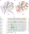
Fig 1
Fig 1. Structure comparison of MAP30 and Momordin.
(A) MAP30 and (B) Momordin as viewed towards their respective active-sites, with key residues discussed in the text highlighted as follows: catalytic-site (green), Mn+2-binding site (yellow), and on the active-site-distal face, the ribosome-binding site (cyan) with the c11-P peptide-interacting Tyr residue (magenta). (C) Sequence alignment of MAP30 and Momordin with residues highlighted as above. Note that the structures for both MAP30 (PDB: 1D8V, 1CF5) and Momordin (PDB: 1MOM, 1AHA) are all missing 23 N-terminal residues. The C-terminal 19 residues are shown in the nuclear magnetic resonance structure of MAP30 (PDB: 1D8V) but not in the X-ray crystal structures of either protein. The N-terminal residues were missing, and C-terminal residues were present, on both proteins employed here.
Fig 2
Fig 2. SARS-CoV-2 inhibition by MAP30 and Momordin.
Viral inhibition and cell viability as a function of concentration of MAP30 and Momordin, either without (A and B) or with (C and D) a C-terminally appended Tat cell penetration peptide, as assessed in the A549 NLRV assay. The results from this and other assays are summarized in Table 1.
Fig 3
Fig 3. Mutation of MAP30.
Assessment of SARS-CoV-2 inhibition by MAP30, MAP30.Y70A, and MAP30.K171, K215 in the A549 NLRV assay. The Y70A active-site mutation abrogates both viral inhibition and cytotoxicity. The MAP30.K171, K215 ribosome-binding-site mutations result in an increase of both the IC50 and CC50 values, while the selectivity index (SI = CC50/IC50) remains the same.
Fig 4
Fig 4. Comparison of MAP30 and Momordin active-site faces.
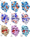
(A-C) MAP30, TCS, and Momordin represented as hydrophobicity surfaces (blue, charged; orange, hydrophobic; white, neutral), (D-F) as charge surfaces (blue, basic; red, acidic; white, neutral), and (G-I) with residues scored as important by Evolutionary Trace shown in red. PDB codes and percent sequence identity to MAP30 as indicated. Ricin (PDB: 1APG, 30.5%) and Saporin (PDB: 1QI7, 23.5%) gave similar patterns (not shown). The binding groove with the catalytic site at the center is located between the two arrows in (A). The regions of the previously identified Mn+2 and Zn+2 binding areas are indicated in (D).
Fig 5
Fig 5. Comparison of MAP30 and Momordin ribosome-binding-site faces.
(A-C) MAP30, TCS, and Momordin represented as hydrophobicity surfaces (blue, charged; orange, hydrophobic; white, neutral), (D-F) as charge surfaces (blue, basic; red, acidic; white, neutral), and (G-I) with residues scored as important by Evolutionary Trace shown in red. PDB codes and percent sequence identity to MAP30 as indicated. Ricin (PDB: 1APG, 30.5%) and Saporin (PDB: 1QI7, 23.5%) gave similar patterns (not shown). The c11-P peptide bound to TCS, and aligned to MAP30 and Momordin, is shown in magenta. The C-terminal end of the peptide is bound in a hydrophobic groove common to all the proteins (A-C) and the N-terminal end, with acidic resides D2, D3, and D4 (indicated by the arrow in D), likely interacts with a basic region on MAP30 but not the corresponding acidic region on Momordin (D and F). The hydrophobic groove (indicated by a dashed arrow in G), and particularly Y164 in MAP30 and Y166 in Momordin, appear to be important (G and I).

Similar articles

Cited by

References

    1. Puri M, Kaur I, Kanwar RK, Gupta RC, Chauhan A, Kanwar JR. Ribosome inactivating proteins (RIPs) from Momordica charantia for anti viral therapy. Curr Mol Med. 2009;9(9):1080–94. doi: 10.2174/156652409789839071 - DOI - PubMed
    1. Puri M, Kaur I, Perugini MA, Gupta RC. Ribosome-inactivating proteins: current status and biomedical applications. Drug Discov Today. 2012;17(13–14):774–83. doi: 10.1016/j.drudis.2012.03.007 - DOI - PubMed
    1. Walsh MJ, Dodd JE, Hautbergue GM. Ribosome-inactivating proteins: potent poisons and molecular tools. Virulence. 2013;4(8):774–84. doi: 10.4161/viru.26399 - DOI - PMC - PubMed
    1. Zhu F, Zhou YK, Ji ZL, Chen XR. The Plant Ribosome-Inactivating Proteins Play Important Roles in Defense against Pathogens and Insect Pest Attacks. Front Plant Sci. 2018;9. - PMC - PubMed
    1. Lu JQ, Zhu ZN, Zheng YT, Shaw PC. Engineering of Ribosome-inactivating Proteins for Improving Pharmacological Properties. Toxins (Basel). 2020;12(3). doi: 10.3390/toxins12030167 - DOI - PMC - PubMed

Publication types

Supplementary concepts