. 2023 Jun 29;19(6):e1011003.
doi: 10.1371/journal.pcbi.1011003. eCollection 2023 Jun.

Laminar specificity of the auditory perceptual awareness negativity: A biophysical modeling study

Affiliations

Laminar specificity of the auditory perceptual awareness negativity: A biophysical modeling study

Carolina Fernandez Pujol et al. PLoS Comput Biol. .

Abstract

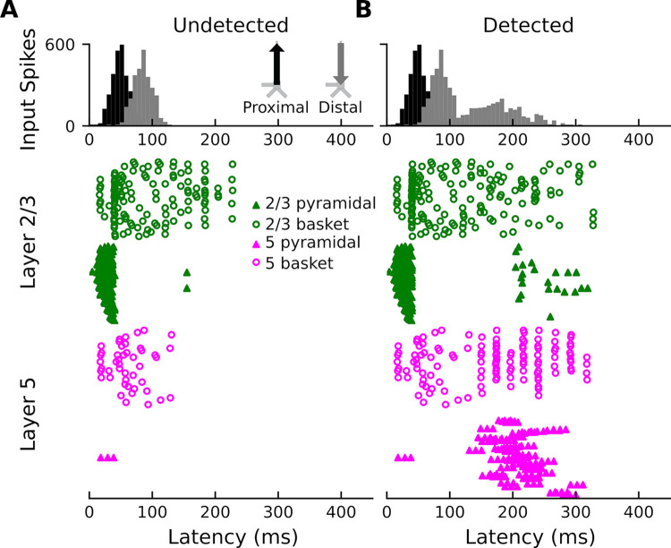
How perception of sensory stimuli emerges from brain activity is a fundamental question of neuroscience. To date, two disparate lines of research have examined this question. On one hand, human neuroimaging studies have helped us understand the large-scale brain dynamics of perception. On the other hand, work in animal models (mice, typically) has led to fundamental insight into the micro-scale neural circuits underlying perception. However, translating such fundamental insight from animal models to humans has been challenging. Here, using biophysical modeling, we show that the auditory awareness negativity (AAN), an evoked response associated with perception of target sounds in noise, can be accounted for by synaptic input to the supragranular layers of auditory cortex (AC) that is present when target sounds are heard but absent when they are missed. This additional input likely arises from cortico-cortical feedback and/or non-lemniscal thalamic projections and targets the apical dendrites of layer-5 (L5) pyramidal neurons. In turn, this leads to increased local field potential activity, increased spiking activity in L5 pyramidal neurons, and the AAN. The results are consistent with current cellular models of conscious processing and help bridge the gap between the macro and micro levels of perception-related brain activity.


Conflict of interest statement

The authors have declared that no competing interests exist.

Figures

Fig 1
Fig 1. Stimulation paradigm and evoked responses.
(A) Trial consisting of a sequence of standard tones (the target) embedded in a “cloud” of tones placed randomly in frequency and time (multitone masking). (B) Source locations of responses shown in C. (C) MEG activity of undetected (blue trace) and detected (orange trace) standard tones for both left and right auditory cortex. (D) Average of undetected and detected responses across hemispheres. (E) Difference waveform between the recorded response to the undetected and the recorded response to detected standard tones. Adapted from [26]. Negative is plotted down.
Fig 2
Fig 2. The Human Neocortical Neurosolver.
Architecture / underlying connectivity of HNN model cortical column and inputs (proximal input in black and distal input in gray) [14].
Fig 3
Fig 3. Current dipoles and laminar profiles.
(A) HNN simulation for the average response to the undetected target tones (dark blue trace). Input spikes are sampled from a Gaussian distribution on each trial for a total of 10 trials (gray traces). A proximal input (47.8 ms) followed by a distal input (84.3 ms) drive the network. R2 between empirical (light blue trace) and simulated data (dark blue) is 0.57 and RMSE is 0.61 nAm. (B) HNN simulation for the average response to detected target tones (dark orange traces). The same proximal and distal inputs used to model the response to undetected target tones, and an additional distal input (169.3 ms), drive the network. R2 between empirical (light orange trace) and simulated data (dark orange) is 0.95 and RMSE is 0.63 nAm. (C-D) Laminar profiles for the responses to the undetected and detected target tones. The contribution of layer 2/3 (which reflects the longitudinal currents in layer-2/3 (L2/3) pyramidal neurons only) is plotted in green and that of layer 5 (which reflects the longitudinal currents in L5 pyramidal neurons only) in magenta. Note that the model traces in panels A and B reflect the aggregate of the green and magenta traces in panels C and D. The corresponding input parameter values are displayed in Fig 4. The network from which the simulated dipole activity arises consists of 60,000 cells.
Fig 4
Fig 4. Driving inputs (and parameters) used to model the responses to the undetected and detected target tones.
The model column used was the calcium model column that consists of a more biologically accurate distribution of Ca2+ channels on L5 pyramidal neurons compared to HNN’s original Jones 2009 model column.
Fig 5
Fig 5. Model cortical column and Local Field Potentials.
(A) HNN provides a model cortical column that consists of a scalable, ten-by-ten pyramidal neuron and basket cell grid (the ratio of pyramidal-neuron-to-basket-cell is 3-to-1). The LFP are estimated from a linear multi-electrode array of 50 electrode contacts. (B) Simulated LFP corresponding to one of the ten undetected target tone responses. (C) Simulated LFP corresponding to one of the ten detected target tone responses.
Fig 6
Fig 6. Network spiking activity.
(A) Network spiking activity for undetected target tones. (B) Network spiking activity for detected target tones.

Update of

References

    1. Crick F, Koch C. Toward a Neurobiological Theory of Consciousness. Seminars in the Neurosciences. 1990;2: 263–275.
    1. Koch C, Massimini M, Boly M, Tononi G. Neural correlates of consciousness: progress and problems. Nat Rev Neurosci. 2016;17: 307–321. doi: 10.1038/nrn.2016.22 - DOI - PubMed
    1. Dykstra AR, Cariani PA, Gutschalk A. A roadmap for the study of conscious audition and its neural basis. Philosophical Transactions of the Royal Society B: Biological Sciences. 2017;372: 20160103. doi: 10.1098/rstb.2016.0103 - DOI - PMC - PubMed
    1. Takahashi N, Oertner TG, Hegemann P, Larkum ME. Active cortical dendrites modulate perception. Science. 2016;354: 1587–1590. doi: 10.1126/science.aah6066 - DOI - PubMed
    1. Takahashi N, Ebner C, Sigl-Glöckner J, Moberg S, Nierwetberg S, Larkum ME. Active dendritic currents gate descending cortical outputs in perception. Nat Neurosci. 2020;23: 1277–1285. doi: 10.1038/s41593-020-0677-8 - DOI - PubMed

Publication types