

Growing thin - How bulk lipid transport drives expansion of the autophagosome membrane but not of its lumen
- PMID: 37385155
- PMCID: PMC10528516
- DOI: 10.1016/j.ceb.2023.102190
Growing thin - How bulk lipid transport drives expansion of the autophagosome membrane but not of its lumen
Abstract
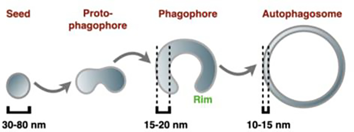
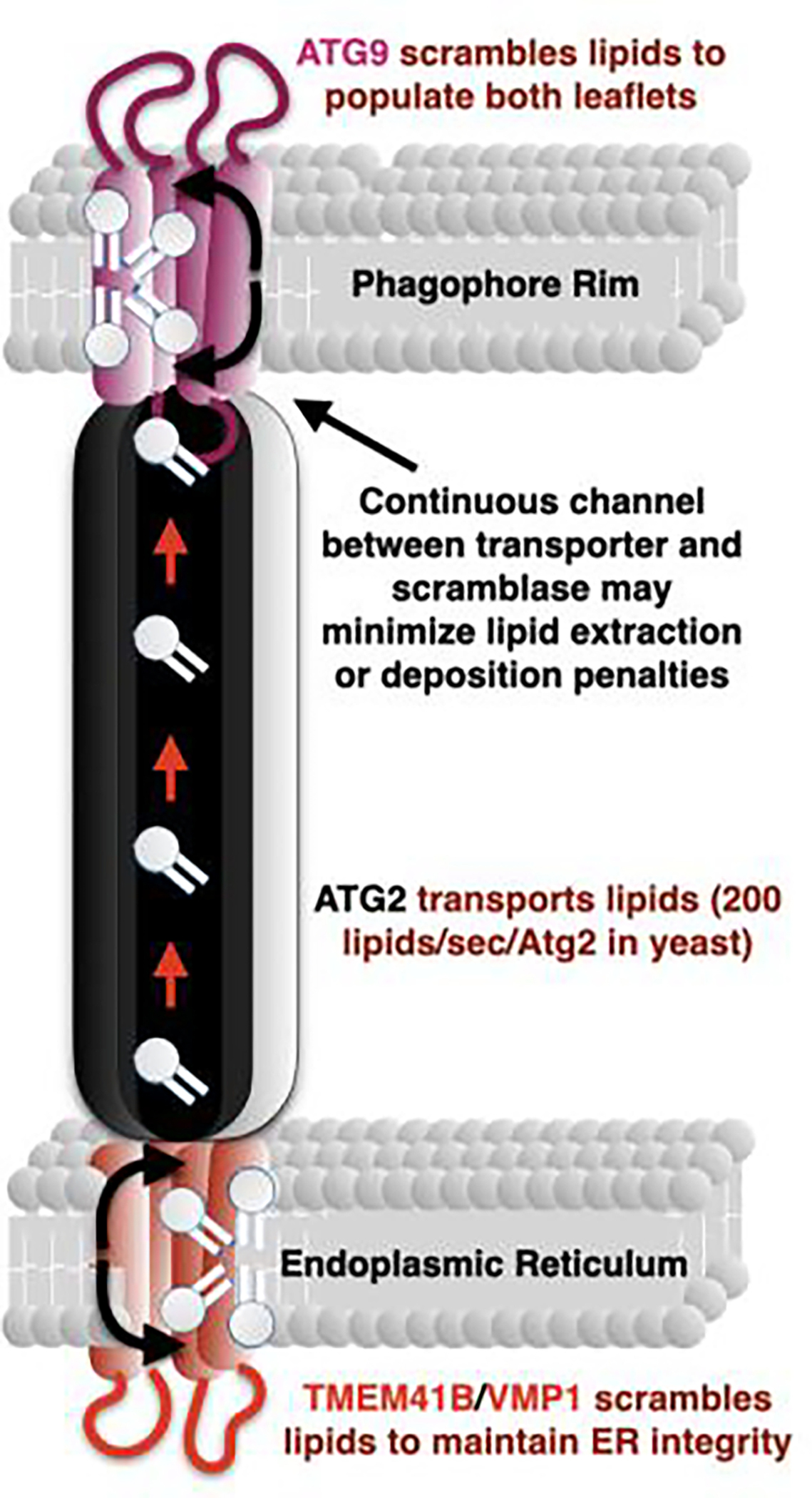
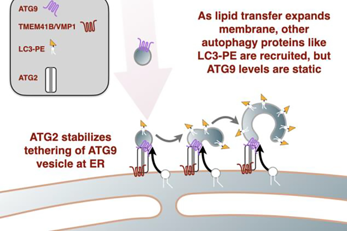
The key event in macroautophagy is the de novo formation of a new organelle called the autophagosome which when complete, will have captured bits of cytoplasm within its double-membrane structure. Eventual fusion with the lysosome allows this captured material to be degraded back to simple molecules which can be recycled to support cell function during starvation. How autophagosomes form has been a challenging question for over 60 years. This review highlights work that forms the basis for an autophagosome membrane expansion model grounded in protein-mediated lipid transport.
Copyright © 2023 Elsevier Ltd. All rights reserved.
Conflict of interest statement
Declaration of competing interest The authors declare that they have no known competing financial interests or personal relationships that could have appeared to influence the work reported in this paper.
Figures
References
-
-
Bieber A, et al., In situ structural analysis reveals membrane shape transitions during autophagosome formation. Proc Natl Acad Sci U S A, 2022. 119(39): p. e2209823119.
*** Here, Wilfling, Baumeister and colleagues follow autophagosome formation in yeast using cryo-ET. Several highlights include describing the organization of the phagophore with respect to other cellular organelles, the capture of small and consistent morphologic changes indicative of the phagophore being pulled into contact sites, measurements of the changing intralumenal distance during phagophore growth, and a thoughtful discussion on the implications of membrane expansion without significant volume increases. In particular, they conclude that direct delivery of lipid must account for over 60% of all expansion and potentially for much more.
-
-
-
Agudo-Canalejo J and Knorr RL, Formation of Autophagosomes Coincides with Relaxation of Membrane Curvature. Methods Mol Biol, 2019. 1880: p. 173–188.
*** Knorr and Mizushima build on previous work from their labs describing how “wetting” along a surface could explain much of the structures forming during membrane expansion. Here they tackle the problem of how autophagosomes capture bits of cytosol and of liquid-liquid like phase separated compartments. In addition to providing a surface along which the membrane can grow, they discuss how the energetics of membrane bending favor a cup-like structure which can mold into the phase-separated compartment.
-
Publication types
MeSH terms
Substances
Grants and funding
LinkOut - more resources
Full Text Sources
