. 2023 Aug 3;62(2):2201374.
doi: 10.1183/13993003.01374-2022. Print 2023 Aug.

Antiviral CD8+ T-cell immune responses are impaired by cigarette smoke and in COPD

Affiliations

Antiviral CD8+ T-cell immune responses are impaired by cigarette smoke and in COPD

Jie Chen et al. Eur Respir J. .

Abstract

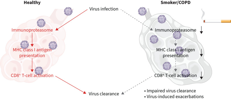
Background: Virus infections drive COPD exacerbations and progression. Antiviral immunity centres on the activation of virus-specific CD8+ T-cells by viral epitopes presented on major histocompatibility complex (MHC) class I molecules of infected cells. These epitopes are generated by the immunoproteasome, a specialised intracellular protein degradation machine, which is induced by antiviral cytokines in infected cells.

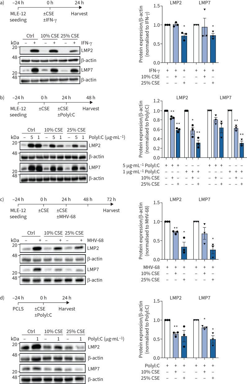
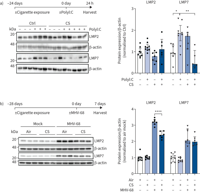
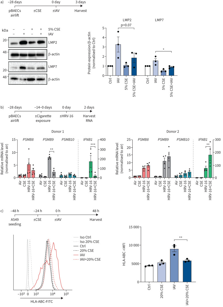
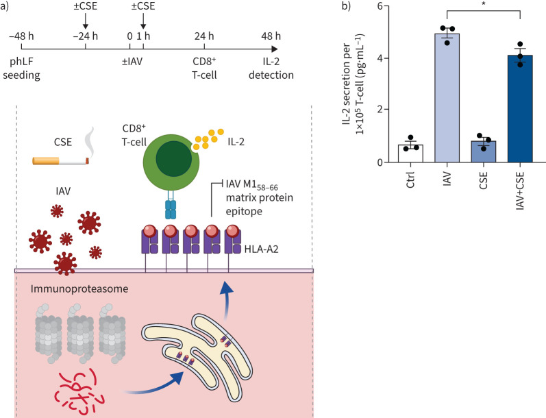
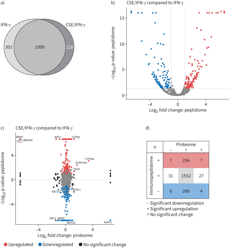
Methods: We analysed the effects of cigarette smoke on cytokine- and virus-mediated induction of the immunoproteasome in vitro, ex vivo and in vivo using RNA and Western blot analyses. CD8+ T-cell activation was determined in co-culture assays with cigarette smoke-exposed influenza A virus (IAV)-infected cells. Mass-spectrometry-based analysis of MHC class I-bound peptides uncovered the effects of cigarette smoke on inflammatory antigen presentation in lung cells. IAV-specific CD8+ T-cell numbers were determined in patients' peripheral blood using tetramer technology.

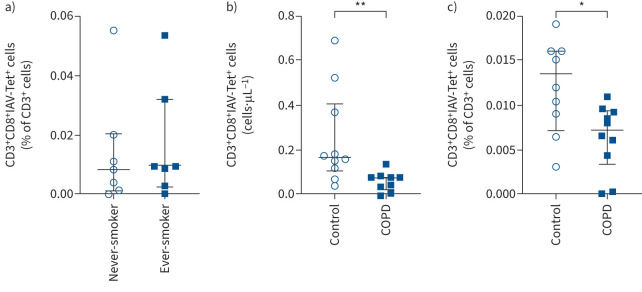
Results: Cigarette smoke impaired the induction of the immunoproteasome by cytokine signalling and viral infection in lung cells in vitro, ex vivo and in vivo. In addition, cigarette smoke altered the peptide repertoire of antigens presented on MHC class I molecules under inflammatory conditions. Importantly, MHC class I-mediated activation of IAV-specific CD8+ T-cells was dampened by cigarette smoke. COPD patients exhibited reduced numbers of circulating IAV-specific CD8+ T-cells compared to healthy controls and asthmatics.

Conclusion: Our data indicate that cigarette smoke interferes with MHC class I antigen generation and presentation and thereby contributes to impaired activation of CD8+ T-cells upon virus infection. This adds important mechanistic insight on how cigarette smoke mediates increased susceptibility of smokers and COPD patients to viral infections.


Conflict of interest statement

Conflict of Interest: H. Ma reports support for the present manuscript from the German Center for Lung Research (DZL). M. Nakayama reports overseas grant from Uehara Memorial Foundation (Japan) and overseas grant from Shiga university of Medical Science, outside the submitted work. A.L. Mora reports support for the present manuscript from NIH (NIH U01 HL1455550-01 and NIH NHLBI R01 HL149825). J.S. Lee reports participation on clinical adjudication committee with Janssen R&D, outside the submitted work. S. Krauss-Etschmann reports support for the present manuscript from the German Center for Lung Research. K. Milger reports consulting fees and lecture honoraria from AstraZeneca, GSK, Janssen, Novartis and Sanofi, outside the submitted work. C.A. Staab-Weijnitz reports support for the present manuscript from Helmholtz Association, German Center for Lung Research (DZL) and Deutsche Forschungsgemeinschaft (DFG) within the Research Training Group GRK2338. K.I. Gaede reports support for the present manuscript from Research Center Borstel – Leibniz Lung Center – BioMaterialBank North, Airway Research Center North, German Center for Lung Research (DZL), PopGen 2.0 Network (P2N). K.I. Gaede also holds a leadership role as member of the Board of Directors of the TMF (www.tmf-ev.de), outside the submitted work. I.E. Kammerl reports support for the present manuscript from ERS (Short Term Fellowship). All other authors have no potential conflicts of interest to declare.

Figures

None
Main findings of the study. Cigarette smoke impairs virus-induced upregulation of the major histocompatibility complex (MHC) class I antigen presentation machinery resulting in reduced activation of antiviral CD8+ T-cells.
FIGURE 1
FIGURE 1
Cigarette smoke impairs interferon (IFN)-γ and virus-induced upregulation of the immunoproteasome in vitro and ex vivo. a) Immunoproteasome subunits low molecular weight protein (LMP)2 and LMP7 expression in MLE-12 treated with 10% or 25% cigarette smoke extract (CSE) and 75 IU·mL−1 IFN-γ for 24 h. Densitometric analysis of LMP2 and LMP7 expression normalised to β-actin with control set to 1 in IFN-γ-treated cells. b) LMP2 and LMP7 expression in MLE-12 treated with 10% or 25% CSE for 24 h and then electroporated with 1 or 5 μg·mL−1 PolyI:C for 24 h. Densitometric analysis of LMP2 and LMP7 expression normalised to β-actin with PolyI:C only group set to 1. c) LMP2 and LMP7 expression in MLE-12 treated with 10% or 25% CSE for 24 h and then infected with murine γ-herpesvirus (MHV)-68 (multiplicity of infection (MOI) 1) for 48 h. Densitometric analysis of LMP2 and LMP7 expression normalised to β-actin with MHV-68 infection group set to 1. d) LMP2 and LMP7 expression in mouse precision-cut lung slices (PCLS) treated with 10% or 25% CSE and co-cultured with 1 μg·mL−1 PolyI:C for 24 h. Densitometric analysis of LMP2 and LMP7 expression normalised to β-actin with PolyI:C only group set to 1. Data are presented as mean±sem, one-sample t-test; *: p<0.05, **: p<0.01.
FIGURE 2
FIGURE 2
Cigarette smoke impairs PolyI:C and virus-induced upregulation of the immunoproteasome in vivo. a) Low molecular-weight protein (LMP)2 and LMP7 expression in mouse lungs 24 h after treatment with 10 μg PolyI:C following 24 days of cigarette smoke (CS) exposure. Densitometric analysis of LMP2 and LMP7 expression normalised to β-actin loading with mean of controls (Ctrl) without PolyI:C treatment set to 1 (median, interquartile range). b) LMP2 and LMP7 expression in mouse lungs after 28 days of cigarette exposure and subsequent infection with murine γ-herpesvirus (MHV)-68 for 7 days. Densitometric analysis of LMP2 and LMP7 expression normalised to β-actin with air mock control set to 1 (median, interquartile range). Kruskal–Wallis test with Dunn's post-test; *: p<0.05, **: p<0.01, ****: p<0.0001.
FIGURE 3
FIGURE 3
Cigarette smoke impairs virus-mediated induction of the immunoproteasome and major histocompatibility complex (MHC) class I in human epithelial cells. a) Low molecular weight protein (LMP)2 and LMP7 expression in primary human bronchial epithelial cells (pBAECs) from three different donors that had been cultured at air–liquid interface conditions with or without cigarette smoke extract (CSE) in their medium [30] and infected with influenza A virus (IAV) for 3 days [60]. Densitometric analysis of LMP2 and LMP7 expression normalised to β-actin with untreated group set to 1. b) pBAECs from two male healthy donors were infected with human rhinovirus (HRV)-16 for 2 days after exposure to two cigarettes per day for 14 days during the air–liquid interface differentiation phase. mRNA levels for PSMB8 (LMP7), PSMB9 (LMP2) and PSMB10 (multicatalytic endopeptidase complex subunit (MECL)-1) and IFNB1 (interferon-β) were determined using reverse transcriptase quantitative PCR and related to housekeeping gene expression (HPRT and RPL32). Data are shown as four replicates per donor (mix of two technical and two independent cultures) with mean±sem. c) Human leukocyte antigen (HLA)-ABC surface expression of A549 cells treated with 20% CSE for 24 h and infected with IAV (multiplicity of infection 1) for 24 h. Fluorescence intensity is shown as mean±sem of three independent experiments. Ctrl: control; Iso: isotype; FITC: fluorescein isothiocyanate; ΔMFI: change in mean fluorescence intensity. One-way ANOVA with Bonferroni post-test. *: p<0.05, **: p<0.01, ***: p<0.001.
FIGURE 4
FIGURE 4
Cigarette smoke impairs activation of influenza A virus (IAV)-specific CD8+ T-cells. a) Schematic depiction of the IAV antigen presentation assay: immunoproteasome-dependent processing of the IAV M1 matrix protein generates the M158–66 peptide, which is loaded onto human leukocyte antigen (HLA)-A2 major histocompatibility complex (MHC) I molecules in the endoplasmic reticulum. HLA-A2/M1-peptide complexes are transported to the cell surface where they activate the specific T-cell hybridoma cell line 4VA1 to secrete interleukin (IL)-2, which can be detected by ELISA. b) IAV-specific CD8+ T-cell activation upon co-culture with IAV-infected HLA-A2+ primary human lung fibroblasts. Primary human lung fibroblasts (phLFs) were treated with 10% cigarette smoke extract (CSE) in 1% fetal bovine serum for 24 h, infected with IAV (multiplicity of infection 1) for 1 h and further cultured for 24 h with or without CSE to be then co-cultured with the CD8+ ifluenza-M1 protein specific T-cells at the ratio of 1:2 for 24 h. IL-2 secreted by the mouse-derived T-cell clone was quantified from the supernatant (mean±sem). Ctrl: control. Significance was analysed with one-way ANOVA with Bonferroni post-test. *: p<0.05.
FIGURE 5
FIGURE 5
Effect of cigarette smoke extract (CSE) on the inflammatory major histocompatibility complex (MHC) class I immunopeptidome. a) Venn diagram of overlapping and unique MHC class I peptides identified in the immunopeptidome of A549 cells (binding rank ≤2%, confidence score ≥4.2, n=3). Cells had either been stimulated with 75 U·mL−1 interferon (IFN)-γ for 24 h or pre-treated with 20% CSE for 48 h and then co-stimulated with 75 U·mL−1 IFN-γ for the past 24 h. b) Volcano plot of the identified MHC class I peptides from a). Significantly upregulated peptides from CSE and IFN-γ-treated cells compared to IFN-γ treatment alone are depicted in red, significantly downregulated peptides are depicted in blue. A dashed horizontal line indicates the significance threshold of 0.05. The two dashed vertical lines indicate the log2 abundance ratio thresholds of −1 and 1. c) Abundance comparison of peptides identified in the immunopeptidome and proteins detected in the proteome from CSE and IFN-γ-treated cells compared to IFN-γ treatment alone. Significantly upregulated MHC class I peptides are depicted in red, while significantly downregulated MHC class I peptides are blue. Proteins significantly regulated in the proteome without significant changes in the peptidome are depicted in black. Peptides significantly regulated in the proteome and the immunopeptidome are labelled with their gene symbol. d) Enumeration of significantly regulated peptides from c).
FIGURE 6
FIGURE 6
Determination of influenza A virus (IAV)-specific CD8+ T-cells in blood of lung-healthy controls and COPD patients. a) Percentage of CD3+CD8+IAV-tetramer(Tet)+ T-cells within the fraction of all CD3+ cells in peripheral blood mononuclear cells (PBMCs) of lung-healthy controls who are never-smokers (n=7, including one Shisha smoker) compared with ex-smokers or current smokers (n=7). b) Absolute numbers of CD3+CD8+IAV-tetramer+ T-cells (cells·μL−1) in isolated PBMCs of COPD patients (n=9) and age-matched lung-healthy controls (n=10). Clinical characteristics can be found in table 1. c) Percentage of CD3+CD8+IAV-tetramer+ T-cells within the fraction of all CD3+ cells in PBMCs of lung healthy controls (n=9) and COPD patients (n=10). Data are presented as median (interquartile range); significance was tested using Mann–Whitney U-test. *: p<0.05, **: p<0.01.

Comment in

References

    1. Decramer M, Janssens W, Miravitlles M. Chronic obstructive pulmonary disease. Lancet 2012; 379: 1341–1351. doi:10.1016/S0140-6736(11)60968-9 - DOI - PMC - PubMed
    1. Soriano JB, Kendrick PJ, Paulson KR, et al. . Prevalence and attributable health burden of chronic respiratory diseases, 1990–2017: a systematic analysis for the Global Burden of Disease Study 2017. Lancet Respir Med 2020; 8: 585–596. doi:10.1016/S2213-2600(20)30105-3 - DOI - PMC - PubMed
    1. Guo-Parke H, Linden D, Weldon S, et al. . Mechanisms of virus-induced airway immunity dysfunction in the pathogenesis of COPD disease, progression, and exacerbation. Front Immunol 2020; 11: 1205. doi:10.3389/fimmu.2020.01205 - DOI - PMC - PubMed
    1. Seemungal T, Harper-Owen R, Bhowmik A, et al. . Respiratory viruses, symptoms, and inflammatory markers in acute exacerbations and stable chronic obstructive pulmonary disease. Am J Respir Crit Care Med 2001; 164: 1618–1623. doi:10.1164/ajrccm.164.9.2105011 - DOI - PubMed
    1. Ritchie AI, Wedzicha JA. Definition, causes, pathogenesis, and consequences of chronic obstructive pulmonary disease exacerbations. Clin Chest Med 2020; 41: 421–438. doi:10.1016/j.ccm.2020.06.007 - DOI - PMC - PubMed

Publication types