. 2023 Jun 29;9(1):198.
doi: 10.1038/s41420-023-01509-8.

Down-regulation of peptidylarginine deiminase type 1 in reconstructed human epidermis disturbs nucleophagy in the granular layer and affects barrier function

Affiliations

Down-regulation of peptidylarginine deiminase type 1 in reconstructed human epidermis disturbs nucleophagy in the granular layer and affects barrier function

Adebayo Candide Alioli et al. Cell Death Discov. .

Abstract

Deimination is a post-translational modification catalyzed by a family of enzymes named peptidylarginine deiminases (PADs). PADs transform arginine residues of protein substrates into citrulline. Deimination has been associated with numerous physiological and pathological processes. In human skin, three PADs are expressed (PAD1-3). While PAD3 is important for hair shape formation, the role of PAD1 is less clear. To decipher the main role(s) of PAD1 in epidermal differentiation, its expression was down-regulated using lentivirus-mediated shRNA interference in primary keratinocytes and in three-dimensional reconstructed human epidermis (RHE). Compared to normal RHEs, down-regulation of PAD1 caused a drastic reduction in deiminated proteins. Whereas proliferation of keratinocytes was not affected, their differentiation was disturbed at molecular, cellular and functional levels. The number of corneocyte layers was significantly reduced, expression of filaggrin and cornified cell envelope components, such as loricrin and transglutaminases, was down-regulated, epidermal permeability increased and trans-epidermal-electric resistance diminished drastically. Keratohyalin granule density decreased and nucleophagy in the granular layer was disturbed. These results demonstrate that PAD1 is the main regulator of protein deimination in RHE. Its deficiency alters epidermal homeostasis, affecting the differentiation of keratinocytes, especially the cornification process, a special kind of programmed cell death.


Conflict of interest statement

The authors declare no competing interests. The funders had no role in the study design, data collection and analysis, decision to publish, or preparation of the manuscript.

Figures

Fig. 1
Fig. 1. Efficiency and specificity of PAD1 down-regulation.
A RT-qPCR of RHEs transduced by sh-ctrl, shPADI1_1, and shPADI1_2. Relative mRNA steady states of PADI1 and PADI3 are reported, other PADI mRNA were undetectable (Cycle threshold, Ct > 35). BD Expression of PAD1, deiminated proteins, and actin. B Western blot analysis of total extract RHEs transduced by sh-ctrl, shPADI1_1, and shPADI1_2. C PAD1 immunofluorescence of RHE sections after transduction by sh-ctrl, shPADI1_1, and shPADI1_2. Representation using a LUT scale reported to highlight the down-regulation of PAD1 in shPADI1_1 and shPADI1_2 compared to sh-ctrl RHEs. D sh-ctrl, shPADI1_1, and shPADI1_2 RHEs were analyzed by indirect immunofluorescence to localize deiminated proteins (AMC staining) in situ. The line at the bottom represents the polycarbonate filter. SC Stratum corneum, LL living cell layers. Scale bars, 10 µm.
Fig. 2
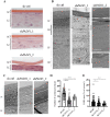
Fig. 2. Tissue morphology and ultrastructural aspects of PAD1 down-regulated RHEs.
A Hematoxylin and eosin staining of sh-ctrl, shPADI1_1, and shPADI1_2 RHEs. B Left to right: full vertical TEM sections of sh-ctrl, shPADI1_1, and shPADI1_2 RHEs. B Right bottom, white arrow: parakeratotic nucleus in shPADI1 RHEs. C Stratum corneum of sh-ctrl, shPADI1_1, and shPADI1_2 RHEs. D Number of corneocyte layers of RHEs (mean ± SD) for sh-ctrl and shPADI1_1, n = 5; shPADI1_2, n = 2. E KHG area of RHEs. (sh-ctrl and shPADI1_1, n = 5; shPADI1_2, n = 2). A, B Black lines: LL living cell layers, SC Stratum corneum, SG S. granulosum (bold line), SS S. spinosum, SB S. basale. Scale bars = 10 µm (A), 5 µm (B), 1 µm (C).
Fig. 3
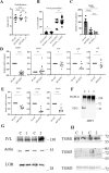
Fig. 3. Effect of PAD1 down-regulation on proliferation, differentiation, and barrier function.
A Proliferation capacity of transduced NHEK by sh-ctrl (n = 11), shPADI1_1 (n = 11), and shPADI1_2 (n = 4). B LY permeability of sh-ctrl and shPADI1_1 RHEs (at 6 h, *p = 0.0317; at 24 h, *p = 0.0163; n = 7, RHEs; 2 banks). C Relative percentage of TEER for sh-ctrl, shPADI1_1, and shPADI1_2 RHEs (n = 6). D, E RT-qPCR analysis for D PADI1, PADI3, (pro)filaggrin (FLG), loricrin (LOR), involucrin (IVL) and for E transglutaminase (TGM) 1, 3 and 5 in sh-ctrl and shPADI1 RHEs. Data were pooled for statistical analysis: shPADI1_1 n = 7 to 13 and shPADI1_2 n = 2 from 5 banks. FH Immunodetections of total protein extracts of sh-ctrl (C), shPADI1_1 (1), and shPADI1_2 (2) RHEs as indicated. Ladder sizes indicated on the right (kDa).
Fig. 4
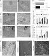
Fig. 4. Effect of PAD1 down-regulation on nuclear shape.
AC Representative illustrations of nuclear shape for sh-ctrl, shPADI1_1, and shPADI1_2 RHEs, respectively. B Deep nuclear deformations (or invaginations) indicated by black arrows. D Nuclear deformation scoring of sh-ctrl (109 nucleus) and shPADI1 (96 nucleus) RHEs, NHEK banks, n = 5; 7 independent experiments, n = 6 for sh-ctrl, shPADI1_1 and 1 for shPADI1_2. E Distribution of nuclear deformation scores for sh-ctrl (gray) and shPADI1_1 (black) RHEs. F Distribution of nuclear deformation scores for sh-ctrl (gray) and shPADI1_2 (black) RHEs. G, H Representative perinuclear vesicles (small circular white area) indicated by white arrows. Also in (B) and (C). I Representative images of perinuclear vesicles (white arrows) and deep invaginations (black arrows) of the nuclear envelope in RHE treated with 400 µM Cl-amidine (ClA-400). Scale bars, 2 µm. N, nucleus.

Similar articles

Cited by

  • Deimination in epidermal barrier and hair formation.
    Méchin MC, Simon M. Méchin MC, et al. Philos Trans R Soc Lond B Biol Sci. 2023 Nov 20;378(1890):20220245. doi: 10.1098/rstb.2022.0245. Epub 2023 Oct 2. Philos Trans R Soc Lond B Biol Sci. 2023. PMID: 37778378 Free PMC article. Review.
  • Neutrophils only die twice.
    Souza FW, Miao EA. Souza FW, et al. Sci Adv. 2023 Dec 22;9(51):eadm8715. doi: 10.1126/sciadv.adm8715. Epub 2023 Dec 20. Sci Adv. 2023. PMID: 38117906 Free PMC article. Review.

References

    1. Chavanas S, Méchin M-C, Takahara H, Kawada A, Nachat R, Serre G, et al. Comparative analysis of the mouse and human peptidylarginine deiminase gene clusters reveals highly conserved non-coding segments and a new human gene, PADI6. Gene. 2004;330:19–27. doi: 10.1016/j.gene.2003.12.038. - DOI - PubMed
    1. Minato T, Unno M, Kitano T. Evolution of S100A3 and PAD3, two important genes for mammalian hair. Gene. 2019;713:143975. doi: 10.1016/j.gene.2019.143975. - DOI - PubMed
    1. Méchin M-C, Sebbag M, Arnaud J, Nachat R, Foulquier C, Adoue V, et al. Update on peptidylarginine deiminases and deimination in skin physiology and severe human diseases. Int J Cosmet Sci. 2007;29:147–68. doi: 10.1111/j.1467-2494.2007.00377.x. - DOI - PubMed
    1. Méchin MC, Takahara H, Simon M. Deimination and peptidylarginine deiminases in skin physiology and diseases. Int J Mol. Sci. 2020;21:566. doi: 10.3390/ijms21020566. - DOI - PMC - PubMed
    1. Briot J, Simon M, Méchin MC. Deimination, intermediate filaments and associated proteins. Int J Mol Sci. 2020;21:8746. doi: 10.3390/ijms21228746. - DOI - PMC - PubMed