. 2023 Jun 29;13(1):10524.
doi: 10.1038/s41598-023-36629-9.

Immunomodulatory response in an experimental model of brain death

Affiliations

Immunomodulatory response in an experimental model of brain death

Alexandre Chagas Santana et al. Sci Rep. .

Abstract

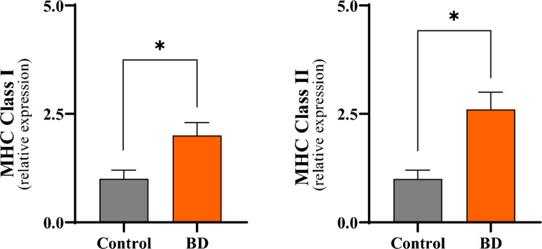
Liver transplantation has come a long way and is now regarded as the gold standard treatment for end-stage liver failure. The great majority of livers utilized in transplantation come from brain-dead donors. A broad inflammatory response characterizes BD, resulting in multiorgan damage. This process is primarily mediated by cytokines, which increase the immunogenicity of the graft. In male Lewis rats, we evaluated the immune response in a BD liver donor and compared it to that of a control group. We studied two groups: Control and BD (rats subjected to BD by increasing intracranial pressure). After the induction of BD, there was an intense rise in blood pressure followed by a fall. There were no significant differences observed between the groups. Blood tissue and hepatic tissue analyzes showed an increase in plasma concentrations of liver enzymes (AST, ALT, LDH and ALP), in addition to pro-inflammatory cytokines and macrophages in liver tissue in animals submitted to BD. The current study found that BD is a multifaceted process that elicits both a systemic immune response and a local inflammatory response in liver tissue. Our findings strongly suggested that the immunogenicity of plasma and liver increased with time following BD.


Conflict of interest statement

The authors declare no competing interests.

Figures

Figure 1
Figure 1
Analysis of cerebral blood flow identified by transcranial color ultrasound. (A) Presence of blood flow in the reverse direction (yellow arrow) before brain death. (B) Presence of flow compatible with brain circulatory collapse (yellow arrow).
Figure 2
Figure 2
Comparative analyzes of the evolution of MBP between the groups during a period of 6 h after the induction of brain death (MBP mean blood pressure).
Figure 3
Figure 3
Immunohistochemistry for detection of M1 macrophages (arrows) in liver tissue 6 h after induction of brain death. (A) Control; (B) Brain death; 400X magnification.
Figure 4
Figure 4
Quantification of M1 macrophage infiltrate 6 h after brain death induction in different experimental groups. *p < 0.05 vs. Control; (BD brain death).
Figure 5
Figure 5
Comparative analyzes of gene expression and concentration of inflammatory meters in liver tissue 6 h after brain death in the different groups. *p < 0.05 vs. Control; #p < 0.05 vs. BD (BD brain death).
Figure 6
Figure 6
Comparative analyzes of Class I MHC and Class II MHC expression in liver tissue 6 h after brain death in the different experimental groups. *p < 0.05 vs. Control; #p < 0.05 vs. BD (BD brain death, MHC major histocompatibility complex).
Figure 7
Figure 7
Comparative analyze s of NFB gene expression in liver tissue 6 h after brain death in different groups. *p < 0.05 vs. Control; #p < 0.05 vs. BD. (BD brain death).
Figure 8
Figure 8
Analysis of the densiometric quantification (arbitrary units) of the NF-αB band in the liver tissue in the different groups. *p < 0.05 vs. Control; #p < 0.05 vs. BD (BD brain death).
Figure 9
Figure 9
Illustrative image of the Super-Shift assay 6 h after brain death induction (BD group), indicating that the dimers present in liver tissue are composed of the p50, p65 and c-Rel subunits (BD brain death).

References

    1. Barker CF, Markmann JF. Historical overview of transplantation. Cold Spring Harb. Perspect. Med. 2013;3:a014977. doi: 10.1101/cshperspect.a014977. - DOI - PMC - PubMed
    1. Pratschke J, Neuhaus P, Tullius SG. What can be learned from brain-death models? Transpl. Int. 2005;18:15–21. doi: 10.1111/j.1432-2277.2004.00018.x. - DOI - PubMed
    1. Floerchinger B, Oberhuber R, Tullius SG. Effects of brain death on organ quality and transplant outcome. Transplant. Rev. 2012;26:54–59. doi: 10.1016/j.trre.2011.10.001. - DOI - PubMed
    1. Danobeitia JS. The brain-dead organ donor: A missed opportunity for early intervention. J. Transplant. Technol. Res. 2012;2:3. doi: 10.4172/2161-0991.1000e117. - DOI
    1. Van der Hoeven JA, et al. Donor brain death reduces survival after transplantation in rat livers preserved for 20 hr. Transplantation. 2001;72:1632–1636. doi: 10.1097/00007890-200111270-00009. - DOI - PubMed