. 2023 Jun 29;14(1):3845.
doi: 10.1038/s41467-023-39575-2.

Immunological imprinting of humoral immunity to SARS-CoV-2 in children

Affiliations

Immunological imprinting of humoral immunity to SARS-CoV-2 in children

Alexander C Dowell et al. Nat Commun. .

Abstract

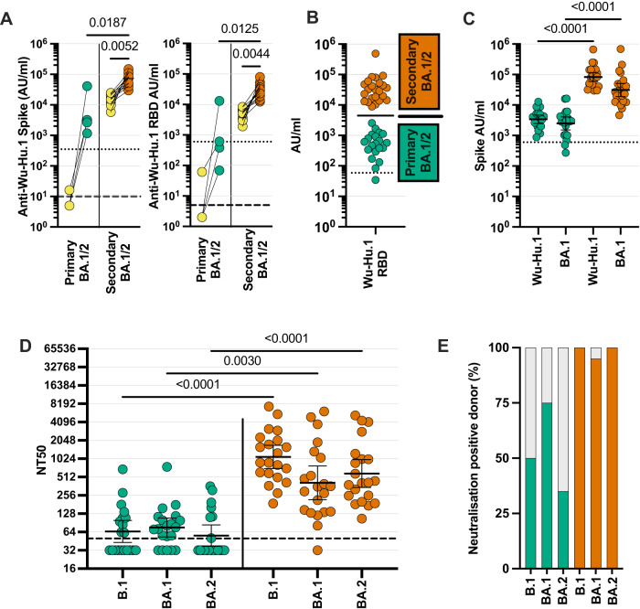
Omicron variants of SARS-CoV-2 are globally dominant and infection rates are very high in children. We measure immune responses following Omicron BA.1/2 infection in children aged 6-14 years and relate this to prior and subsequent SARS-CoV-2 infection or vaccination. Primary Omicron infection elicits a weak antibody response with poor functional neutralizing antibodies. Subsequent Omicron reinfection or COVID-19 vaccination elicits increased antibody titres with broad neutralisation of Omicron subvariants. Prior pre-Omicron SARS-CoV-2 virus infection or vaccination primes for robust antibody responses following Omicron infection but these remain primarily focussed against ancestral variants. Primary Omicron infection thus elicits a weak antibody response in children which is boosted after reinfection or vaccination. Cellular responses are robust and broadly equivalent in all groups, providing protection against severe disease irrespective of SARS-CoV-2 variant. Immunological imprinting is likely to act as an important determinant of long-term humoral immunity, the future clinical importance of which is unknown.


Conflict of interest statement

The authors declare no competing interests.

Figures

Fig. 1
Fig. 1. Low titre antibodies and neutralisation develop following primary Omicron infection.
SARS-CoV-2-specific antibody responses were determined in children with recent Omicron BA.1/2 infection. A Wuhan-Hu-1 (Wu-Hu-1) spike antibody binding in samples from children who were SARS-CoV-2 seronegative (n = 4) or seropositive (n = 11) prior to Omicron infection (yellow). Dotted lines indicate seropositive cut-off for Wuhan-Hu-1-specific antibody response; dashed lines indicate below the limit of detection. Data were presented as arbitrary units (AU)/ml. One-way Friedman Test with Dunn’s multiple comparisons test. B Wuhan-Hu-1 RBD-specific antibody binding in the total cohort of 43 children with recent Omicron BA.1/2 infection shows a bimodal distribution of primary (green; n = 20) or secondary (orange; n = 23) SARS-CoV-2 infection status. Data were presented as arbitrary units (AU)/ml. Bar indicates geometric mean; representative of primary or secondary Omicron infection. C Wuhan-Hu-1 and Omicron BA.1 spike-specific antibody binding in children with primary (green; n = 20) or secondary (orange; n = 23) Omicron BA.1/2 infection. The dotted line indicates seropositive cut-offs for Wuhan-Hu-1. Data were presented as arbitrary units (AU)/ml. One-way Kruskal–Wallis test with Dunn’s multiple comparisons test, bars indicate geometric mean ±95% CI. D Neutralisation of pseudovirus-bearing B.1 (Wuhan-Hu-1 and D614G) ancestral or Omicron BA.1 or BA.2 sequence spike protein in children following primary (green; n = 20) or secondary (orange; n = 21) Omicron infection. Dashed lines indicate the limit of detection. Data were presented as NT50, the titre at which infectivity had been reduced by 50% relative to controls. One-way Kruskal–Wallis test with Dunn’s multiple comparisons test, bars indicate geometric mean ±95% CI. E Bar graph indicating the percentage of children with detectable neutralising titres to B.1 or Omicron BA.1 or BA.2 following primary (green, n = 20) or secondary (orange, n = 21) Omicron infection. Source data are provided as a Source Data file.
Fig. 2
Fig. 2. Secondary antigenic challenge enhances antibody levels and neutralisation following primary Omicron infection.
A Antibody levels specific for spike from Wuhan-Hu-1 and Omicron BA.1, BA.2, BA.4, and BA.5 were determined in children following primary BA.1/2 Omicron infection (green; n = 19), Omicron reinfection (purple; n = 13) and COVID-19 vaccination (blue; n = 11) after primary BA.1/2 infection. B Neutralisation of pseudovirus-bearing ancestral B.1 or Omicron variant spike protein in children following primary Omicron BA.1/2 infection (green; n = 19), Omicron BA.4/5 reinfection (purple; n = 13) or following COVID-19 vaccination after primary BA.1/2 infection (blue; n = 11). Data were presented as NT50, the titre at which infectivity had been reduced by 50% relative to controls. Dashed lines indicate the limit of detection. Kruskal–Wallis test with Dunn’s multiple comparisons test, bars indicate geometric mean ±95% CI. Data were presented as arbitrary units (AU)/ml. Source data are provided as a Source Data file.
Fig. 3
Fig. 3. COVID-19 vaccine induces strong antibody responses that are not enhanced following breakthrough infection.
A Antibody levels specific for spike from Wuhan-Hu-1, and Omicron BA.1, BA.2, BA.4, and BA.5 were determined in children following vaccination (pink, n = 15) or BA.1/2 Omicron breakthrough infection (black, n = 6). Data were presented as arbitrary units (AU)/ml. B Neutralisation of pseudovirus-bearing ancestral B.1 or Omicron variant spike protein in children following vaccination (pink, n = 15) or BA.1/2 Omicron breakthrough infection (black, n = 6). Dashed lines indicate the limit of detection, bars indicate the geometric mean. Data were presented as NT50, the titre at which infectivity had been reduced by 50% relative to controls. Source data are provided as a Source Data file.
Fig. 4
Fig. 4. Relative antibody binding to Omicron or ancestral spike protein in relation to primary virus or vaccine challenge.
A Antibody binding against ancestral Wuhan-Hu-1 or Omicron BA.1 spike in children following three different forms of initial primary antigenic challenge. (i) Initial primary Omicron infection (green; n = 19) and subsequent Omicron reinfection (purple; n = 13) or vaccination (blue; n = 11); (ii) initial primary pre-Omicron infection (yellow; n = 54), followed by secondary Omicron BA.1/2 infection (orange; n = 23); iii) primary vaccination (pink; n = 15) followed by breakthrough Omicron infection (black; n = 6). Inset percentages indicate the geometric mean BA.1 antibody level in respect to the Wuhan-Hu-1-specific antibody level. Data were presented as arbitrary units (AU)/ml. Two-tailed paired Wilcoxon test, bars indicate geometric mean ±95% CI. B Ratio of antibody binding against Omicron or ancestral Wuhan-Hu-1 spike (Omicron/ancestral) in children following initial primary Omicron or pre-Omicron infection, or vaccination as (A). Kruskal–Wallis test with Dunn’s multiple comparisons test, bars indicate median ±95% CI. Source data are provided as a Source Data file.
Fig. 5
Fig. 5. SARS-CoV-2-specific T cell responses are induced reliably following primary Omicron infection and not boosted further by reinfection or vaccination.
IFNγ ELISpot was used to assess cellular response to ancestral and Omicron sequence spike peptide pools. Peptides from Influenza (Flu) were included as a control. Children with primary Omicron infection (A, green, n = 17), Omicron secondary infection (B, orange, n = 10), pre-Omicron infection sampled prior to the Omicron wave (C, yellow, n = 5), or vaccinated children (D, pink, n = 9). Negative control samples including pre-pandemic paediatric blood samples and DMSO alone incubation were used to define the background level of 32 sfc/106 PBMC (16), dotted line. The dashed line indicates the limit of detection. Results are presented as spot-forming cells (sfc) per 106 PBMC. Source data are provided as a Source Data file.
Fig. 6
Fig. 6. Relative magnitude of omicron-specific antibody response following single or dual SARS-CoV-2 infection or vaccination.
Values are given in relation to the magnitude following primary Omicron infection. Values for vaccination show response after one or two vaccine doses.

References

    1. Tang J, et al. Cross-reactive immunity against the SARS-CoV-2 Omicron variant is low in pediatric patients with prior COVID-19 or MIS-C. Nat. Commun. 2022;13:2979. doi: 10.1038/s41467-022-30649-1. - DOI - PMC - PubMed
    1. Sievers BL, et al. Antibodies elicited by SARS-CoV-2 infection or mRNA vaccines have reduced neutralizing activity against Beta and Omicron pseudoviruses. Sci. Transl. Med. 2022;14:eabn7842. doi: 10.1126/scitranslmed.abn7842. - DOI - PMC - PubMed
    1. Koelle K, Martin MA, Antia R, Lopman B, Dean NE. The changing epidemiology of SARS-CoV-2. Science. 2022;375:1116–1121. doi: 10.1126/science.abm4915. - DOI - PMC - PubMed
    1. Elliott P, et al. Twin peaks: the Omicron SARS-CoV-2 BA.1 and BA.2 epidemics in England. Science. 2022;376:eabq4411. doi: 10.1126/science.abq4411. - DOI - PMC - PubMed
    1. Elliott P, et al. Rapid increase in Omicron infections in England during December 2021: REACT-1 study. Science. 2022;375:1406–1411. doi: 10.1126/science.abn8347. - DOI - PMC - PubMed

Publication types

Substances

Supplementary concepts