

A new perspective during laryngo-tracheal surgery: the use of an ultra-thin endotracheal tube (Tritube®) and flow-controlled ventilation-a retrospective case series and a review of the literature
- PMID: 37386531
- PMCID: PMC9411832
- DOI: 10.1186/s44158-022-00066-3
A new perspective during laryngo-tracheal surgery: the use of an ultra-thin endotracheal tube (Tritube®) and flow-controlled ventilation-a retrospective case series and a review of the literature
Abstract
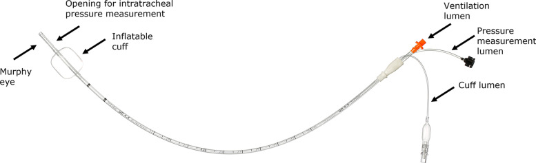
Background: Upper airway surgery often poses a challenge to both anesthesiologists and surgeons, as airway access, mechanical ventilation, and surgical difficulties may occur in a tricky combination. To fulfill the need for a tubeless surgery, techniques such as apneic oxygenation or jet ventilation may be used, which carry the risk of several complications. The ultrathin cuffed endotracheal tube Tritube can be used with flow-controlled ventilation (FCV) to provide adequate surgical field and ventilation. To assess the feasibility, safety, and effectiveness of this technique, we describe a series of 21 patients, with various lung conditions, undergoing laryngo-tracheal surgery with FCV delivered via Tritube. Moreover, we perform a narrative systematic review to summarize clinical data on the use of Tritube during upper airway surgery.
Results: All patients were successfully intubated in one attempt with Tritube. The median (interquartile range [IQR]) tidal volume was 6.7 (6.2-7.1) mL/kg of ideal body weight, the median end-expiratory pressure was 5.3 (5.0-6.4) cmH2O, and the median peak tracheal pressure was 16 (15-18) cmH2O. The median minute volume was 5.3 (5.0-6.4) L/min. Median global alveolar driving pressure was 8 (7-9) cmH2O. The median maximum level of end-tidal CO2 was 39 (35-41) mmHg. During procedures involving laser, the maximum fraction of inspired oxygen was 0.3, with the median lowest peripheral oxygen saturation of 96% (94-96%). No complications associated with intubation or extubation occurred. In one patient, the ventilator needed to be rebooted for a software issue. In two (10%) patients, Tritube needed to be flushed with saline to remove secretions. In all patients, optimal visualization and accessibility of the surgical site were obtained, according to the surgeon in charge. Thirteen studies (seven case reports, two case series, three prospective observational studies, and one randomized controlled trial) were included in the narrative systematic review and described.
Conclusions: Tritube in combination with FCV provided adequate surgical exposure and ventilation in patients undergoing laryngo-tracheal surgery. While training and experience with this new method is needed, FCV delivered with Tritube may represent an ideal approach that benefits surgeons, anesthesiologists, and patients with difficult airways and compromised lung mechanics.
Keywords: Airway management; FCV; Flow controlled ventilation; Laryngeal surgery; Optimized ventilation; Tritube.
© 2022. The Author(s).
Conflict of interest statement
The authors declare that they have no competing interests.
Figures


References
LinkOut - more resources
Full Text Sources