. 2023 Jun 30;21(1):427.
doi: 10.1186/s12967-023-04270-9.

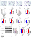
Inhibition of DRP1-dependent mitochondrial fission by Mdivi-1 alleviates atherosclerosis through the modulation of M1 polarization

Affiliations

Inhibition of DRP1-dependent mitochondrial fission by Mdivi-1 alleviates atherosclerosis through the modulation of M1 polarization

Ze-da-Zhong Su et al. J Transl Med. .

Abstract

Background: Inflammation and immune dysfunction with classically activated macrophages(M1) infiltration are important mechanisms in the progression of atherosclerosis (AS). Dynamin-related protein 1 (DRP1)-dependent mitochondrial fission is a novel target for alleviating inflammatory diseases. This study aimed to investigate the effects of DRP1 inhibitor Mdivi-1 on AS.

Methods: ApoE-/- mice were fed with a high-fat diet supplemented with or without Mdivi-1. RAW264.7 cells were stimulated by ox-LDL, pretreated with or without MCC950, Mito-TEMPO, or Mdivi-1. The burden of plaques and foam cell formation were determined using ORO staining. The blood lipid profles and inflammatory cytokines in serum were detected by commercial kits and ELISA, respectively. The mRNA expression of macrophage polarization markers, activation of NLRP3 and the phosphorylation state of DRP1 were detected. Mitochondrial reactive oxygen species (mito-ROS), mitochondrial staining, ATP level and mitochondrial membrane potential were detected by mito-SOX, MitoTracker, ATP determination kit and JC-1 staining, respectively.

Results: In vivo, Mdivi-1 reduced the plaque areas, M1 polarization, NLRP3 activation and DRP1 phosphorylation at Ser616. In vitro, oxidized low-density lipoprotein (ox-LDL) triggered M1 polarization, NLRP3 activation and abnormal accumulation of mito-ROS. MCC950 and Mito-TEMPO suppressed M1 polarization mediated foam cell formation. Mito-TEMPO significantly inhibited NLRP3 activation. In addition, Mdivi-1 reduced foam cells by inhibiting M1 polarization. The possible mechanisms responsible for the anti-atherosclerotic effects of Mdivi-1 on reducing M1 polarization were associated with suppressing mito-ROS/NLRP3 pathway by inhibiting DRP1 mediated mitochondrial fission. In vitro, similar results were observed by DRP1 knockdown.

Conclusion: Inhibition of DRP1-dependent mitochondrial fission by Mdivi-1 alleviated atherogenesis via suppressing mito-ROS/NLRP3-mediated M1 polarization, indicating DRP1-dependent mitochondrial fission as a potential therapeutic target for AS.

Keywords: Atherosclerosis; DRP1; M1 polarization; Mdivi-1; Mitochondrial fission; NLRP3 inflammasome.


Conflict of interest statement

The authors declare that the research was conducted without any commercial or financial relationships that could be construed as a potential competing interest.

Figures

Fig. 1
Fig. 1
Mdivi-1 protected against atherosclerosis in HFD ApoE−/− mice. The size of the aortic lesion (A, B) and the lipid deposition in the aortic sinus (C, D) determined by ORO staining (scale bar: 200 μm. 40 × magnification), n = 4. (E) Body weight determined by electronic scale, n = 6. The levels of TC (F), LDL-C (G) and HDL-C (H) in serum detected by the corresponding kits, n = 6. * p < 0.05, ** p < 0.01, *** p < 0.001, and ns p > 0.05
Fig. 2
Fig. 2
Mdivi-1 regulated M1 polarization in aortic tissue from HFD ApoE−/− mice. The levels of inflammatory cytokines including IL-6 (A), TNF-α (B), and IL-10 (C) in the serum measured by ELISA, n = 6. (D) Representative immunohistochemical staining showing the expression of CD86 and CD206 in aortic sections (scale bar: 100 μm. 200 × magnification). (E,F) The mean density of CD86 and CD206 was analyzed, n = 3. The M1 markers mRNA, including CD86 (G), iNOS (H), IL-6 (I), TNF-α (J), MCP-1 (K) and M2 markers mRNA, such as CD206 (L), CD163 (M), IL-10 (N) detected by qRT-PCR, n = 3. * p < 0.05, ** p < 0.01, and *** p < 0.001
Fig. 3
Fig. 3
Mdivi-1 reduced phosphorylation of DRP1 (Ser616) and NLRP3 activation in the aortic tissues of HFD ApoE−/− mice. Western blot showing the protein level of phosphorylation-DRP1 (Ser616), total DRP-1, NLRP3, pro-caspase-1, and cleaved-caspase-1 (A). The relative protein expression levels of (B) phosphorylation-DRP1 (Ser616)/β-actin, (C) DRP1/β-actin, (D) NLRP3/β-actin (E) pro-caspase-1/β-actin and (F) cleaved-caspase-1/β-actin in aortic tissues. Three independent replications were performed. ** p < 0.01, *** p < 0.001, and ns p > 0.05
Fig. 4
Fig. 4
ox-LDL induced M1 polarization, NLRP3 activation, and abnormal accumulation of mito-ROS. qRT-PCR showing M1 polarization markers mRNA expression, including CD86 (A) and iNOS (B), and pro-inflammatory factors mRNAs such as IL-6 (C), TNF-α (D), and MCP-1 (E). RT-PCR indicating M2 markers mRNA expression including CD206(F), CD163(G) and IL-10(H). Western blot demonstrating the protein expression of NLRP3, pro-caspase-1, and cleaved-caspase-1 (I). The relative protein expression levels of (J) NLRP3/β-actin, (K) pro-caspase-1/β-actin and (L) cleaved-caspase-1/β-actin in RAW264.7 cells. Using mito-SOX to detect mito-ROS (M,N) (scale bar: 25 μm. 200 × magnification). The SOD activity and level of MDA detected by corresponding kits (O,P). Three independent replications were performed. * p < 0.05, ** p < 0.01, *** p < 0.001, and ns p > 0.05
Fig. 5
Fig. 5
Mito-ROS/NLRP3 pathway mediated M1 polarization and foam cell formation. A, B ORO staining detecting lipid deposition in macrophages (scale bar: 100 μm. 200 × magnification). The mRNA expression of M1 markers, including CD86 (C) and iNOS (D), IL-6 (E), TNF-α (F), MCP-1 (G) and M2 markers, including CD206 (H) and IL-10 (I) detected by qRT-PCR. The protein level of NLRP3, pro-caspase-1, and cleaved-caspase-1 detected by western blot (J). The relative protein expression levels of (K) NLRP3/β-actin, (L) pro-caspase-1/β-actin and (M) cleaved-caspase-1/β-actin in RAW264.7 cells. Three independent replications were performed. * p < 0.05, ** p < 0.01, *** p < 0.001, and ns p > 0.05
Fig. 6
Fig. 6
Mdivi-1 reduced M1 polarization mediated foam cell formation by inhibiting mito-ROS/NLRP3 activation. A, B ORO staining to detect lipid deposition in macrophages (200 × magnification). The mRNA expression of M1 polarization markers, including CD86 (C) and iNOS (D), IL-6 (E), TNF-α (F), MCP-1 (G) and M2 polarization markers, including CD206 (H), CD163(I) and IL-10 (J) detected by qRT-PCR. K, L Mito-ROS was detected by mito-SOX (scale bar: 25 μm. 200 × magnification). M, N The SOD activity and level of MDA detected by corresponding kits. The protein level of NLRP3, pro-caspase-1, and cleaved-caspase-1 detected by western blot (O). The relative protein expression levels of (P) NLRP3/β-actin (Q) pro-caspase-1/β-actin and (R) cleaved-caspase-1/β-actin in RAW264.7 cells. Three independent replications were performed. * p < 0.05, ** p < 0.01, *** p < 0.001, and ns p > 0.05
Fig. 7
Fig. 7
Mitochondrial fission inhibition by Mdivi-1 improved mitochondrial dysfunction caused by ox-LDL. A MitoTracker staining to detect the number of mitochondrial fragments (scale bar: 5 μm. 600 × magnification). B Analysis of the relative number of mitochondria. C, D JC-1 staining to detect mitochondrial membrane potential (scale bar: 50 μm. 200 × magnification). E Detecting ATP level by an ATP determination kit. F The protein level of phosphorylation-DRP1 (Ser616) and total DRP-1 detected by western blot. The relative protein expression levels of (G) DRP1/β-actin, and (H) phosphorylation-DRP1 (Ser616)/β-actin in RAW264.7 cells. (I) Western blot to detect the translocation of DRP1. The relative protein expression levels of (J) DRP1/VDAC1 in the mitochondria fractions and (K) DRP1/β-actin in cytosol fractions. Three independent replications were performed. * p < 0.05, ** p < 0.01, *** p < 0.001, and ns p > 0.05
Fig. 8
Fig. 8
DRP1 knockdown reduced M1 polarization mediated foam cell formation by inhibiting mito-ROS/NLRP3 activation. A The protein level of DRP1 detected by western blot. The relative protein expression levels of (B) DRP1/β-actin in RAW264.7 cells. C, D ORO staining to detect lipid deposition in macrophages (scale bar: 100 μm. 200 × magnification). qRT-PCR to detect the mRNA expression of M1 polarization markers, including (E) iNOS, (F) IL-6 and M2 markers, including (G) CD206, and (H) IL-10. I, J mito-ROS detected by mito-SOX (scale bar: 25 μm. 200 × magnification). (K, L) The SOD activity and level of MDA detected by corresponding kits. M The protein level of NLRP3, pro-caspase-1, and cleaved-caspase-1 detected by western blot. The relative protein expression levels of (N) NLRP3/β-actin (O) pro-caspase-1/β-actin and (P) cleaved-caspase-1/β-actin in RAW264.7 cells. Three independent replications were performed. * p < 0.05, ** p < 0.01, *** p < 0.001, and ns p > 0.05
Fig. 9
Fig. 9
DRP1 knockdown improved mitochondrial fission associated mitochondrial dysfunction caused by ox-LDL. A MitoTracker staining to detect the number of mitochondrial fragments (scale bar: 5 μm. 600 × magnification). B Analysis of the relative number of mitochondria. C, D JC-1 staining to detect mitochondrial membrane potential (scale bar: 50 μm. 200 × magnification). E Detecting ATP level by an ATP determination kit. Three independent replications were performed. ** p < 0.01, *** p < 0.001, and ns p > 0.05
Fig. 10
Fig. 10
Mechanism diagram of the anti-atherosclerotic effect of DRP1-dependent mitochondrial fission inhibition by Mdivi-1

References

    1. Bäck M, Yurdagul A, Jr, Tabas I, Öörni K, Kovanen PT. Inflammation and its resolution in atherosclerosis: mediators and therapeutic opportunities. Nat Rev Cardiol. 2019;16(7):389–406. - PMC - PubMed
    1. Chen W, Schilperoort M, Cao Y, Shi J, Tabas I, Tao W. Macrophage-targeted nanomedicine for the diagnosis and treatment of atherosclerosis. Nat Rev Cardiol. 2022;19(4):228–249. doi: 10.1038/s41569-021-00629-x. - DOI - PMC - PubMed
    1. Colin S, Chinetti-Gbaguidi G, Staels B. Macrophage phenotypes in atherosclerosis. Immunol Rev. 2014;262(1):153–166. doi: 10.1111/imr.12218. - DOI - PubMed
    1. Sica A, Mantovani A. Macrophage plasticity and polarization: in vivo veritas. J Clin Invest. 2012;122(3):787–795. doi: 10.1172/JCI59643. - DOI - PMC - PubMed
    1. Peled M, Fisher EA. Dynamic aspects of macrophage polarization during atherosclerosis progression and regression. Front Immunol. 2014;12(5):579. - PMC - PubMed

Publication types