Case Reports
. 2023 Jun 30;17(1):296.
doi: 10.1186/s13256-023-03993-z.

A rare primary hepatic neuroendocrine tumour with laparoscopic resection: a case report

Affiliations
Case Reports

A rare primary hepatic neuroendocrine tumour with laparoscopic resection: a case report

Hanan M Alghamdi. J Med Case Rep. .

Abstract

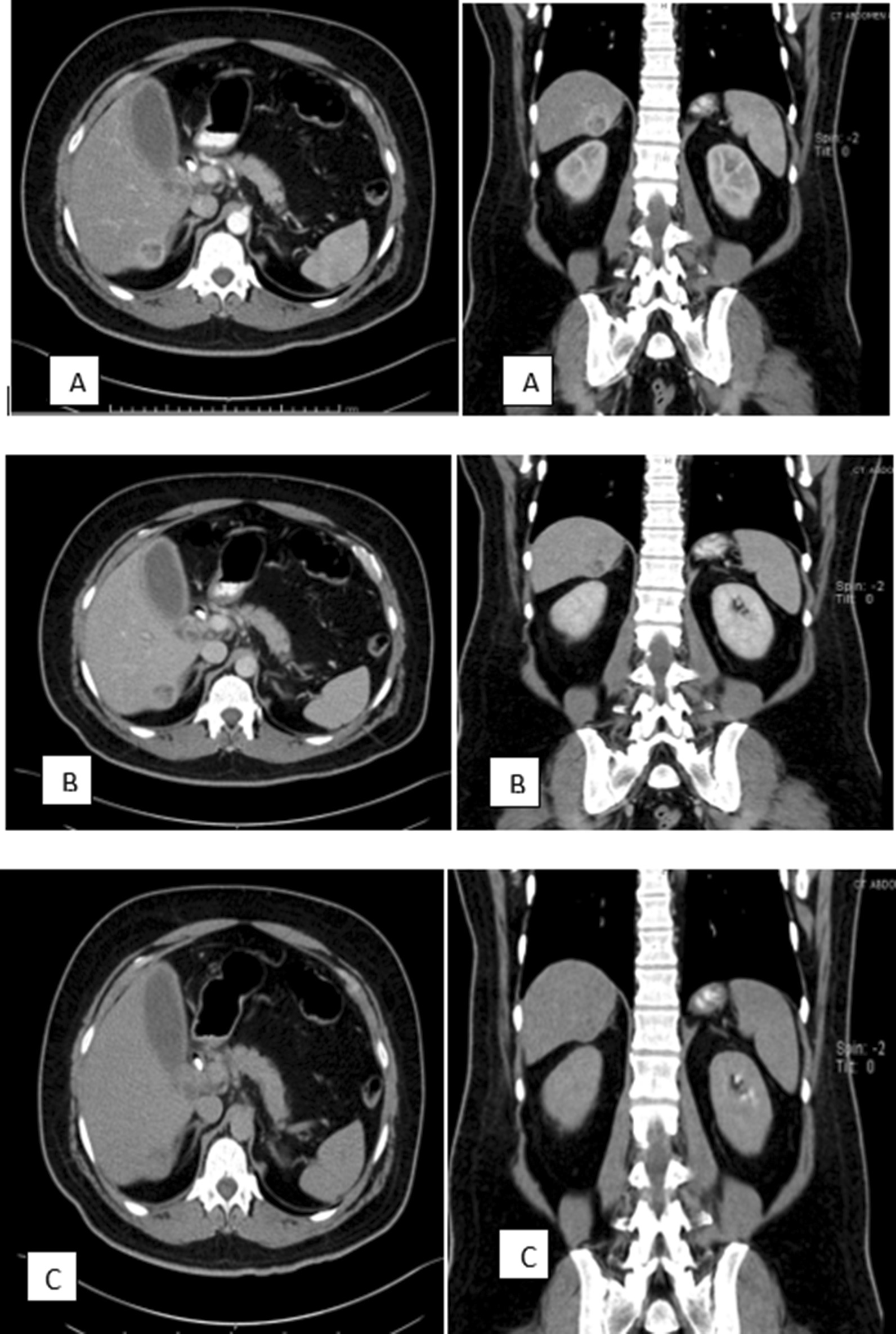
Introduction: Primary hepatic neuroendocrine tumours (PHNETs) are a rare form of hepatic neoplasms, and it is difficult to differentiate them from common hepatic malignancies in routine imaging studies.

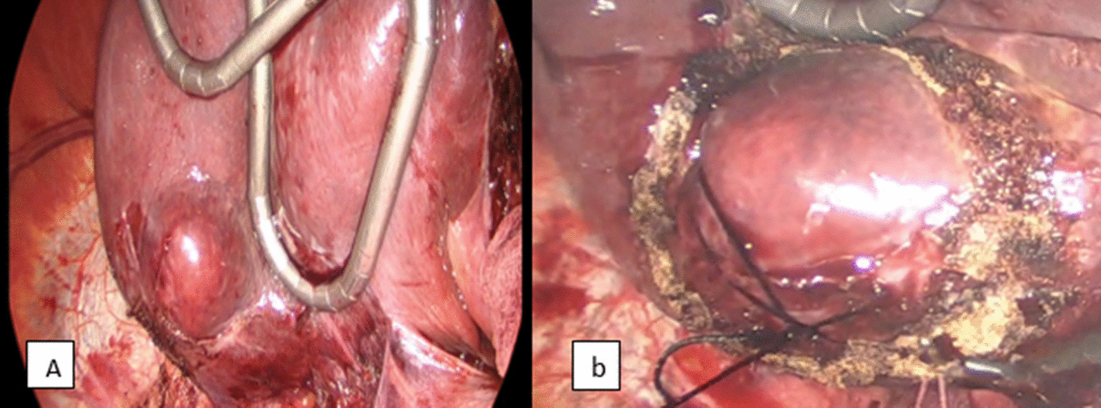
Presentation of the case: We describe the case of a 60-year-old Indian male patient with a tentative preoperative diagnosis of hepatocellular carcinoma (HCC). Nevertheless, the definitive post-operative diagnosis was made by Histopathological and immunohistochemical assessment, which revealed a grade II neuroendocrine tumour (NET) of moderate differentiation. Surgical resection was performed through a minimally invasive approach with a favourable postoperative course and a short hospital stay. One-month Post-operative Octreotide scan showed no extrahepatic primary origin of the tumour.

Discussion: PHNET is a rare entity, and multi modalities investigations, including imaging, serology, endoscopy series, and histopathology findings, aside from long-term follow-up to rule out another primary origin, are essential for the final diagnosis of PHNET. Surgical resection stands as the mainstay of treatment of PHNETs.

Conclusion: The absence of primary liver diseases should expand our possible differential diagnosis. Laparoscopic surgical resection of PHNETs carries a favourable outcome.

Keywords: Liver neuroendocrine tumour; Liver primary neuroendocrine tumour; Neuroendocrine tumour.


Conflict of interest statement

The author has no conflict of interest to declare or commercial associations.

Figures

Fig. 1
Fig. 1
Computerized tomography scan of abdomen and pelvis with contrast; a arterial phase, b venous phase, c delayed phase (axial view on the left side and coronal view on the right side). The mass is seen At segment VI of the RT liver lobe, causing a contour bulge of the liver capsule inferiorly with a clear fat plane between the mass and the RT kidney and is enhancing at the arterial and venous phases (A, B) with washout at the delayed phase (C)
Fig. 2
Fig. 2
F18 FDG Positron emission tomography-computed tomography images of the coronal liver fusion (left side), Axial fusion (right side). A single hypometabolic hepatic lesion was seen in the right liver lobe (4 × 3 × 3.5 cm3). No hypermetabolic lesions were identified in other organs
Fig. 3
Fig. 3
Shows the exophytic component of the tumour at the undersurface of the cephalad retracted liver (a), silk suture, a figure of eight fashion was taken deeply to the tumour to aid the counter-traction (b)
Fig. 4
Fig. 4
Microscopic picture of the tumour H&E shows tumour cells arranged as solid nests, cells with atypia, varying sizes, multiple mitosis rates, and with large, round, or oval nuclei, fine, granular chromatin, and lying to one side, neuroendocrine tumour-infiltrating the hepatic parenchyma. Magnification: D = × 40 (a), × 100 (b), × 200 (c), × 400 (d)

References

    1. Maggard MA, O'Connell JB, Ko CY. Updated population-based review of carcinoid tumors. Ann Surg. 2004;240(1):117–122. doi: 10.1097/01.sla.0000129342.67174.67. - DOI - PMC - PubMed
    1. Meng XF, Pan YW, Wang ZB, Duan WD. Primary hepatic neuroendocrine tumor case with a preoperative course of 26 years: a case report and literature review. World J Gastroenterol. 2018;24(24):2640–2646. doi: 10.3748/wjg.v24.i24.2640. - DOI - PMC - PubMed
    1. Edmondson HA. Tumors of the liver and intrahepatic bile ducts. 1958; Armed Forces Institute of Pathology, Washington, DC: 216, Vol. section 7, fasc. 25.
    1. Fenoglio LM, Severini S, Ferrigno D, Gollè G, Serraino C, Bracco C, Castagna E, Brignone C, Pomero F, Migliore E, David E, Salizzoni M. Primary hepatic carcinoid: a case report and literature review. World J Gastroenterol. 2009;15(19):2418–2422. doi: 10.3748/wjg.15.2418. - DOI - PMC - PubMed
    1. Xin Q, Lv R, Lou C, Ma Z, Liu GQ, Zhang Q, Yu HB, Zhang CS. Primary hepatic neuroendocrine carcinoma coexisting with distal cholangiocarcinoma: a case report and review of the literature. Medicine (Baltimore) 2020;99(26):e20854. doi: 10.1097/MD.0000000000020854. - DOI - PMC - PubMed

Publication types