Epibulbar osseous choristoma: a case report
- PMID: 37388506
- PMCID: PMC10299902
- DOI: 10.1093/jscr/rjad371
Epibulbar osseous choristoma: a case report
Abstract
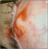
Peribulbar osseous choristoma is a benign, solid nodule; it is a subtype of epibulbar choristomas (belongs to single tissue choristomas), consisting of pure bony tissues. Epibulbar osseous choristoma is the rarest subtype of epibulbar choristomas, with only 65 cases reported since the mid-19th century; so, the rarity of the disease drove me to report it. A 7-year-old female presented with a painless left ocular superotemporal mass, which was present since birth and located under the conjunctiva. The primary diagnoses included lipodermoid and subconjunctival foreign bodies. Ocular interventions included a B-scan, examination under anesthesia and surgical excision of the mass in toto, and the histopathological examination showed that it was an osseous choristoma.
Keywords: choristoma; epibulbar.
Published by Oxford University Press and JSCR Publishing Ltd. © The Author(s) 2023.
Conflict of interest statement
None declared.
Figures


Similar articles
-
Epibulbar osseous choristoma: A case report.Am J Ophthalmol Case Rep. 2016 Oct 13;5:4-6. doi: 10.1016/j.ajoc.2016.10.002. eCollection 2017 Apr. Am J Ophthalmol Case Rep. 2016. PMID: 29503936 Free PMC article.
-
Epibulbar osseous choristoma: a clinicopathological case series and review of the literature.Klin Monbl Augenheilkd. 2012 Apr;229(4):420-3. doi: 10.1055/s-0031-1299256. Epub 2012 Apr 11. Klin Monbl Augenheilkd. 2012. PMID: 22496017 Review.
-
Epibulbar osseous choristoma: case report and review of the literature.Ophthalmic Surg Lasers. 2002 Sep-Oct;33(5):410-5. Ophthalmic Surg Lasers. 2002. PMID: 12358295 Review.
-
Epibulbar osseous choristoma: Two case reports.World J Clin Cases. 2022 Jan 21;10(3):1093-1098. doi: 10.12998/wjcc.v10.i3.1093. World J Clin Cases. 2022. PMID: 35127924 Free PMC article.
-
A case of epibulbar osseous choristoma with review of literature.Int Ophthalmol. 2014 Oct;34(5):1145-8. doi: 10.1007/s10792-014-9952-6. Epub 2014 May 6. Int Ophthalmol. 2014. PMID: 24799346 Review.
References
-
- Shanthala PR, Khandige S. Epibulbar osseous choristoma: a rare entity. J Evol Med Dent Sci 2013;2:5717–9.
-
- Balci O, Oduncu A. A case of epibulbar osseous choristoma with review of literature. Int Ophthalmol 2014;34:1145–8. - PubMed
-
- Parihar A, Verma S, Trehan A, Vashisht P. A case of epibulbar osseous choristoma. J Mar Med Soc 2018;20:157–8.
Publication types
LinkOut - more resources
Full Text Sources

