

Intraoral angiosarcoma with unusual clinical presentation: A case report
- PMID: 37389059
- PMCID: PMC10300316
- DOI: 10.1016/j.heliyon.2023.e17056
Intraoral angiosarcoma with unusual clinical presentation: A case report
Abstract
Introduction: Angiosarcoma is a rare and highly aggressive soft tissue malignancy originating from vascular and lymphatic endothelial cells. Epithelioid angiosarcoma is the rarest subtype of angiosarcoma, characterized by the proliferation of large polygonal cells with an epithelioid feature. The occurrence of these tumors in the oral cavity is highly uncommon, and immunohistochemistry staining is essential to differentiate epithelioid angiosarcoma from mimicking lesions.
Aim: To present a case of intraoral angiosarcoma with an unusual clinical presentation and behavior and to report, to the best of our knowledge, a first primary appendix epithelioid angiosarcoma with metastasis foci in the oral cavity.
Objectives: To discuss the clinical, histological, and immunochemical features of an unusual case of intraoral angiosarcoma.
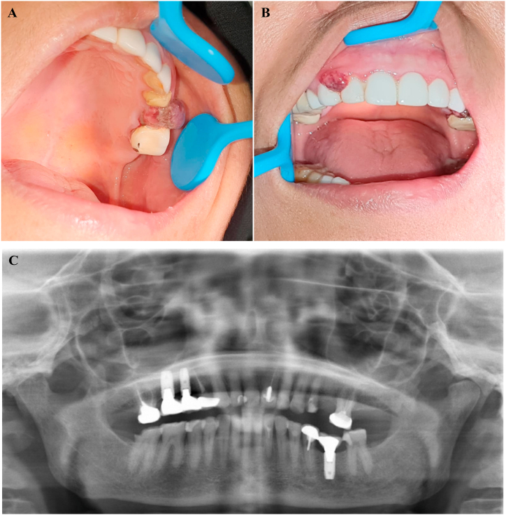
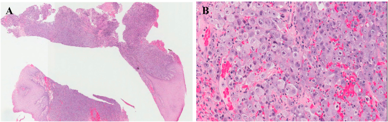
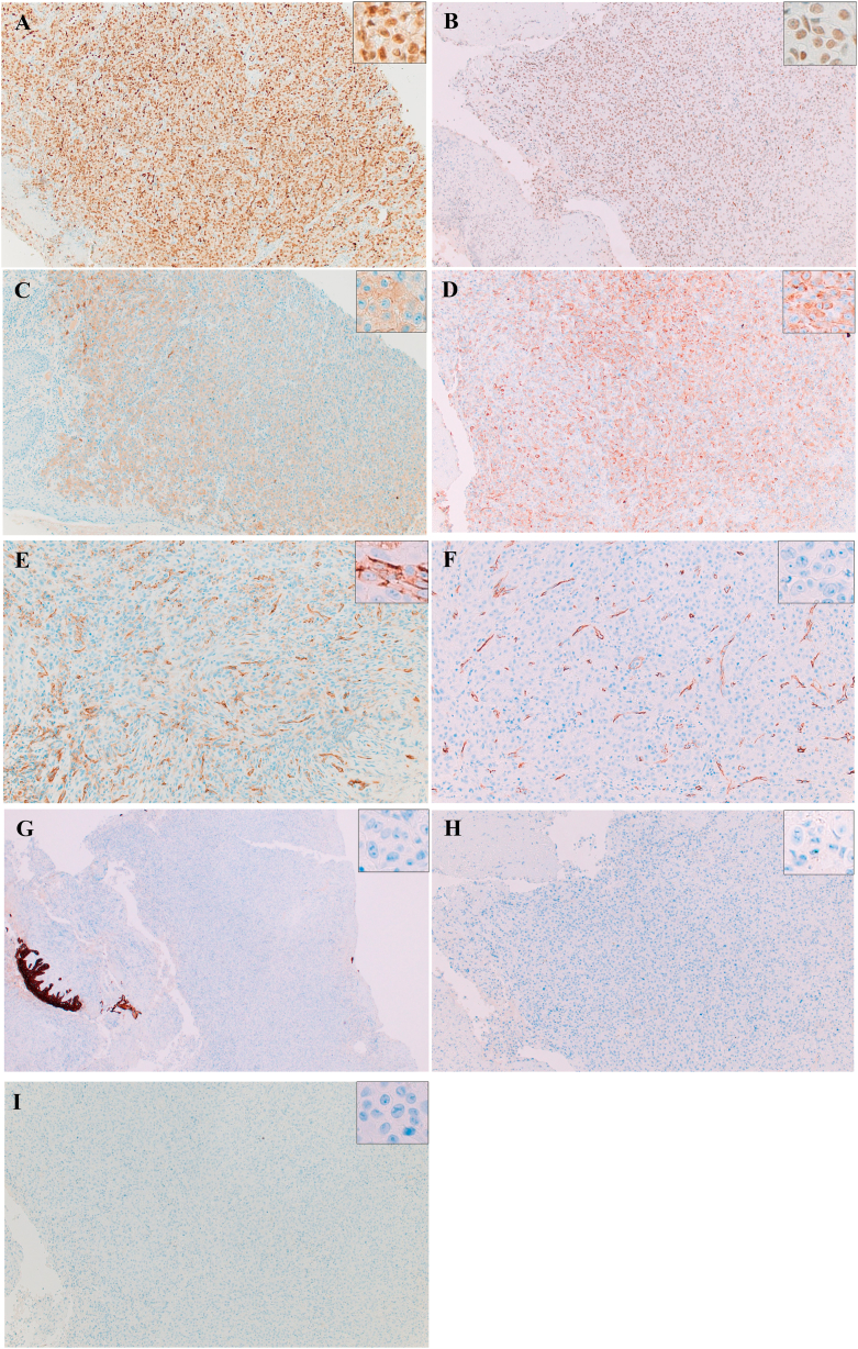
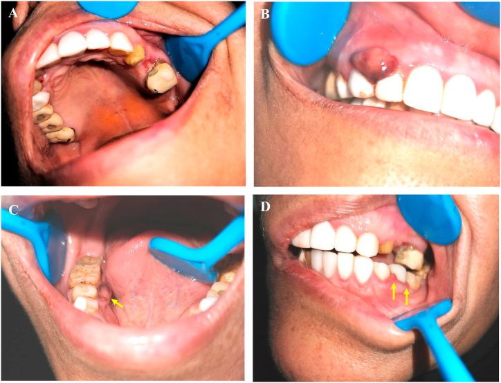
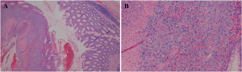
Case report: A 53-year-old Saudi female with an uncommon clinical presentation of intraoral angiosarcoma. The patient reported the lesion being painless, slowly growing, and of a six-month duration. The microscopic examination and immunohistochemical evaluation showed epithelioid angiosarcoma. The tumor cells were positive to ERG, FLI 1, and CD31 (focal) and negative to CK HMW, CD45, S100, HMB 45, D2-4, and CD 34.
Discussion: Due to the extremely rare occurrence and non-characteristic presentation of angiosarcoma in the oral cavity, many lesions maybe included in the differential diagnosis. Thus, making the diagnosis of intraoral angiosarcoma difficult.
Keywords: Angiosarcoma; Appendicitis; Epithelioid; Intraoral.
© 2023 The Authors. Published by Elsevier Ltd.
Conflict of interest statement
The authors declare that they have no known competing financial interests or personal relationships that could have appeared to influence the work reported in this paper.
Figures
Similar articles
-
Epithelioid angiosarcoma of bone and soft tissue: a report of seven cases with emphasis on morphologic diversity, immunohistochemical features and clinical outcome.Tumori. 2011 Sep-Oct;97(5):585-9. doi: 10.1177/030089161109700508. Tumori. 2011. PMID: 22158488
-
Epithelioid angiosarcoma of the chest wall with atypical morphology: report of one case.Int J Clin Exp Pathol. 2019 Oct 1;12(10):3944-3948. eCollection 2019. Int J Clin Exp Pathol. 2019. PMID: 31933787 Free PMC article.
-
Epithelioid angiosarcoma of the duodenum: a case report.Surg Case Rep. 2022 Feb 28;8(1):35. doi: 10.1186/s40792-022-01391-z. Surg Case Rep. 2022. PMID: 35224706 Free PMC article.
-
Pleural epithelioid angiosarcoma with lymphatic differentiation arisen after radiometabolic therapy for thyroid carcinoma: immunohistochemical findings and review of the literature.Diagn Pathol. 2017 Aug 15;12(1):60. doi: 10.1186/s13000-017-0652-1. Diagn Pathol. 2017. PMID: 28810922 Free PMC article. Review.
-
Primary epithelioid angiosarcoma originating from the mandibular gingiva: a case report of an extremely rare oral lesion.World J Surg Oncol. 2020 Oct 3;18(1):260. doi: 10.1186/s12957-020-01999-1. World J Surg Oncol. 2020. PMID: 33010804 Free PMC article. Review.
Cited by
-
Primary epithelioid angiosarcoma of the mandibular gingiva: diagnostic pitfalls, about an unusual entity.J Surg Case Rep. 2024 May 24;2024(5):rjae323. doi: 10.1093/jscr/rjae323. eCollection 2024 May. J Surg Case Rep. 2024. PMID: 38800505 Free PMC article.
References
-
- Fujisawa Y., Yoshino K., Fujimura T., Nakamura Y., Okiyama N., Ishitsuka Y., Watanabe R., Fujimoto M. vol. 8. 2018 Mar 2. p. 46. (Front Oncol. Cutaneous Angiosarcoma: the Possibility of New Treatment Options Especially for Patients with Large Primary Tumor). PMID: 29552543; PMCID: PMC5840142. - DOI - PMC - PubMed
-
- Smrke A., Hamm J., Karvat A., Simmons C., Srikanthan A. A retrospective review of 145 patients with angiosarcoma: Radiation therapy, extent of resection and chemotherapy are important predictors of survival. Mol. Clin. Oncol. 2020 Aug;13(2):179–185. doi: 10.3892/mco.2020.2055. Epub 2020 Jun 2. PMID: 32714543; PMCID: PMC7366222. - DOI - PMC - PubMed
-
- Muñoz M., Monje F., Alonso del Hoyo J.R., Martín-Granizo R. Oral angiosarcoma misdiagnosed as a pyogenic granuloma. J. Oral Maxillofac. Surg. 1998 Apr;56(4):488–491. 10.1016/s0278-2391(98)90719-4. PMID: 9580135. - PubMed
Publication types
LinkOut - more resources
Full Text Sources
Research Materials
Miscellaneous
