

White matter changes in fetal brains with ventriculomegaly
- PMID: 37389403
- PMCID: PMC10303118
- DOI: 10.3389/fnana.2023.1160742
White matter changes in fetal brains with ventriculomegaly
Abstract
Introduction: Ventriculomegaly (VM) is a fetal brain malformation which may present independently (isolated form) or in association with different cerebral malformations, genetic syndromes or other pathologies (non-isolated form).
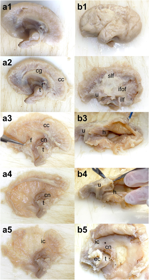
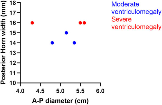
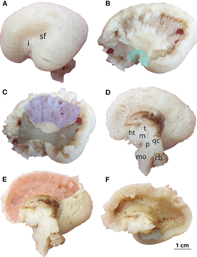
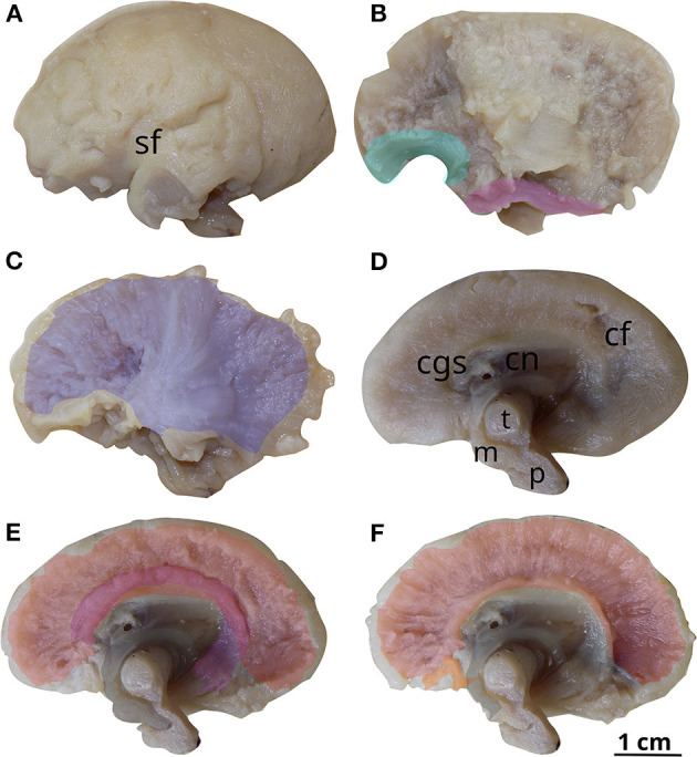
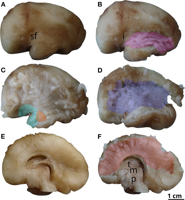
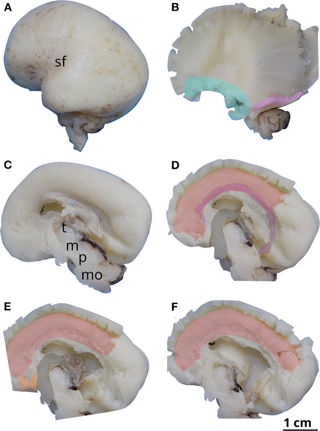
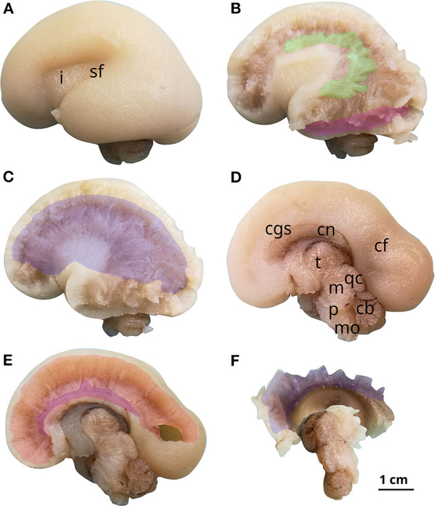
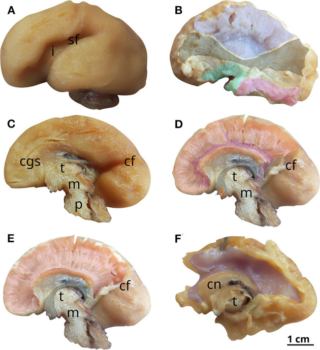
Methods: This paper aims to study the effect of ventriculomegaly on the internal tridimensional architecture of fetal brains by way of Klingler's dissection. Ventriculomegaly was diagnosed using fetal ultrasonography during pregnancy and subsequently confirmed by necropsy. Taking into consideration the diameter of the lateral ventricle (measured at the level of the atrium), the brains were divided into two groups: moderate ventriculomegaly (with atrial diameter between 13 and 15 mm) and severe ventriculomegaly (with atrial diameter above 15 mm).
Results and discussion: The results of each dissection were described and illustrated, then compared with age-matched reference brains. In the pathological brains, fascicles in direct contact with the enlarged ventricles were found to be thinner and displaced inferiorly, the opening of the uncinate fasciculus was wider, the fornix was no longer in contact with the corpus callosum and the convexity of the corpus callosum was inverted. We have studied the prevalence of neurodevelopmental delay in children born with ventriculomegaly in the literature and discovered that a normal developmental outcome was found in over 90% of the mild VM cases, approximately 75% of the moderate and 60% in severe VM, with the correlated neurological impairments ranging from attention deficits to psychiatric disorders.
Keywords: fetal brain; fiber dissection; human brain development; ventriculomegaly; white matter tracts.
Copyright © 2023 Horgos, Mecea, Boer, Buruiana, Ciortea, Mihu, Florian, Florian, Stamatian, Szabo, Albu, Susman and Pascalau.
Conflict of interest statement
The authors declare that the research was conducted in the absence of any commercial or financial relationships that could be construed as a potential conflict of interest.
Figures
Similar articles
-
White Matter Dissection of the Fetal Brain.Front Neuroanat. 2020 Sep 25;14:584266. doi: 10.3389/fnana.2020.584266. eCollection 2020. Front Neuroanat. 2020. PMID: 33071763 Free PMC article.
-
[Fetal ventriculomegaly: diagnosis using magnetic resonance imaging and its prognosis].Zhonghua Fu Chan Ke Za Zhi. 2010 Jan;45(1):22-5. Zhonghua Fu Chan Ke Za Zhi. 2010. PMID: 20367921 Chinese.
-
Clinical outcome of mild fetal ventriculomegaly.Am J Obstet Gynecol. 1998 Feb;178(2):218-22. doi: 10.1016/s0002-9378(98)80003-3. Am J Obstet Gynecol. 1998. PMID: 9500477
-
Mild ventriculomegaly from fetal consultation to neurodevelopmental assessment: A single center experience and review of the literature.Eur J Paediatr Neurol. 2018 Nov;22(6):919-928. doi: 10.1016/j.ejpn.2018.04.001. Epub 2018 Apr 12. Eur J Paediatr Neurol. 2018. PMID: 29709429 Review.
-
Role of magnetic resonance imaging in fetuses with mild or moderate ventriculomegaly in the era of fetal neurosonography: systematic review and meta-analysis.Ultrasound Obstet Gynecol. 2019 Aug;54(2):164-171. doi: 10.1002/uog.20197. Epub 2019 Jul 11. Ultrasound Obstet Gynecol. 2019. PMID: 30549340
References
LinkOut - more resources
Full Text Sources