Review
. 2024 Jun;26(3):382-390.
doi: 10.1007/s11307-023-01836-3. Epub 2023 Jun 30.

Towards Characterization of Skin Melanoma in the Clinic by Electron Paramagnetic Resonance (EPR) Spectroscopy and Imaging of Melanin

Affiliations
Review

Towards Characterization of Skin Melanoma in the Clinic by Electron Paramagnetic Resonance (EPR) Spectroscopy and Imaging of Melanin

Mohammad Wehbi et al. Mol Imaging Biol. 2024 Jun.

Abstract

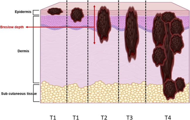
The incidence of melanoma is continuously increasing over time. Melanoma is the most aggressive skin cancer, significantly reducing quality of life and survival rates of patients at advanced stages. Therefore, early diagnosis remains the key to change the prognosis of patients with melanoma. In this context, advanced technologies are under evaluation to increase the accuracy of the diagnostic, to better characterize the lesions and visualize their possible invasiveness in the epidermis. Among the innovative methods, because melanin is paramagnetic, clinical low frequency electron paramagnetic resonance (EPR) that characterizes the melanin content in the lesion has the potential to be an adjunct diagnostic method of melanoma. In this review, we first summarize the challenges faced by dermatologists and oncologists in melanoma diagnostic and management. We also provide a historical perspective on melanin detection with a focus on EPR spectroscopy/imaging of melanomas. We describe key elements that allow EPR to move from in vitro studies to in vivo and finally to patients for melanoma studies. Finally, we provide a critical view on challenges to meet to make EPR operational in the clinic to characterize pigmented lesions.

Keywords: In vivo; Cancer; Clinical EPR; EPR; ESR; Low frequency; Melanin; Melanoma; Mice; Patients.


Conflict of interest statement

The authors declare no competing interests.

Figures

Fig. 1
Fig. 1
Illustration of different patterns of skin invasion by melanoma cells. Skin melanoma staging is based on the TNM (tumor, node, metastasis) characterization. The tumor category T is determined based on Breslow tumor thickness and the presence or absence of ulceration. The stage of the skin lesion is dependent on the tumor thickness, with T1, T2, T3, and T4 lesions presenting a thickness smaller than 1 mm, between 1 and 2 mm, between 2 and 4 mm, and larger than 4 mm, respectively. Melanomas with a Breslow thickness lower than 0.8 mm have an excellent prognostic. Thicker the melanomas, higher the risk of migration of melanoma cells to lymph nodes (N) and higher the risk of distant metastases (M)
Fig. 2
Fig. 2
Representative EPR spectra (9 GHz) of natural eu- and pheomelanins detected in human hair samples. Bottom: spectrum of eumelanin signal recorded from black hairs (semiquinone free radicals). Top: spectrum of pheomelanin from blond hairs (note the presence of a shoulder at low field due to the presence of o-semiquinone-imine free radicals)
Fig. 3
Fig. 3
Comparison between anatomopathological samples (top row) and their corresponding EPR images obtained on human melanoma samples with a 9 GHz spectrometer. Adapted from results described in studies [23, 26]
Fig. 4
Fig. 4
Influence of tumor growth (B16 melanoma model) on the EPR signal recorded in vivo in mice using a clinical EPR spectrometer operating at 1 GHz. Adapted from results described in studies [32]
Fig. 5
Fig. 5
Experimental set-up used in the clinical studies in Brussels on lesions suspect of melanoma (from studies described in [34]. A Person installed in low field magnet with a surface coil put at the surface of the analyzed lesion on the arm. B Focus on the surface coil resonator
Fig. 6
Fig. 6
Data recorded noninvasively from a melanoma lesion in a patient enrolled in the clinical EPR study described in [34]. A Average of first derivative EPR spectra recorded from the skin. The melanin signal is in the center (highlighted by green arrows). For field positioning and quantification, a reference spectrum is taken using a sample of 15N-perdeuterated nitroxide (15N-PDT) fixed along the axis of the resonator. The signal of the reference appears as a doublet (highlighted by the blue arrows). B First integration of this signal
Fig. 7
Fig. 7
Main results from the clinical EPR study described in [34]. Left: the EPR signal of melanin was significantly higher in melanoma lesions (n=26) compared to the signal recorded in benign atypical nevi (n=62) (****, p < 0.0001). Right: a trend toward a higher signal intensity (though not significant) was observed in high Breslow depth melanomas than in low Breslow lesions

References

    1. Halpern AC, Marghoob AA, Sober AJ, Mar V, Marchetti MA, et al. Clinical presentations of melanoma. In: Balch CM, et al., editors. Cutaneous Melanoma. 6. Springer; 2020. pp. 108–145.
    1. Dinnes J, Deeks JJ, Chuchu N, et al. Dermoscopy, with and without visual inspection, for diagnosing melanoma in adults. Cochrane Database Syst Rev. 2018;12(12):CD011902. - PMC - PubMed
    1. Dinnes J, Deeks JJ, Grainge MJ, et al. Visual inspection for diagnosing cutaneous melanoma in adults. Cochrane Database Syst Rev. 2018;12(12):CD013194. - PMC - PubMed
    1. Piccolo D, Ferrari A, Peris K, Daidone R, Ruggeri B, Chimenti S. Dermoscopic diagnosis by a trained clinician vs. a clinician with minimal dermoscopy training vs. computer-aided diagnosis of 341 pigmented skin lesions: A comparative study. Br J Dermatol. 2002;147(3):481–486. doi: 10.1046/j.1365-2133.2002.04978.x. - DOI - PubMed
    1. Calzavara-Pinton P, Longo C, Venturini M, Sala R, Pellacani G. Reflectance confocal microscopy for in vivo skin imaging. Photochem Photobiol. 2008;84(6):1421–1430. doi: 10.1111/j.1751-1097.2008.00443.x. - DOI - PubMed