. 2023 Jun 30;18(6):e0287634.
doi: 10.1371/journal.pone.0287634. eCollection 2023.

Introducing a new type of alternative laryngeal mucosa model

Affiliations

Introducing a new type of alternative laryngeal mucosa model

Tanja Grossmann et al. PLoS One. .

Abstract

Research of human vocal fold (VF) biology is hampered by several factors. The sensitive microstructure of the VF mucosa is one of them and limits the in vivo research, as biopsies carry a very high risk of scarring. A laryngeal organotypic model consisting of VF epithelial cells and VF fibroblasts (VFF) may overcome some of these limitations. In contrast to human VFF, which are available in several forms, availability of VF epithelial cells is scarce. Buccal mucosa might be a good alternative source for epithelial cells, as it is easily accessible, and biopsies heal without scarring. For this project, we thus generated alternative constructs consisting of immortalized human VF fibroblasts and primary human buccal epithelial cells. The constructs (n = 3) were compared to native laryngeal mucosa at the histological and proteomic level. The engineered constructs reassembled into a mucosa-like structure after a cultivation period of 35 days. Immunohistochemical staining confirmed a multi-layered stratified epithelium, a collagen type IV positive barrier-like structure resembling the basement membrane, and an underlying layer containing VFF. Proteomic analysis resulted in a total number of 1961 identified and quantified proteins. Of these, 83.8% were detected in both native VF and constructs, with only 53 proteins having significantly different abundance. 15.3% of detected proteins were identified in native VF mucosa only, most likely due to endothelial, immune and muscle cells within the VF samples, while 0.9% were found only in the constructs. Based on easily available cell sources, we demonstrate that our laryngeal mucosa model shares many characteristics with native VF mucosa. It provides an alternative and reproducible in vitro model and offers many research opportunities ranging from the study of VF biology to the testing of interventions (e.g. drug testing).


Conflict of interest statement

The authors have declared that no competing interests exist.

Figures

Fig 1
Fig 1. Schematic illustration of the 3D co-cultivation procedure.
Fibroblast-specific culture medium (CnT-PR-F), fully defined co-culture medium (CnT-PR-FTAL).
Fig 2
Fig 2. Representative phase-contrast images of isolated hBEC colonies.
Cobblestone-like cells 24 hours after start of the isolation (A) and confluent cell layer after additional 14 days of cultivation (B). (both 4x magnification).
Fig 3
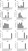
Fig 3. Descriptive hBEC cell characterization.
Relative mRNA levels of cell-type specific marker genes cytokeratin 5 (A), cytokeratin 14 (B), cytokeratin 13 (C), involucrin (D), cadherin 1 (E), tumor protein p63 (F), Thy-1 cell surface antigen (G), vimentin (H), von Willebrand factor (I), and C-C motif chemokine receptor (J) in isolated hBEC (n = 4), HaCaT, HUVEC, THP-1, and hVFF cells.
Fig 4
Fig 4. Representative H&E staining images of embedded hVFF.
hVFF after a cultivation period of 14 days seeded at a density of 5x10⁴ cells/insert (A), 1x10⁵ cells/insert (B), and 2x10⁵ cells/insert (C). (all 20x magnification).
Fig 5
Fig 5. Contraction of rat tail collagen matrix with or without embedded hVFF.
Representative schematics and photographs illustrating the matrix contraction following seeding of immortalized hVFF within the rat tail collagen, type I matrix.
Fig 6
Fig 6. Co-cultivated constructs resemble a multi-layered epithelium and an underlying lamina propria.
Representative H&E staining of vocal folds (A), buccal mucosa (B) and co-cultivated construct (C), vimentin staining of vocal folds (D), buccal mucosa (E) and co-cultivated construct (F). All formalin-fixed, paraffin-embedded sections, 20x magnification.
Fig 7
Fig 7. Co-cultivated constructs show adherent cell junctions and multiple epithelial layers.
Representative staining for E-cadherin staining of vocal folds (A), buccal mucosa (B) and co-cultivated construct (C); CK5/14 staining of vocal folds (D), buccal mucosa (E) and co-cultivated construct (F); p63 staining of vocal folds (G), buccal mucosa (H) and co-cultivated construct (I); and IVL staining of vocal folds (J), buccal mucosa (K) and co-cultivated construct (L). All formalin-fixed, paraffin-embedded sections, 20x magnification.
Fig 8
Fig 8. Co-cultivated constructs resemble basement membrane-like structure.
Basal collagen type IV (COL IV) staining of formalin-fixed, paraffin-embedded section of vocal folds (A), buccal mucosa (B) and co-cultivated construct (C). (scale bar: 100 μm, all 10x magnification).
Fig 9
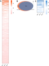
Fig 9. Proteomic profiling of vocal fold and co-cultivated construct samples.
Heatmap showing proteins detected solely in VF tissue samples (A). Venn diagram depicting the number of proteins detected solely in VF tissue samples, solely in CC samples, and in both sample types (B). Heatmap showing proteins detected solely in CC samples (C). Proteins are sorted in a decreasing manner according to their mean LFQ intensity values (A,C). Protein IDs, protein and gene names are listed in S4 and S5 Tables. Data from 3 biological replicates of VF and CC samples.
Fig 10
Fig 10. Differentially expressed proteins in vocal fold and co-cultivated construct samples.
Volcano plot showing 893 identified proteins including 53 proteins with significantly differential protein abundance (data from 3 biological replicates of VF and CC samples, p = 0.005) after Benjamini-Hochberg correction for multiple testing between co-cultivated construct (CC, left side, blue dots) and vocal fold (VF, right side, red dots) samples.

Similar articles

References

    1. Fukahori M, Chitose S, Sato K, Sueyoshi S, Kurita T, Umeno H, et al.. Regeneration of Vocal Fold Mucosa Using Tissue-Engineered Structures with Oral Mucosal Cells. PLoS One. 2016;11(1):e0146151. doi: 10.1371/journal.pone.0146151 - DOI - PMC - PubMed
    1. Ling C, Li Q, Brown ME, Kishimoto Y, Toya Y, Devine EE, et al.. Bioengineered vocal fold mucosa for voice restoration. Sci Transl Med. 2015;7(314):314ra187. doi: 10.1126/scitranslmed.aab4014 - DOI - PMC - PubMed
    1. Chen X, Lungova V, Zhang H, Mohanty C, Kendziorski C, Thibeault SL. Novel immortalized human vocal fold epithelial cell line: In vitro tool for mucosal biology. FASEB J. 2021;35(2):e21243. doi: 10.1096/fj.202001423R - DOI - PMC - PubMed
    1. Grossmann T, Darnhofer B, Birner-Gruenberger R, Kirsch A, Gugatschka M. Descriptive proteomics of paired human vocal fold and buccal mucosa tissue. Proteomics Clin Appl. 2022;16(2):e2100050. doi: 10.1002/prca.202100050 - DOI - PMC - PubMed
    1. Chen X, Thibeault SL. Novel isolation and biochemical characterization of immortalized fibroblasts for tissue engineering vocal fold lamina propria. Tissue Eng Part C Methods. 2009;15(2):201–12. doi: 10.1089/ten.tec.2008.0390 - DOI - PMC - PubMed

Publication types