

Wound healing responses of urinary extravasation after urethral injury
- PMID: 37391520
- PMCID: PMC10313654
- DOI: 10.1038/s41598-023-37610-2
Wound healing responses of urinary extravasation after urethral injury
Abstract
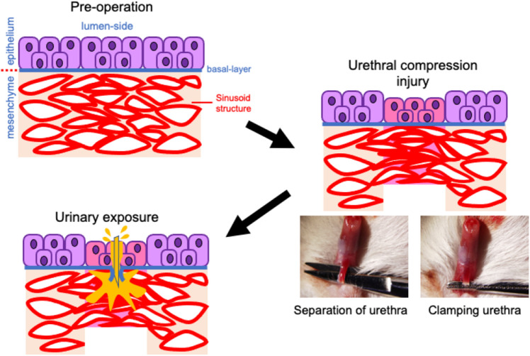
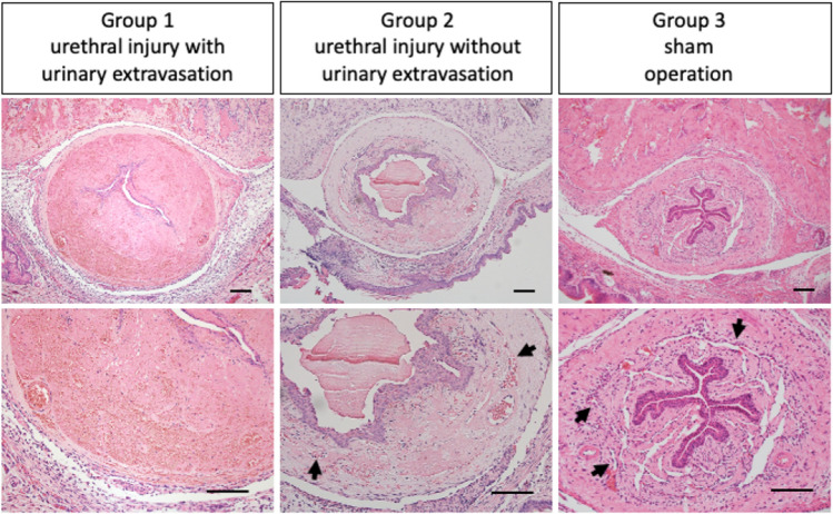
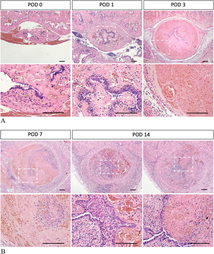
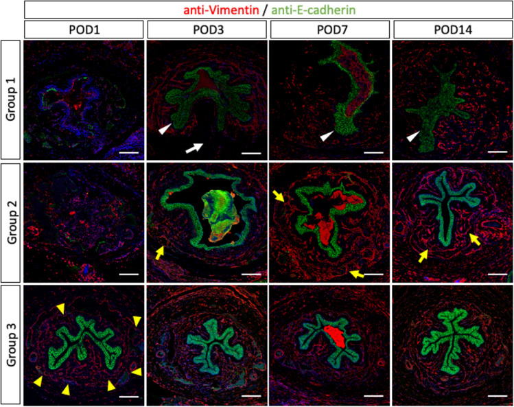
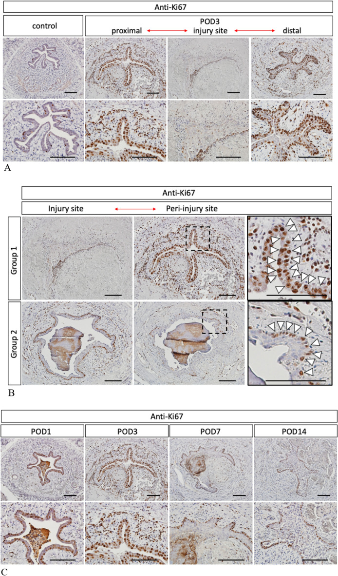
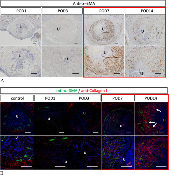
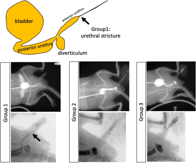
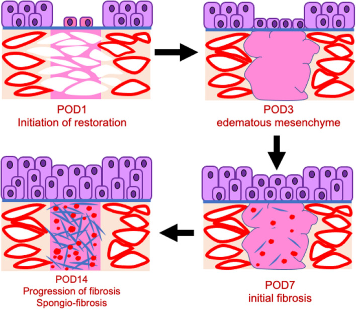
The post-surgical fluid leakage from the tubular tissues is a critical symptom after gastrointestinal or urinary tract surgeries. Elucidating the mechanism for such abnormalities is vital in surgical and medical science. The exposure of the fluid such as peritonitis due to urinary or gastrointestinal perforation has been reported to induce severe inflammation to the surrounding tissue. However, there have been no reports for the tissue responses by fluid extravasation and assessment of post-surgical and injury complication processes is therefore vital. The current model mouse study aims to investigate the effect of the urinary extravasation of the urethral injuries. Analyses on the urinary extravasation affecting both urethral mesenchyme and epithelium and the resultant spongio-fibrosis/urethral stricture were performed. The urine was injected from the lumen of urethra exposing the surrounding mesenchyme after the injury. The wound healing responses with urinary extravasation were shown as severe edematous mesenchymal lesions with the narrow urethral lumen. The epithelial cell proliferation was significantly increased in the wide layers. The mesenchymal spongio-fibrosis was induced by urethral injury with subsequent extravasation. The current report thus offers a novel research tool for surgical sciences on the urinary tract.
© 2023. The Author(s).
Conflict of interest statement
The authors declare no competing interests.
Figures
Similar articles
-
Evaluation of surgical procedures of mouse urethra by visualization and the formation of fistula.Sci Rep. 2020 Oct 26;10(1):18251. doi: 10.1038/s41598-020-75184-5. Sci Rep. 2020. PMID: 33106510 Free PMC article.
-
Postoperative urinary extravasation does not impact anterior urethroplasty surgical outcomes: a Latin American large cohort study.Int Urol Nephrol. 2020 Oct;52(10):1899-1905. doi: 10.1007/s11255-020-02497-9. Epub 2020 May 21. Int Urol Nephrol. 2020. PMID: 32440837
-
miR-21 modification enhances the performance of adipose tissue-derived mesenchymal stem cells for counteracting urethral stricture formation.J Cell Mol Med. 2018 Nov;22(11):5607-5616. doi: 10.1111/jcmm.13834. Epub 2018 Sep 4. J Cell Mol Med. 2018. PMID: 30179296 Free PMC article.
-
Problems of the urethra. Surgical approaches.Probl Vet Med. 1989 Jan-Mar;1(1):17-35. Probl Vet Med. 1989. PMID: 2520098 Review.
-
Cells Involved in Urethral Tissue Engineering: Systematic Review.Cell Transplant. 2019 Sep-Oct;28(9-10):1106-1115. doi: 10.1177/0963689719854363. Epub 2019 Jun 25. Cell Transplant. 2019. PMID: 31237144 Free PMC article.
Cited by
-
Ultrasound imaging of male urethral stricture disease: a narrative review of the available evidence, focusing on selected prospective studies.World J Urol. 2024 Jan 13;42(1):32. doi: 10.1007/s00345-023-04760-x. World J Urol. 2024. PMID: 38217706 Free PMC article. Review.
-
IFRD1 is required for maintenance of bladder epithelial homeostasis.iScience. 2024 Oct 28;27(12):111282. doi: 10.1016/j.isci.2024.111282. eCollection 2024 Dec 20. iScience. 2024. PMID: 39628564 Free PMC article.
-
A novel vascularized urethra-on-a-chip model.Sci Rep. 2025 Mar 7;15(1):8062. doi: 10.1038/s41598-025-92485-9. Sci Rep. 2025. PMID: 40055501 Free PMC article.
-
A new role for IFRD1 in regulation of ER stress in bladder epithelial homeostasis.bioRxiv [Preprint]. 2024 Jan 10:2024.01.09.574887. doi: 10.1101/2024.01.09.574887. bioRxiv. 2024. PMID: 38260387 Free PMC article. Preprint.
References
Publication types
MeSH terms
LinkOut - more resources
Full Text Sources
