. 2023 Jun 30;14(1):3886.
doi: 10.1038/s41467-023-39623-x.

Drug reinforcement impairs cognitive flexibility by inhibiting striatal cholinergic neurons

Affiliations

Drug reinforcement impairs cognitive flexibility by inhibiting striatal cholinergic neurons

Himanshu Gangal et al. Nat Commun. .

Abstract

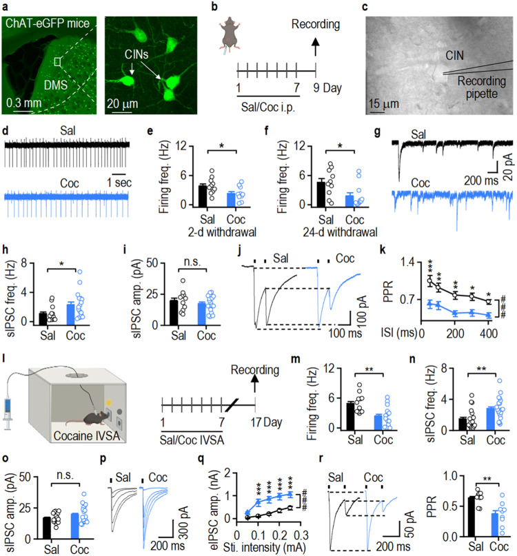
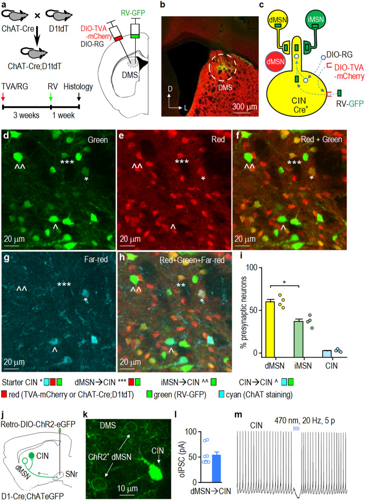
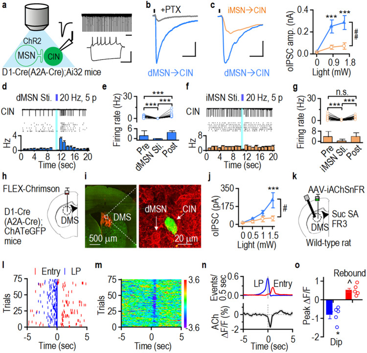
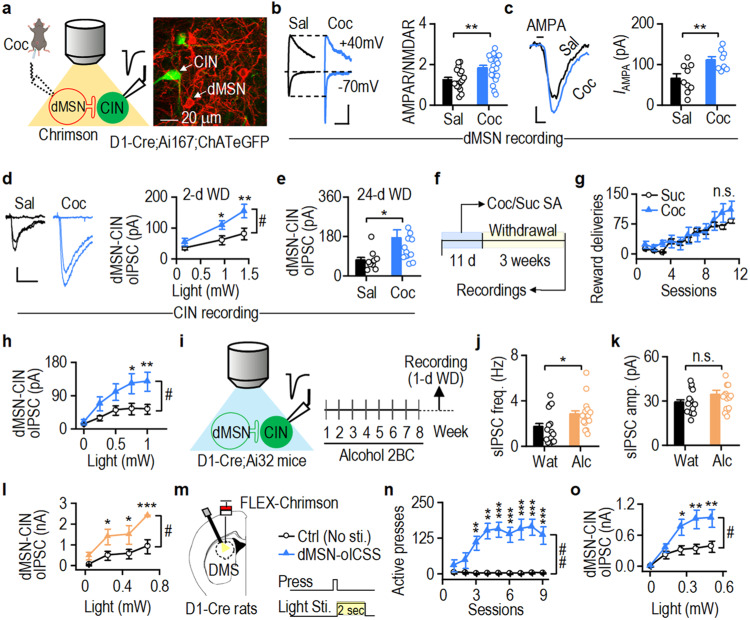
Addictive substance use impairs cognitive flexibility, with unclear underlying mechanisms. The reinforcement of substance use is mediated by the striatal direct-pathway medium spiny neurons (dMSNs) that project to the substantia nigra pars reticulata (SNr). Cognitive flexibility is mediated by striatal cholinergic interneurons (CINs), which receive extensive striatal inhibition. Here, we hypothesized that increased dMSN activity induced by substance use inhibits CINs, reducing cognitive flexibility. We found that cocaine administration in rodents caused long-lasting potentiation of local inhibitory dMSN-to-CIN transmission and decreased CIN firing in the dorsomedial striatum (DMS), a brain region critical for cognitive flexibility. Moreover, chemogenetic and time-locked optogenetic inhibition of DMS CINs suppressed flexibility of goal-directed behavior in instrumental reversal learning tasks. Notably, rabies-mediated tracing and physiological studies showed that SNr-projecting dMSNs, which mediate reinforcement, sent axonal collaterals to inhibit DMS CINs, which mediate flexibility. Our findings demonstrate that the local inhibitory dMSN-to-CIN circuit mediates the reinforcement-induced deficits in cognitive flexibility.


Conflict of interest statement

The authors declare no competing interest.

Figures

Fig. 1
Fig. 1. Cocaine administration compromises reversal learning.
a Schematic of intraperitoneal (i.p.) injections of saline (Sal) or cocaine (Coc; 15 mg/kg) in wild-type rats. b Repeated cocaine injections caused hyperlocomotion in rats; ***p < 0.001 versus Sal, ###p < 0.001. c Initial instrumental training of action (A)-outcome (O) contingency: rats were trained to press the left lever (A1) to receive sucrose solution in a central magazine (O1; A1→O1) and the right lever (A2) to receive food pellets in an adjacent magazine (O2; A2→O2). The establishment of A-O contingencies was assessed via a devaluation test. d Reversal training (A2→O1, A1→O2) and 2nd devaluation. e Cocaine did not alter lever press rate during initial training. f Saline (left) and cocaine (right) groups were sensitive to outcome devaluation; ***p < 0.001. g Cocaine did not alter the lever-press rate during reversal training. h The saline, but not cocaine, group was sensitive to outcome devaluation after reversal training; ***p < 0.001. i Schematic of i.p. injections of saline or cocaine (15 mg/kg) in wild-type mice and subsequent initial and reversal training followed by devaluation tests. j Saline and cocaine groups were sensitive to outcome devaluation after initial training; **p = 0.004, ^^p = 0.006. k The saline, but not cocaine, group was sensitive to outcome devaluation after reversal training; *p = 0.037. Val valued, Dev devalued, LP lever presses, n.s (not significant; e, g, h, k). Two-way RM ANOVA followed by Tukey post-hoc test (b, e, g), mixed model ANOVA followed by simple effects test (f, h, j, k). n = 10 (a–h Sal), 8 (a–h Coc), 11 (i–k Sal), 12 (i–k Coc). Data are presented as mean values ± SEM. Source data are provided as a Source Data file.
Fig. 2
Fig. 2. Cocaine exposure potentiates GABAergic inputs onto DMS CINs.
a Confocal images of cholinergic interneurons (CINs) in the dorsomedial striatum (DMS) in a ChAT-eGFP mouse. b–e Repeated cocaine (Coc, blue) but not saline (Sal, black) intraperitoneal (i.p.) injections reduced the spontaneous firing frequency (freq.) of DMS CINs 2 d after the last injection; *p = 0.0328. f Cocaine-induced inhibition of DMS CIN activity persisted 24 d after the last injection; *p = 0.0257. gi, Sample spontaneous IPSCs (sIPSCs) traces (g). Cocaine increased the frequency (h; *p = 0.0337), but not amplitude (amp.) (i), of sIPSCs in DMS CINs. j, k Cocaine decreased paired-pulse ratios (PPR) of electrically evoked IPSCs (eIPSCs) in CINs; *p < 0.05, **p < 0.01, ***p < 0.001 versus Coc; ###p < 0.001. l ChAT-eGFP mice were trained with cocaine intravenous self-administration (IVSA) for 7 d; DMS CINs were recorded 10 d after the last training session. m, Cocaine IVSA inhibited DMS CIN firing 10 d after the last exposure; **p = 0.003. n, o Cocaine IVSA increased the frequency (n; **p = 0.006), but not amplitude (o), of sIPSCs. p, q Cocaine IVSA potentiated eIPSCs in DMS CINs; ***p < 0.001 versus Sal, ###p < 0.001. r Cocaine IVSA decreased the eIPSC PPR in DMS CINs; **p = 0.002. Sti.: Stimulation, n.s. (not significant; i, o). Unpaired t test (e, f, h, i, m, n, o, r), two-way RM ANOVA followed by Tukey post-hoc test (k, q). n = 11 neurons from 3 mice (11/3) (e, Sal), 10/3 (e, Coc; f, Sal), 11/4 (f, Coc), 18/4 (h, i; Sal), 20/4 (h, i; Coc), 26/3 (k, Sal), 23/3 (k, Coc), 12/4 (m, Sal), 14/4 (m, Coc), 13/4 (n, o; Sal), 17/4 (n, o, Coc), 9/4 (q, Sal & Coc; r, Sal & Coc). Data are presented as mean values ± SEM. Source data are provided as a Source Data file.
Fig. 3
Fig. 3. CINs receive striatal inputs primarily from dMSNs.
a Schematic of experimental design. Cre-dependent helper viruses (AAV-DIO-TVA (EnvA receptor)-mCherry and AAV-DIO-RG (rabies glycoprotein)) were infused into the dorsomedial striatum (DMS) of ChAT-Cre;D1-tdTomato (tdT) mice, followed by rabies virus (RV-GFP) infusion at the same site 3 weeks later. Coronal sections were prepared 1 week after rabies virus infusion and were stained for choline acetyltransferase (ChAT; far-red, pseudo colored with cyan). b Confocal micrograph showing the injection site and viral expression in the DMS. L lateral, D dorsal. c Schematic showing viral expression and retrograde spread of RV-GFP. AAV-DIO-TVA-mCherry and AAV-DIO-RG infected Cre-positive cholinergic interneurons (CINs). RV-GFP infected TVA-positive CINs (starter cells expressed GFP and mCherry), labeling their presynaptic neurons with GFP. Since direct-pathway medium spiny neurons (dMSNs) were labeled red (from D1-tdT), dMSNs presynaptic to the starter CINs were yellow (red and green overlap), whereas putative indirect pathway MSNs (iMSNs) were green. dh Confocal micrographs of a DMS section; *Starter CINs, ***dMSN→CIN, ^^iMSN→CIN, ^CIN→CIN. i Summarized data showing that significantly more dMSNs (yellow) than iMSNs (green only) project to CINs; *p = 0.036. j AAVretro-DIO-ChR2-eGFP (Channelrhodopsin-2) was infused into the substantia nigra pars reticulata (SNr) of D1-Cre;ChAT-eGFP mice and DMS CINs were recorded. k Image of the DMS demonstrating ChR2-eGFP expression in dMSNs (cell membrane) and eGFP expression in CINs (cytoplasm). l Summarized optically-induced inhibitory postsynaptic currents (oIPSCs) recorded from CINs. m A burst of light stimulation (470 nm, 20 Hz, 5 pulses) of SNr-projecting dMSNs inhibited CIN firing. Unpaired t test (i). n = 4 mice (i) and 8/3 (l). Data are presented as mean values ± SEM. Source data are provided as a Source Data file.
Fig. 4
Fig. 4. CINs receive stronger functional GABAergic inputs from dMSNs than from iMSNs.
a Optogenetic stimulation of ChR2-expressing direct pathway (dMSNs) or indirect pathway MSNs (iMSNs) in D1-Cre;Ai32 or A2A-Cre;Ai32 mice respectively (left). Spontaneous firing (top right) and characteristic sag (bottom right) in DMS CINs. b Picrotoxin (PTX; gray)-sensitive optically evoked dMSN→CIN inhibitory postsynaptic current (oIPSC, blue). c oIPSCs were greater at dMSN→CIN (blue) than at iMSN→CIN synapses (orange); ***p < 0.001 versus iMSN→CIN, ##p = 0.002. d dMSN stimulation (Sti.; 20 Hz, 5 ms, 5 pulses) induced pause-rebound firing in a CIN. Top, firing; middle, multiple sweeps; bottom, corresponding histogram. e Summarized data comparing CIN firing before (pre), during and after (post) dMSN-burst stimulation; ***p < 0.001. f Representative histogram demonstrating a pause but no rebound of CIN firing on iMSNs stimulation (20 Hz, 5 ms, 5 pulses). g Summarized data demonstrating iMSN-mediated inhibition of CIN firing; ***p < 0.001. h Infusion of AAV-FLEX-Chrimson-tdTomato in the DMS of D1-Cre;ChAT-eGFP and A2A-Cre;ChAT-eGFP mice. i Micrograph demonstrating Chrimson-tdTomato expression in dMSNs and GFP in CINs in D1-Cre;ChAT-eGFP mice. j Greater oIPSCs at dMSN→CIN than at iMSN→CIN synapses; ***p < 0.001 versus iMSN→CIN, #p = 0.039. k Wild-type (WT) rats infused with AAV-iAChSnFR in the DMS were trained to self-administer sucrose (Suc SA) using a fixed ratio 3 (FR3) schedule. l Sample lever pressing (LP, blue) and magazine entry (Entry, red) events, trials aligned to reward delivery. m Corresponding heat map of ACh activity. n Averaged ACh signal. o Summarized data demonstrating a significant dip (blue) and rebound (red) in ACh activity; *p = 0.014, ^p = 0.015 versus baseline. n.s. (not significant; g). Scale bars: 1 s (a, top right); 100 ms, 50 mV (a, bottom right); 50 ms, 200 pA (b); 10 ms, 250 pA (c). Two-way RM ANOVA followed by Tukey post-hoc test (c, j), paired t test (e, g, o). n = 13/5 (c, dMSNs), 16/5 (c, iMSNs), 44 sweeps/8 neurons (i), 32 sweeps/8 neurons (g), 10/3 (j, dMSNs), 14/5 (j, iMSNs) and 5 rats (o). Data are presented as mean values ± SEM. Source data are provided as a Source Data file.
Fig. 5
Fig. 5. Drug reinforcement potentiates GABAergic dMSN→CIN transmission in the DMS.
a, b Repeated cocaine (Coc) but not saline (Sal) injections increased AMPAR/NMDAR ratios in DMS dMSNs of D1-Cre;Ai167;ChAT-eGFP mice, wherein dMSNs expressed Chrimson-tdTomato and CINs expressed eGFP; **p = 0.003. c Cocaine potentiated AMPA-induced currents (IAMPA) in dMSNs; **p = 0.008. d, e Cocaine potentiated dMSN→CIN oIPSCs after 2 d (d; *p = 0.056, **p < 0.01 versus Sal; #p = 0.012) and 24 d (e; *p = 0.037) withdrawal (WD). f, g D1-Cre;Ai167;ChAT-eGFP mice self-administered (SA) cocaine or sucrose and were recorded from after 3 weeks. h Cocaine self-administration caused greater potentiation of dMSN→CIN oIPSCs than sucrose self-administration; *p < 0.05, **p < 0.01 versus Suc; #p = 0.043. i D1-Cre;Ai32 and ChAT-eGFP mice consumed alcohol (Alc; orange), controls received only water (Wat; black). j, k Alcohol consumption increased sIPSC frequency (freq.) (j; *p = 0.030), but not amplitude (amp.) (k), in DMS CINs of ChAT-eGFP mice. l Alcohol potentiated dMSN→CIN oIPSCs in D1-Cre;Ai32 mice; *p < 0.05, ***p < 0.001 versus Wat; #p = 0.011. m, n D1-Cre rats infused with AAV-FLEX-Chrimson-tdTomato in the DMS pressed levers for optical intracranial self-stimulation (oICSS; blue) of dMSNs (2-s constant), controls (Ctrl; black) did not receive stimulation; **p < 0.01, ***p < 0.001 versus Ctrl; ##p = 0.001. o dMSN self-stimulation potentiated dMSN→CIN oIPSCs 1 d after the last training session; *p < 0.05, **p < 0.01 versus Ctrl; #p = 0.021. n.s. (not significant; g, k). Scale bars: 100 ms, 50 pA (b); 50 s, 30 pA (c); 50 ms, 30 pA (d). Two-way RM ANOVA followed by Tukey post-hoc test (d, g, h, l, n, o), unpaired t test (b, c, e, j, k). n = 18/4 (b, Sal), 22/5 (b, Coc), 9/3 (c, Sal), 8/3 (c, Coc), 22/5 (d, Sal), 22/7 (d, Coc), 10/3 (e, Sal), 14/4 (e, Coc), 7 mice/group (g), 20/5 (h, Suc), 22/5 (h, Coc), 15/3 (j, k; Wat), 16/4 (j, k; Alc), 6/4 (l, Wat), 5/3 (l, Alc), 6 rats/group (n), 14/5 (o, Ctrl), and 13/6 (o, dMSN-oICSS). Data are presented as mean values ± SEM. Source data are provided as a Source Data file.
Fig. 6
Fig. 6. Time-locked optogenetic inhibition of DMS CINs impairs reversal and extinction learning.
ac AAV-DIO-eNpHR-eYFP (halorhodopsin; blue) or AAV-DIO-eYFP (Ctrl: control; black) was infused into the dorsomedial striatum (DMS) of ChAT-Cre;tdTomato (tdT) rats (a). tdT-positive cholinergic interneurons (CINs) expressed eNpHR-eYFP (b) and were inhibited by yellow light stimulation of eNpHR in slice recordings (c). d Initial action (A)-outcome (O) contingency training (A1→O1; A2→O2) (left) and the initial devaluation test (right). e eYFP and eNpHR groups were sensitive to outcome devaluation after initial training; ***p < 0.001. f The initial devaluation (Dev.) index did not differ between groups. g Reversal training (A1→O2; A2→O1) (left) and the devaluation test after reversal (right). Reward deliveries were paired with laser stimulation. h The eYFP (left), but not the eNpHR (right), group was sensitive to outcome devaluation after reversal training (left); **p = 0.009. i The devaluation index was lower in the eNpHR group than in controls; *p = 0.034. j Extinction training: lever presses did not lead to reward deliveries (Ø). Reward omissions were paired with laser stimulation. k Lever-press rate was higher in the eNpHR group than in controls during the first extinction session; **p = 0.006 versus eYFP. l Averaged lever pressing for the two groups during the first extinction session (Ses. 1). m Summarized data showing that lever presses during 10–20 min of the extinction session were higher in the eNpHR group than the eYFP controls; **p = 0.009. Val valued, Dev devalued, LP lever presses, BL baseline, n.s. (not significant; f, h). Scale bar: 1 s, 20 pA (c). Unpaired t test (f, i, m), mixed model ANOVA followed by simple effects test (e, h), two-way RM ANOVA followed by Tukey post-hoc test (k). n = 9 rats/group. Data are presented as mean values ± SEM. Source data are provided as a Source Data file.

References

    1. Cui G, et al. Concurrent activation of striatal direct and indirect pathways during action initiation. Nature. 2013;494:238–242. doi: 10.1038/nature11846. - DOI - PMC - PubMed
    1. Mink JW. The Basal Ganglia and involuntary movements: impaired inhibition of competing motor patterns. Arch. Neurol. 2003;60:1365–1368. doi: 10.1001/archneur.60.10.1365. - DOI - PubMed
    1. Wang W, et al. Striatal mu-opioid receptor activation triggers direct-pathway GABAergic plasticity and induces negative affect. Cell Rep. 2023;7:112089. doi: 10.1016/j.celrep.2023.112089. - DOI - PMC - PubMed
    1. Everitt BJ, Robbins TW. Neural systems of reinforcement for drug addiction: from actions to habits to compulsion. Nat. Neurosci. 2005;8:1481–1489. doi: 10.1038/nn1579. - DOI - PubMed
    1. Schultz W. Predictive reward signal of dopamine neurons. J. Neurophysiol. 1998;80:1–27. doi: 10.1152/jn.1998.80.1.1. - DOI - PubMed

Publication types

Substances