

Oxidative Stress-Involved Mitophagy of Retinal Pigment Epithelium and Retinal Degenerative Diseases
- PMID: 37391574
- PMCID: PMC10477140
- DOI: 10.1007/s10571-023-01383-z
Oxidative Stress-Involved Mitophagy of Retinal Pigment Epithelium and Retinal Degenerative Diseases
Abstract
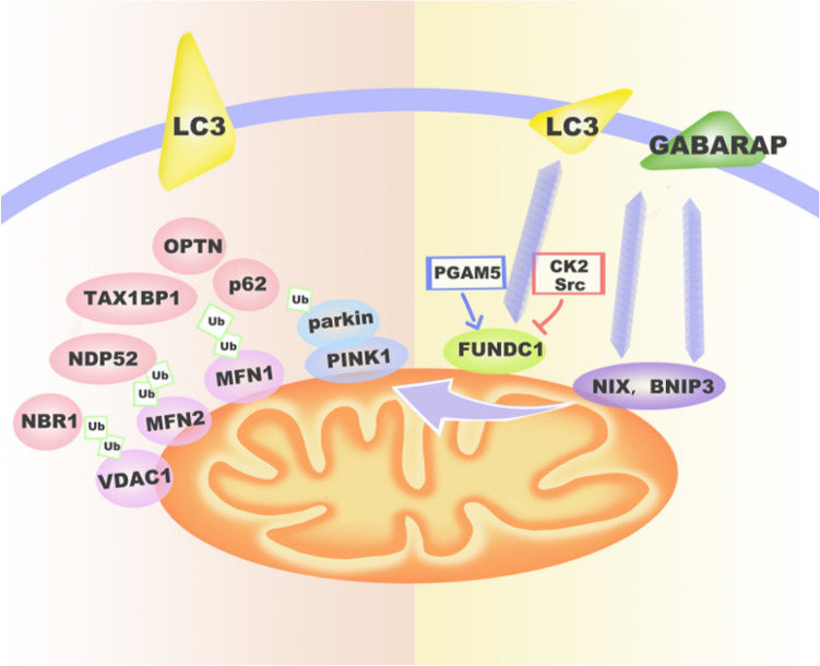
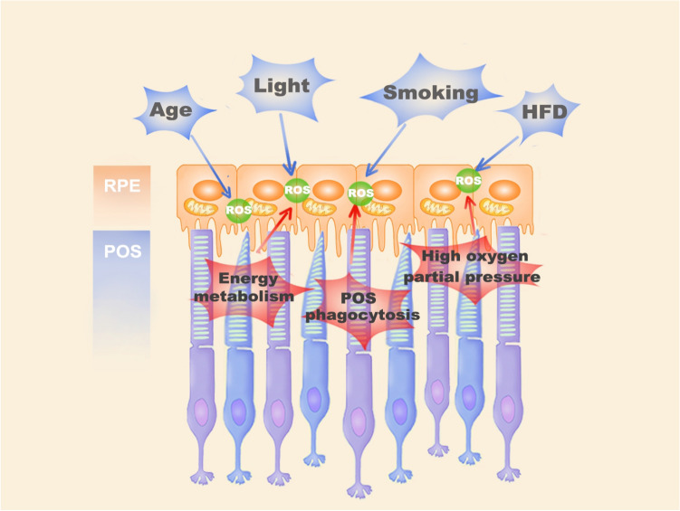
The retinal pigment epithelium (RPE) is a highly specialized and polarized epithelial cell layer that plays an important role in sustaining the structural and functional integrity of photoreceptors. However, the death of RPE is a common pathological feature in various retinal diseases, especially in age-related macular degeneration (AMD) and diabetic retinopathy (DR). Mitophagy, as a programmed self-degradation of dysfunctional mitochondria, is crucial for maintaining cellular homeostasis and cell survival under stress. RPE contains a high density of mitochondria necessary for it to meet energy demands, so severe stimuli can cause mitochondrial dysfunction and the excess generation of intracellular reactive oxygen species (ROS), which can further trigger oxidative stress-involved mitophagy. In this review, we summarize the classical pathways of oxidative stress-involved mitophagy in RPE and investigate its role in the progression of retinal diseases, aiming to provide a new therapeutic strategy for treating retinal degenerative diseases. The role of mitophagy in AMD and DR. In AMD, excessive ROS production promotes mitophagy in the RPE by activating the Nrf2/p62 pathway, while in DR, ROS may suppress mitophagy by the FOXO3-PINK1/parkin signaling pathway or the TXNIP-mitochondria-lysosome-mediated mitophagy.
Keywords: Age-related macular degeneration; Diabetic retinopathy; Mitophagy; Oxidative stress; Retinal pigment epithelium.
© 2023. The Author(s).
Conflict of interest statement
The authors declare no conflict of interest. The authors declare that the research was conducted in the absence of any commercial or financial relationships that could be construed as a potential conflict of interest. The funders had no role in the design of the study; in the collection, analyses, or interpretation of data; in the writing of the manuscript; or in the decision to publish the results.
Figures
Similar articles
-
Mitophagy initiates retrograde mitochondrial-nuclear signaling to guide retinal pigment cell heterogeneity.Autophagy. 2023 Mar;19(3):966-983. doi: 10.1080/15548627.2022.2109286. Epub 2022 Aug 13. Autophagy. 2023. PMID: 35921555 Free PMC article.
-
High-glucose induces retinal pigment epithelium mitochondrial pathways of apoptosis and inhibits mitophagy by regulating ROS/PINK1/Parkin signal pathway.Biomed Pharmacother. 2019 Mar;111:1315-1325. doi: 10.1016/j.biopha.2019.01.034. Epub 2019 Jan 15. Biomed Pharmacother. 2019. PMID: 30841445
-
Investigating mitochondrial fission, fusion, and autophagy in retinal pigment epithelium from donors with age-related macular degeneration.Sci Rep. 2022 Dec 16;12(1):21725. doi: 10.1038/s41598-022-26012-5. Sci Rep. 2022. PMID: 36526679 Free PMC article.
-
Mechanisms of mitochondrial dysfunction and their impact on age-related macular degeneration.Prog Retin Eye Res. 2020 Nov;79:100858. doi: 10.1016/j.preteyeres.2020.100858. Epub 2020 Apr 13. Prog Retin Eye Res. 2020. PMID: 32298788 Free PMC article. Review.
-
Mitochondrial quality control in AMD: does mitophagy play a pivotal role?Cell Mol Life Sci. 2018 Aug;75(16):2991-3008. doi: 10.1007/s00018-018-2843-7. Epub 2018 May 18. Cell Mol Life Sci. 2018. PMID: 29777261 Free PMC article. Review.
Cited by
-
Exposure to volatile organic compounds is a risk factor for diabetes retinopathy: a cross-sectional study.Front Public Health. 2024 Jan 30;12:1347671. doi: 10.3389/fpubh.2024.1347671. eCollection 2024. Front Public Health. 2024. PMID: 38351959 Free PMC article.
-
Lemon Peel Water Extract: A Novel Material for Retinal Health, Protecting Retinal Pigment Epithelial Cells against Dynamin-Related Protein 1-Mediated Mitochondrial Fission by Blocking ROS-Stimulated Mitogen-Activated Protein Kinase/Extracellular Signal-Regulated Kinase Pathway.Antioxidants (Basel). 2024 Apr 27;13(5):538. doi: 10.3390/antiox13050538. Antioxidants (Basel). 2024. PMID: 38790643 Free PMC article.
-
Epigenetic Modifications in the Retinal Pigment Epithelium of the Eye During RPE-Related Regeneration or Retinal Diseases in Vertebrates.Biomedicines. 2025 Jun 25;13(7):1552. doi: 10.3390/biomedicines13071552. Biomedicines. 2025. PMID: 40722628 Free PMC article. Review.
-
Oxidative Stress and Inflammation-Related mRNAs Are Elevated in Serum of a Finnish Wet AMD Cohort.Invest Ophthalmol Vis Sci. 2024 Nov 4;65(13):30. doi: 10.1167/iovs.65.13.30. Invest Ophthalmol Vis Sci. 2024. PMID: 39546296 Free PMC article.
-
Evaluation of retinal structure changes with AI-based OCT image segmentation for sodium iodate induced retinal degeneration.Front Cell Neurosci. 2025 Jun 18;19:1605639. doi: 10.3389/fncel.2025.1605639. eCollection 2025. Front Cell Neurosci. 2025. PMID: 40606272 Free PMC article.
References
-
- Aizu Y, Oyanagi K, Hu J, Nakagawa H (2002) Degeneration of retinal neuronal processes and pigment epithelium in the early stage of the streptozotocin-diabetic rats. Neuropathology 22(3):161–170. 10.1046/j.1440-1789.2002.00439.x - PubMed
-
- Antonetti DA, Barber AJ, Bronson SK, Freeman WM, Gardner TW, Jefferson LS, Kester M, Kimball SR, Krady JK, LaNoue KF, Norbury CC, Quinn PG, Sandirasegarane L, Simpson IA (2006) Diabetic retinopathy: seeing beyond glucose-induced microvascular disease. Diabetes 55(9):2401–2411. 10.2337/db05-1635 - PubMed
Publication types
MeSH terms
Substances
Grants and funding
LinkOut - more resources
Full Text Sources
Medical
Research Materials
