. 2023 Jul 1;15(14):6641-6657.
doi: 10.18632/aging.204825. Epub 2023 Jul 1.

Human senescent fibroblasts trigger progressive lung fibrosis in mice

Affiliations

Human senescent fibroblasts trigger progressive lung fibrosis in mice

Fernanda Hernandez-Gonzalez et al. Aging (Albany NY). .

Abstract

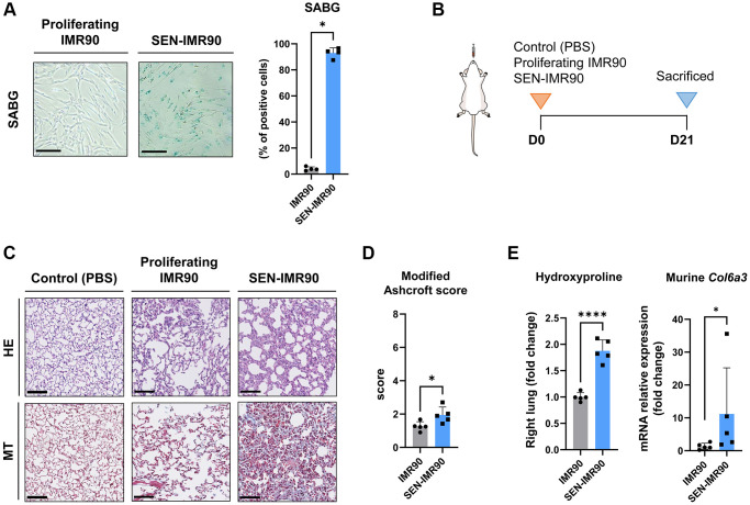
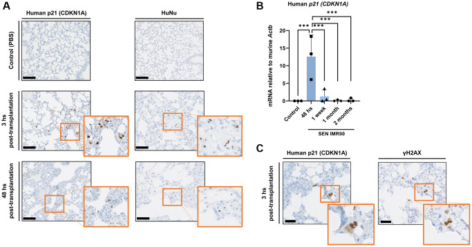
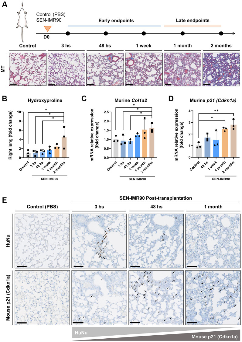
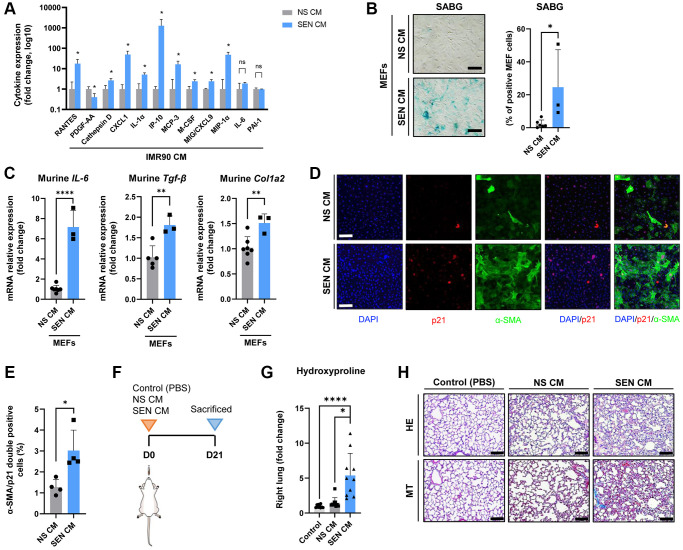
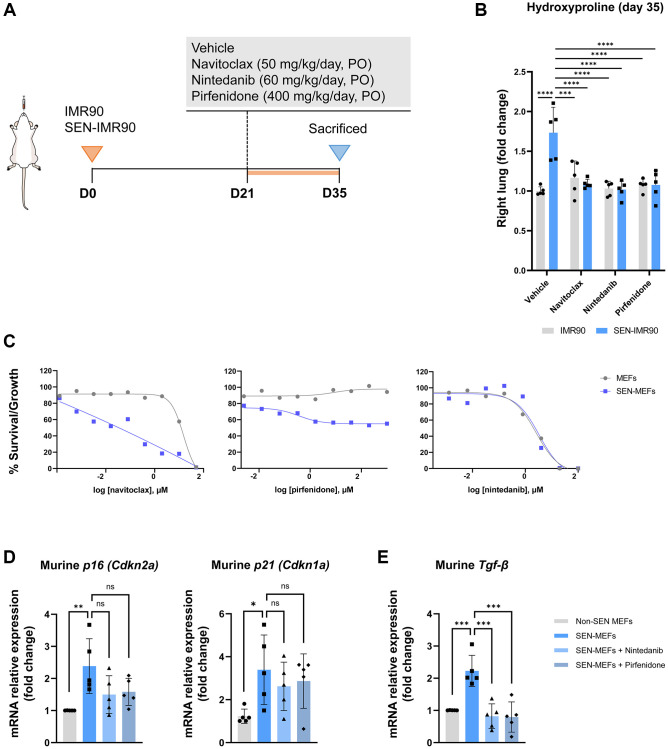
Cell senescence has recently emerged as a potentially relevant pathogenic mechanism in fibrosing interstitial lung diseases (f-ILDs), particularly in idiopathic pulmonary fibrosis. We hypothesized that senescent human fibroblasts may suffice to trigger a progressive fibrogenic reaction in the lung. To address this, senescent human lung fibroblasts, or their secretome (SASP), were instilled into the lungs of immunodeficient mice. We found that: (1) human senescent fibroblasts engraft in the lungs of immunodeficient mice and trigger progressive lung fibrosis associated to increasing levels of mouse senescent cells, whereas non-senescent fibroblasts do not trigger fibrosis; (2) the SASP of human senescent fibroblasts is pro-senescence and pro-fibrotic both in vitro when added to mouse recipient cells and in vivo when delivered into the lungs of mice, whereas the conditioned medium (CM) from non-senescent fibroblasts lacks these activities; and, (3) navitoclax, nintedanib and pirfenidone ameliorate lung fibrosis induced by senescent human fibroblasts in mice, albeit only navitoclax displayed senolytic activity. We conclude that human senescent fibroblasts, through their bioactive secretome, trigger a progressive fibrogenic reaction in the lungs of immunodeficient mice that includes the induction of paracrine senescence in the cells of the host, supporting the concept that senescent cells actively contribute to disease progression in patients with f-ILDs.

Keywords: antifibrotics; cellular senescence; mouse model; pulmonary fibrosis; senolytic.


Conflict of interest statement

CONFLICTS OF INTEREST: M.S. is a shareholder of Senolytic Therapeutics, Life Biosciences, Rejuveron Senescence Therapeutics and Altos Labs, and is an advisor of Rejuveron Senescence Therapeutics and Altos Labs. The funders had no role in the study design, data collection and analysis, decision to publish, or manuscript preparation. F.H.-G. and J.S. report honoraria for lectures, presentations, or educational events, and support for attending meetings and/or travel received from Roche and Boehringer Ingelheim (disclosures made outside the submitted work). F.P., N.P., V.R., J.A.L.-D., K.M., M.A., M.I.M.M., D.M., A.A., R.F. do not have conflicts of interest to declare in relation to this study.

Figures

Figure 1
Figure 1
Senescent human lung fibroblasts induce lung fibrosis in mice. (A) IMR90 lung fibroblasts were exposed to γ-irradiation (20 Gy). Fourteen days later, senescence was confirmed by SABG staining (scale bar, 100 μm). (B) Immunodeficient (nude) mice were randomized to receive intratracheal instillation of proliferating human lung fibroblasts (IMR90) or senescent IMR90 (SEN-IMR90). PBS was used as a negative control. (C) Representative images of lung sections stained with Hematoxylin Eosin (HE) and Masson’s Trichrome (MT) from mice injected with IMR90 cells, SEN-IMR90 cells or PBS at 21 days post-injection. Scale bar 100 μm). (D) Modified Ashcroft score of MT staining in sections from mice injected with IMR90 or SEN-IMR90 cells at 21 days post-injection; n = 5. These data are part of a larger experiment presented in Triana-Martinez F, et al. [27]. (E) Hydroxyproline content in the right lung tissue of mice injected with SEN-IMR90 compared with IMR90 group at 21 days post-injection; n = 5 (left panel). These data are part of a larger experiment presented in Triana-Martinez F, et al. [27]. Relative expression of the mRNA coding for Col6a3 relative to Actin-b levels in lung cell extracts from mice injected with IMR90 and SEN-IMR90 cells at 21 days post-injection; n = 5 (right panel). All values are expressed as fold change relative to IMR90 group. Statistical significance was assessed by U-Mann Whitney test: *p < 0.05, ****p < 0.0001. For further explanations, see text.
Figure 2
Figure 2
Senescent human lung fibroblasts infiltration and engraftment in mice lungs. (A) Images of lung sections showing IHC staining for CDKN1A/p21Cip1/Waf1 (left panel) and HuNu (right panel) from mice injected with SEN-IMR90 or control. Engraftment of senescent cells (arrows) in mice sacrificed after 3 and 48 hours post-transplantation. Positive cells were confirmed using histology at low magnification (20×, scale bar 100 μm), and high magnification (40×, orange box). (B) RT–PCR expression of CDKN1A/p21Cip1/Waf1 was measured relative to murine Actin-b to demonstrate the engraftment of SEN-IMR90 in the lungs of nude mice after 48 hours post-transplantation, and their presence at later endpoints compared to control; n = 3 each group. Statistical significance was assessed by the one-way ANOVA with Tukey test: ***p < 0.001. (C) Serial section and staining with antibodies against human CDKN1A/p21Cip1/Waf1 and Phospho-H2AX (γ-H2AX) to demonstrate senescence state of the engrafted cells. For further explanations, see text.
Figure 3
Figure 3
Lung fibrosis induced by senescent human lung fibroblasts is progressive. (A) Diagram showing the experimental plan to evaluate the dynamics of the development of pulmonary fibrosis in nude mice, as well as representative images of lung sections stained with Masson’s Trichrome (MT) (20×, scale bar 100 μm). These animals received intratracheal instillation of irradiated SEN-IMR90, compared with PBS-exposed mice at early endpoints, and bleomycin-challenged mice (single injection, dose of 3 UI/kg) at late endpoints; n = 3 each group. (B) Hydroxyproline content in the right lung of mice injected with SEN-IMR90 compared with control (PBS). (C) Relative expression of the mRNA coding for murine Col1a2 in the lungs of the same mice as in panel B. (D) Relative expression of the mRNA coding for murine Cdkn1a/p21Cip1/Waf1 in the same mice as in panel B. For panels B, C and D, n = 3 for each experimental group and statistical significance was assessed by the one-way ANOVA with Tukey test: **p < 0.01; *p < 0.05. For panels B, C and D, the group labelled SEN-IMR90 (2 months) is the same group labelled SEN-IMR90 in Supplementary Figure 1A–1C, and the data are the same. (E) Images of lung sections showing positive cells using IHC staining for HuNu and mouse p21 (Cdkn1a/p21Cip1/Waf1) from mice injected with SEN-IMR90 or control. Engraftment of senescent cells (arrows) in mice sacrificed after different time points (3 hours, 48 hours, and 1 month), showing their dramatic reduction after 48 hours post-transplantation, and the gradually increase of mouse p21 over time (20×, scale bar 100 μm).
Figure 4
Figure 4
The secretome of senescent human lung fibroblasts as mediator of murine lung fibrosis. (A) Diagram showing cytokine concentrations in conditioned media (CM) corresponding to 0.5 million cells collected from irradiated SEN-IMR90 (SEN-CM) compared with proliferating IMR90-derived CM (NS-CM) as control, were quantified by using human cytokine arrays; n = 4 each group, independent experiments. Statistical significance was assessed by the two-tailed Student’s test: *p < 0.05. (B) Mouse Embryo Fibroblasts (MEF) incubated with SEN-CM or NS-CM as control for 6 days. Senescence was confirmed by SABG staining (scale bar, 100 μm). (C) Transcriptional upregulation of the profibrotic secretome components (IL-6, Tgf-β and Col1a2) was confirmed by RT-PCR at 2 days post-exposure to the indicated CM; n = 6 SEN-CM and n = 3 NS-CM group, independent experiments. Statistical significance was assessed by the two-tailed Student’s test: *p < 0.05. (D) Representative immunofluorescence images showing double staining of Cdkn1a/p21Cip1/Waf1 (red), and α-smooth muscle actin (α-SMA) (green) (10×, scale bar, 100 μm). (E) Quantification of the average number of α-SMA/p21 double positive cells as observed in the images shown in panel D using ImageJ. Quantification was performed from 4 experiments with >25 cells quantified for each condition. Statistical significance was assessed by the two-tailed Student’s t-test: *p < 0.05. (F) Diagram showing the experimental plan to evaluate the effect of the secretome in nude mice lung. These animals were intratracheally delivered SEN-CM or NS-CM, normalized by the number of cells (corresponding to 5 x 10⁵ cells each group), using PBS as negative control; n = 10 each group. (G) Hydroxyproline content in the right lung tissues of mice injected with SEN-CM or NS-CM, compared with control; n = 10 each group. (H) Representative images of lung sections of nude mice 21 days after instillation of SEN-CM or NS-CM, or PBS as negative control, stained with Hematoxylin Eosin (HE) and Masson’s Trichrome (MT) (40×, scale bar 100 μm) showing that SEN-CM initiated a cascade of the events that induced mild fibrosis. Statistical significance was assessed by the one-way ANOVA with Tukey test: ***p < 0.001; *p < 0.05. For further explanations, see text.
Figure 5
Figure 5
Effects of antifibrotic and senolytic drugs. (A) Scheme showing the experimental design to assess the effect of antifibrotic or senolytic drugs. Nude mice were randomized after 21 days post-injection of irradiated SEN-IMR90 cells or IMR90 cells as negative control, to either the two approved antifibrotics drugs (nintedanib or pirfenidone), a senolytic drug (navitoclax), or vehicle, for two weeks. (B) Hydroxyproline content in the right lung tissues of mice treated with navitoclax, nintedanib or pirfenidone, compared with control; n = 5 each group. Statistical significance was assessed by the one-way ANOVA with Tukey test: ***p < 0.001; ****p < 0.0001. (C) Senolytic activity of navitoclax (left panel), pirfenidone (middle panel) or nintedanib (right panel). Diagram showing the senolytic activity of these agents after exposure of senescent MEFs (SEN-MEFs) or non-senescent MEFs (NS-MEFs) to increasing concentration of navitoclax, pirfenidone, nintedanib or vehicle for 72 hours, as confirmed by relative expression of the mRNA coding for murine senescence markers (Cdkn2a/p16INK4a and Cdkn1a/p21Cip1/Waf1), measured relative to Actin-b levels in lung cell extracts of nintedanib or pirfenidone group compared to control (D); n = 5 each group, independent experiments. Statistical significance was assessed by the one-way ANOVA with Tukey test: **p < 0.01; *p < 0.05. (E) Relative expression of the mRNA coding for Tgf-β (transforming growth factor-β) was measured relative to Actin-b levels in SEN-MEFs treated with pirfenidone or nintenadib, compared with control; n = 5 each group, independent experiments. Statistical significance was assessed by the one-way ANOVA with Tukey test: ***p < 0.001. For further explanations, see text.

References

    1. Wijsenbeek M, Cottin V. Spectrum of Fibrotic Lung Diseases. N Engl J Med. 2020; 383:958–68. 10.1056/NEJMra2005230 - DOI - PubMed
    1. Raghu G, Remy-Jardin M, Myers JL, Richeldi L, Ryerson CJ, Lederer DJ, Behr J, Cottin V, Danoff SK, Morell F, Flaherty KR, Wells A, Martinez FJ, et al., and American Thoracic Society, European Respiratory Society, Japanese Respiratory Society, and Latin American Thoracic Society. Diagnosis of Idiopathic Pulmonary Fibrosis. An Official ATS/ERS/JRS/ALAT Clinical Practice Guideline. Am J Respir Crit Care Med. 2018; 198:e44–68. 10.1164/rccm.201807-1255ST - DOI - PubMed
    1. Lederer DJ, Martinez FJ. Idiopathic Pulmonary Fibrosis. N Engl J Med. 2018; 378:1811–23. 10.1056/NEJMra1705751 - DOI - PubMed
    1. López-Otín C, Blasco MA, Partridge L, Serrano M, Kroemer G. The hallmarks of aging. Cell. 2013; 153:1194–217. 10.1016/j.cell.2013.05.039 - DOI - PMC - PubMed
    1. Faner R, Rojas M, Macnee W, Agustí A. Abnormal lung aging in chronic obstructive pulmonary disease and idiopathic pulmonary fibrosis. Am J Respir Crit Care Med. 2012; 186:306–13. 10.1164/rccm.201202-0282PP - DOI - PubMed

Publication types