Review
. 2023 Jul 1;21(1):60.
doi: 10.1186/s12958-023-01112-7.

Connecting G protein-coupled estrogen receptor biomolecular mechanisms with the pathophysiology of preeclampsia: a review

Affiliations
Review

Connecting G protein-coupled estrogen receptor biomolecular mechanisms with the pathophysiology of preeclampsia: a review

Allan Kardec Nogueira Alencar et al. Reprod Biol Endocrinol. .

Abstract

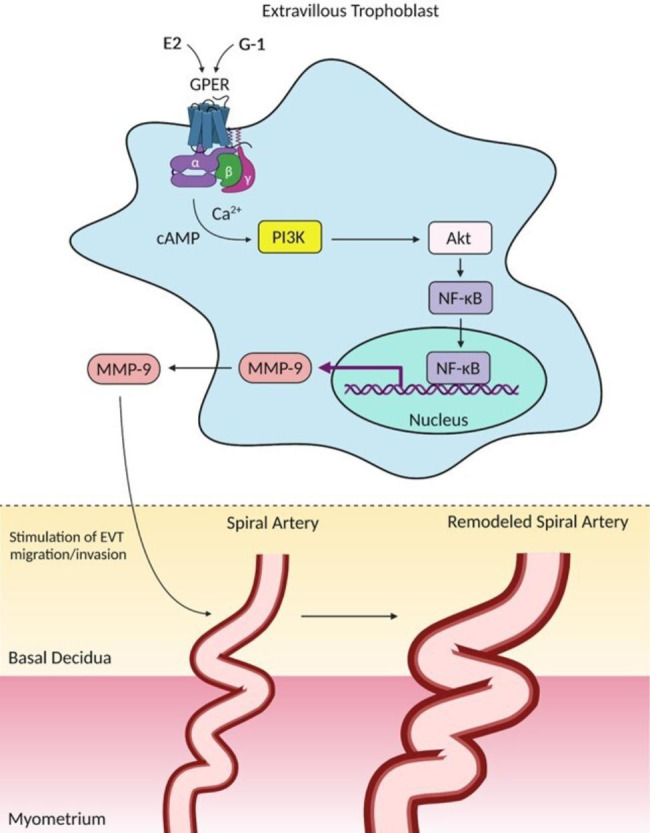
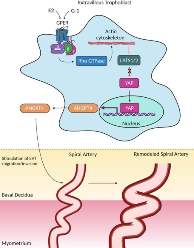
Background: Throughout the course of pregnancy, small maternal spiral arteries that are in contact with fetal tissue undergo structural remodeling, lose smooth muscle cells, and become less responsive to vasoconstrictors. Additionally, placental extravillous trophoblasts invade the maternal decidua to establish an interaction between the fetal placental villi with the maternal blood supply. When successful, this process enables the transport of oxygen, nutrients, and signaling molecules but an insufficiency leads to placental ischemia. In response, the placenta releases vasoactive factors that enter the maternal circulation and promote maternal cardiorenal dysfunction, a hallmark of preeclampsia (PE), the leading cause of maternal and fetal death. An underexplored mechanism in the development of PE is the impact of membrane-initiated estrogen signaling via the G protein-coupled estrogen receptor (GPER). Recent evidence indicates that GPER activation is associated with normal trophoblast invasion, placental angiogenesis/hypoxia, and regulation of uteroplacental vasodilation, and these mechanisms could explain part of the estrogen-induced control of uterine remodeling and placental development in pregnancy.

Conclusion: Although the relevance of GPER in PE remains speculative, this review provides a summary of our current understanding on how GPER stimulation regulates some of the features of normal pregnancy and a potential link between its signaling network and uteroplacental dysfunction in PE. Synthesis of this information will facilitate the development of innovative treatment options.

Keywords: Angiogenesis; Estrogen; Extravillous trophoblast; GPER; Hypoxia; Preeclampsia; Pregnancy; Spiral arteries; Uteroplacental circulation.


Conflict of interest statement

The authors declare that they have no competing interests.

Figures

Fig. 1
Fig. 1
Overview of GPER signaling involved in the modulation of EVT migration/invasion through the PI3K/Akt-MMP-9 axis. Pharmacological modulation of GPER by E2 or its selective agonist G-1 stimulates distinct subunits of heterotrimeric G proteins. Gαq and Gαs are examples of subunits stimulated by GPER, which augment the intracellular levels of second messengers (Ca2+ and cAMP) to promote activation of PI3K/Akt enzymes. Once activated, PI3K/Akt cascade triggers NF-κB translocation to the nucleus, where it encodes the synthesis of MMP-9, a downstream effector of GPER-regulated EVT cell migration/invasion and subsequent spiral artery remodeling. Additional mechanisms involved in the GPER/PI3K/Akt/MMP-9 downstream signaling pathway are provided in this figure and have been published elsewhere [140, 141]. GPER, G protein-coupled estrogen receptor; EVT, extravillous trophoblast; PI3K, phosphoinositide 3-kinase; Akt, protein kinase B; MMP-9, matrix metalloproteinase 9; E2, estrogen; cAMP, cyclic adenosine monophosphate; NF-κB, nuclear factor-κB. This artwork was created using the BioRender software
Fig. 2
Fig. 2
Molecular mechanisms by which GPER stimulates EVT migration/invasion through the Hippo pathway. When GPER is activated by E2 or G-1, its Gαq subunit stimulates Rho GTPase, which in turn causes actin cytoskeleton organization, a crucial regulator of the Hippo pathway. Actin cytoskeleton inhibits LATS1/2 activity, thus increasing the translocation of YAP protein to the nucleus. Disruption of actin cytoskeleton or inhibition of Rho GTPase facilitate the phosphorylation/activation of LATS1/2 and subsequent inhibition of YAP nuclear translocation and activity. This results in cytoplasmic retention of YAP and its proteolytic degradation. However, the inhibition of LATS1/2 by the actin cytoskeleton is a crucial mechanism responsible by YAP transcriptional activity in the nucleus, where this protein encodes the synthesis of ANGPTL4. When ANGPTL4 is produced, it modulates the EVT cell migration/invasion and subsequent spiral artery remodeling. Additional mechanisms involved in the GPER-induced ANGPTL4 synthesis by the Hippo pathway are provided in this figure and have been published elsewhere [142]. GPER, G protein-coupled estrogen receptor; EVT, extravillous trophoblast; Hippo pathway, Hippo tumor-suppressor pathway; LATS1/2, large tumor suppressor 1 and 2 kinases; YAP, Yes-associated protein; ANGPTL4, angiopoietin-like 4. This artwork was created using the BioRender software

References

    1. Abalos E, Cuesta C, Grosso AL, Chou D, Say L. Global and regional estimates of preeclampsia and eclampsia: a systematic review. Eur J Obstet Gynecol Reprod Biol. 2013;170(1):1–7. doi: 10.1016/j.ejogrb.2013.05.005. - DOI - PubMed
    1. Ananth CV, Keyes KM, Wapner RJ. Pre-eclampsia rates in the United States, 1980–2010: age-period-cohort analysis. BMJ. 2013;347:f6564. doi: 10.1136/bmj.f6564. - DOI - PMC - PubMed
    1. Duley L. The global impact of pre-eclampsia and eclampsia. Semin Perinatol. 2009;33(3):130–7. doi: 10.1053/j.semperi.2009.02.010. - DOI - PubMed
    1. Ananth CV, Lavery JA, Friedman AM, Wapner RJ, Wright JD. Serious maternal complications in relation to severe pre-eclampsia: a retrospective cohort study of the impact of hospital volume. BJOG. 2017;124(8):1246–53. doi: 10.1111/1471-0528.14384. - DOI - PubMed
    1. Hypertension in pregnancy Report of the American College of Obstetricians and Gynecologists’ Task Force on hypertension in pregnancy. Obstet Gynecol. 2013;122(5):1122–31. doi: 10.1097/01.AOG.0000437382.03963.88. - DOI - PubMed

LinkOut - more resources